|
Rectus Abdominis Muscle
The rectus abdominis muscle, () also known as the "abdominal muscle" or simply better known as the "abs", is a pair of segmented skeletal muscle on the ventral aspect of a person's abdomen. The paired muscle is separated at the midline by a band of dense connective tissue called the linea alba, and the connective tissue defining each lateral margin of the rectus abdominus is the linea semilunaris. The muscle extends from the pubic symphysis, pubic crest and pubic tubercle inferiorly, to the xiphoid process and costal cartilages of the 5th–7th ribs superiorly. The rectus abdominis muscle is contained in the rectus sheath, which consists of the aponeuroses of the lateral abdominal muscles. Each rectus abdominus is traversed by bands of connective tissue called the tendinous intersections, which interrupt it into distinct muscle bellies. Structure The rectus abdominis is a very long flat muscle, which extends along the whole length of the front of the abdomen, and is s ... [...More Info...] [...Related Items...] OR: [Wikipedia] [Google] [Baidu] |
Pubic Crest
Medial to the pubic tubercle is the pubic crest, which extends from this process to the medial end of the pubis (bone), pubic bone. It gives attachment to the conjoint tendon, the rectus abdominis, the abdominal external oblique muscle, and the pyramidalis muscle. The point of junction of the crest with the medial border of the bone is called the ''angle'' to it, as well as to the symphysis, the superior crus of the subcutaneous inguinal ring is attached. References External links * Bones of the pelvis Pubis (bone) {{musculoskeletal-stub ... [...More Info...] [...Related Items...] OR: [Wikipedia] [Google] [Baidu] |
Aponeurosis
An aponeurosis (; : aponeuroses) is a flattened tendon by which muscle attaches to bone or fascia. Aponeuroses exhibit an ordered arrangement of collagen fibres, thus attaining high tensile strength in a particular direction while being vulnerable to tensional or shear forces in other directions. They have a shiny, whitish-silvery color, are histologically similar to tendons, and are very sparingly supplied with blood vessels and nerves. When dissected, aponeuroses are papery and peel off by sections. The primary regions with thick aponeuroses are in the ventral abdominal region, the dorsal lumbar region, the ventriculus in birds, and the palmar (palms) and plantar (soles) regions. Anatomy Anterior abdominal aponeuroses The anterior abdominal aponeuroses are located just superficial to the rectus abdominis muscle. It has for its borders the external oblique, pectoralis muscles, and the latissimus dorsi. Posterior lumbar aponeuroses The posterior lumbar aponeuroses are sit ... [...More Info...] [...Related Items...] OR: [Wikipedia] [Google] [Baidu] |
Costoxiphoid Ligaments
The costoxiphoid ligaments (chondroxiphoid ligaments) are inconstant strand-like fibrous bands that connect the anterior and posterior surfaces of the seventh costal cartilage, and sometimes those of the sixth, to the front and back of the xiphoid process the sternum The sternum (: sternums or sterna) or breastbone is a long flat bone located in the central part of the chest. It connects to the ribs via cartilage and forms the front of the rib cage, thus helping to protect the heart, lungs, and major bl .... They vary in length and breadth in different subjects; those on the back of the joint are less distinct than those in front. References Ligaments of the torso {{ligament-stub ... [...More Info...] [...Related Items...] OR: [Wikipedia] [Google] [Baidu] |
Human Body
The human body is the entire structure of a Human, human being. It is composed of many different types of Cell (biology), cells that together create Tissue (biology), tissues and subsequently Organ (biology), organs and then Organ system, organ systems. The external human body consists of a human head, head, hair, neck, torso (which includes the thorax and abdomen), Sex organ, genitals, arms, Hand, hands, human leg, legs, and Foot, feet. The internal human body includes organs, Human tooth, teeth, bones, muscle, tendons, ligaments, blood vessels and blood, lymphatic vessels and lymph. The study of the human body includes anatomy, physiology, histology and embryology. The body Anatomical variation, varies anatomically in known ways. Physiology focuses on the systems and organs of the human body and their functions. Many systems and mechanisms interact in order to maintain homeostasis, with safe levels of substances such as sugar, iron, and oxygen in the blood. The body is st ... [...More Info...] [...Related Items...] OR: [Wikipedia] [Google] [Baidu] |
Sternalis Muscle
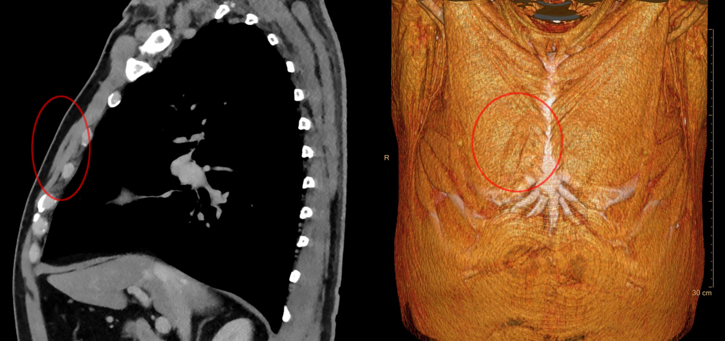
The rectus sternalis muscle is an anatomical variation that lies in front of the sternal end of the pectoralis major parallel to the margin of the sternum. The sternalis muscle may be a variation of the pectoralis major or of the rectus abdominis. Structure The sternalis is a muscle that runs along the anterior aspect of the body of the sternum. It lies superficially and parallel to the sternum. Its origin and insertion are variable. The sternalis muscle often originates from the upper part of the sternum and can display varying insertions such as the pectoral fascia, lower ribs, costal cartilages, rectus sheath, aponeurosis of the abdominal external oblique muscle. It may be present unilaterally or bilaterally. There is still a great deal of disagreement about its innervation and its embryonic origin. In a review, it was reported that the muscle was innervated by the external or internal thoracic nerves in 55% of the cases, by the intercostal nerves in 43% of the cases, whi ... [...More Info...] [...Related Items...] OR: [Wikipedia] [Google] [Baidu] |
Spinal Nerves
A spinal nerve is a mixed nerve, which carries motor, sensory, and autonomic signals between the spinal cord and the body. In the human body there are 31 pairs of spinal nerves, one on each side of the vertebral column. These are grouped into the corresponding cervical, thoracic, lumbar, sacral and coccygeal regions of the spine. There are eight pairs of cervical nerves, twelve pairs of thoracic nerves, five pairs of lumbar nerves, five pairs of sacral nerves, and one pair of coccygeal nerves. The spinal nerves are part of the peripheral nervous system. Structure Each spinal nerve is a mixed nerve, formed from the combination of nerve root fibers from its dorsal and ventral roots. The dorsal root is the afferent sensory root and carries sensory information to the brain. The ventral root is the efferent motor root and carries motor information from the brain. The spinal nerve emerges from the spinal column through an opening (intervertebral foramen) between adjacen ... [...More Info...] [...Related Items...] OR: [Wikipedia] [Google] [Baidu] |
Intercostal Nerves
The intercostal nerves are part of the somatic nervous system, and arise from the anterior rami of the thoracic spinal nerves from T1 to T11. The intercostal nerves are distributed chiefly to the thoracic pleura and abdominal peritoneum, and differ from the anterior rami of the other spinal nerves in that each pursues an independent course without plexus formation. The first two nerves supply fibers to the upper limb and thorax; the next four distribute to the walls of the thorax; the lower five supply the walls of the thorax and abdomen. The 7th intercostal nerve ends at the xyphoid process of the Human sternum, sternum. The 10th intercostal nerve terminates at the navel. The 12th (subcostal nerve, subcostal) thoracic is distributed to the walls of the abdomen and groin. Each of these fibers contains around 1300 axons. Unlike the nerves from the autonomic nervous system that innervate the visceral pleura of the thoracic cavity, the intercostal nerves arise from the somatic nerv ... [...More Info...] [...Related Items...] OR: [Wikipedia] [Google] [Baidu] |
Intercostal Artery
The intercostal arteries are a group of arteries passing within an intercostal space (the space between two adjacent ribs). There are 9 anterior and 11 posterior intercostal arteries on each side of the body. The anterior intercostal arteries are branches of the internal thoracic artery and its terminal branchthe musculophrenic artery. The posterior intercostal arteries are branches of the supreme intercostal artery and thoracic aorta. Each anterior intercostal artery anastomoses with the corresponding posterior intercostal artery arising from the thoracic aorta. Anterior intercostal arteries Origin The upper six anterior intercostal arteries are branches of the internal thoracic artery (anterior intercostal branches of internal thoracic artery). The internal thoracic artery then divides into its two terminal branches, one of which - the musculophrenic artery - proceeds to issue anterior intercostal arteries to the remaining 7th, 8th, and 9th intercostal spaces; th ... [...More Info...] [...Related Items...] OR: [Wikipedia] [Google] [Baidu] |
Internal Thoracic Artery
The internal thoracic artery (ITA), also known as the internal mammary artery, is an artery that supplies the anterior chest wall and the breasts. It is a paired artery, with one running along each side of the sternum, to continue after its bifurcation as the superior epigastric and musculophrenic arteries. Structure The internal thoracic artery arises from the anterior surface of the subclavian artery near its origin. It has a width of between 1-2 mm. It travels downward on the inside of the rib cage, approximately 1 cm from the sides of the sternum, and thus medial to the nipple. It is accompanied by the internal thoracic vein. It runs deep to the abdominal external oblique muscle, but superficial to the vagus nerve. In adults, the internal thoracic artery lies closest to the sternum at the first intercostal space. The gap between the artery and lateral border of the sternum increases when going downwards, up to 1.1 cm to 1.3 cm at the sixth intercost ... [...More Info...] [...Related Items...] OR: [Wikipedia] [Google] [Baidu] |
Superior Epigastric Artery
In human anatomy, the superior epigastric artery is a terminal branch of the internal thoracic artery that provides arterial supply to the abdominal wall, and upper rectus abdominis muscle. It enters the rectus sheath to descend upon the inner surface of the rectus abdominis muscle. It ends by anastomosing with the inferior epigastric artery. Structure Origin The superior epigastric artery arises from the internal thoracic artery (referred to as the internal mammary artery in the accompanying diagram). Course and relations The superior epigastric artery pierces the diaphragm to enter the rectus sheath and descend upon the deep surface of the rectus abdominis. Along its course, it is accompanied by a similarly named vein, the superior epigastric vein. Anastomoses It anastomoses with the inferior epigastric artery within the rectus abdominis muscle at the umbilicus. Distribution Where it anastomoses, the superior epigastric artery supplies the anterior par ... [...More Info...] [...Related Items...] OR: [Wikipedia] [Google] [Baidu] |
Arcuate Line (anterior Abdominal Wall)
The arcuate line of rectus sheath (the arcuate line or the semicircular line of Douglas) is a line of demarcation corresponding to the free inferior margin of the posterior layer of the rectus sheath inferior to which only the anterior layer of the rectus sheath is present and the rectus abdominis muscle is therefore in direct contact with the transversalis fascia. The arcuate line is concave inferior-wards. The arcuate line is visible upon the inner surface of the abdominal wall. The arcuate line may be a well-defined, or may be represented by a gradual waning of the aponeurotic fibres with concomitant increasing prominence of the transversalis fascia. The arcuate line occurs about midway between the umbilicus and pubic symphysis, however, this varies from person to person. The inferior epigastric artery and vein pass across the arcuate line to enter the rectus sheath. Anatomy Superior to the arcuate line, the internal oblique muscle aponeurosis splits to envelop the rec ... [...More Info...] [...Related Items...] OR: [Wikipedia] [Google] [Baidu] |


