|
Plesiopterys Wildi
''Plesiopterys'' (“plesio” meaning “near,” and “pterys” meaning “wing” or “pterygoid bone”) is an extinct genus of plesiosaur originating from the Posidonia Shale, Posidonienschiefer of Holzmaden, Germany, and lived during the Early Jurassic period. The type species, type and monotypic taxon, only species is ''P. wildi'', known from two immature specimens with the subadult measuring long. It possesses a unique combination of both primitive and derived characters, and is currently displayed at the State Museum of Natural History Stuttgart, State Museum of Natural History and the Hauff Museum, Germany. Discovery and naming In 2004, Frank Robin O'Keefe named the species ''Plesiopterys wildi''. The generic name is a combination of Greek ''plesios'', "near", and ''pterys'', "wing", the latter also referring to the pterygoid bones (apart from the fins). The specific name honors the German paleontologist Rupert Wild for his contributions to the Mesozoic vertebrate p ... [...More Info...] [...Related Items...] OR: [Wikipedia] [Google] [Baidu] |
Early Jurassic
The Early Jurassic Epoch (geology), Epoch (in chronostratigraphy corresponding to the Lower Jurassic series (stratigraphy), Series) is the earliest of three epochs of the Jurassic Period. The Early Jurassic starts immediately after the Triassic–Jurassic extinction event, 201.3 Ma (million years ago), and ends at the start of the Middle Jurassic 174.7 ±0.8 Ma. Certain rocks of marine origin of this age in Europe are called "Lias Group, Lias" and that name was used for the period, as well, in 19th-century geology. In southern Germany rocks of this age are called Black Jurassic. Origin of the name Lias There are two possible origins for the name Lias: the first reason is it was taken by a geologist from an England, English quarryman's dialect pronunciation of the word "layers"; secondly, sloops from north Cornwall, Cornish ports such as Bude would sail across the Bristol Channel to the Vale of Glamorgan to load up with rock from coastal limestone quarries (lias and Carbonif ... [...More Info...] [...Related Items...] OR: [Wikipedia] [Google] [Baidu] |
Dorsal Vertebrae
In vertebrates, thoracic vertebrae compose the middle segment of the vertebral column, between the cervical vertebrae and the lumbar vertebrae. In humans, there are twelve thoracic vertebra (anatomy), vertebrae of intermediate size between the cervical and lumbar vertebrae; they increase in size going towards the lumbar vertebrae. They are distinguished by the presence of Zygapophysial joint, facets on the sides of the bodies for Articulation (anatomy), articulation with the head of rib, heads of the ribs, as well as facets on the transverse processes of all, except the eleventh and twelfth, for articulation with the tubercle (rib), tubercles of the ribs. By convention, the human thoracic vertebrae are numbered T1–T12, with the first one (T1) located closest to the skull and the others going down the spine toward the lumbar region. General characteristics These are the general characteristics of the second through eighth thoracic vertebrae. The first and ninth through twelfth v ... [...More Info...] [...Related Items...] OR: [Wikipedia] [Google] [Baidu] |
Ichthyosaur
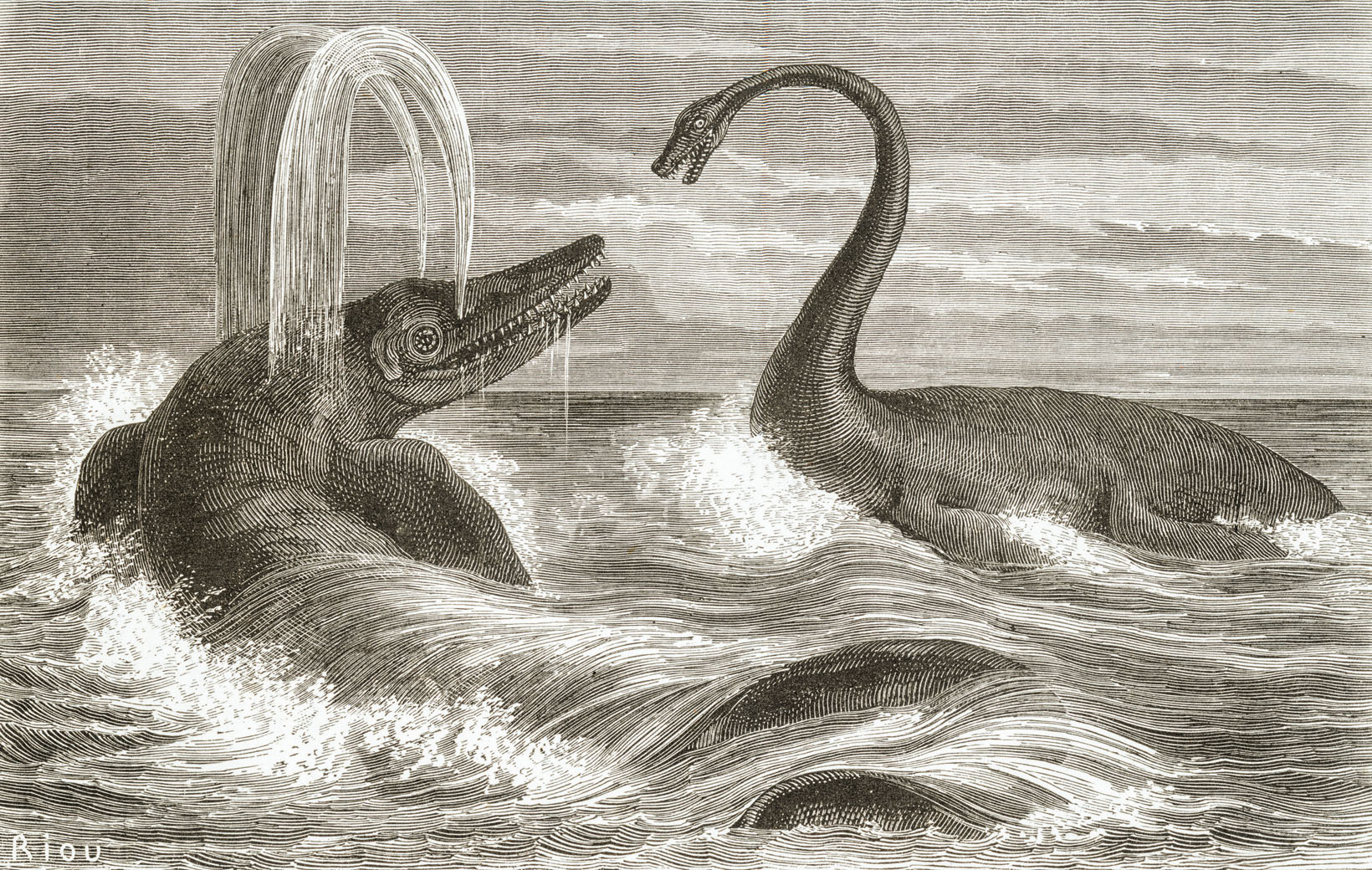
Ichthyosauria is an order of large extinct marine reptiles sometimes referred to as "ichthyosaurs", although the term is also used for wider clades in which the order resides. Ichthyosaurians thrived during much of the Mesozoic era; based on fossil evidence, they first appeared around 250 million years ago ( Ma) and at least one species survived until about 90 million years ago, into the Late Cretaceous. During the Early Triassic epoch, ichthyosaurs and other ichthyosauromorphs evolved from a group of unidentified land reptiles that returned to the sea, in a development similar to how the mammalian land-dwelling ancestors of modern-day dolphins and whales returned to the sea millions of years later, which they gradually came to resemble in a case of convergent evolution. Ichthyosaurians were particularly abundant in the Late Triassic and Early Jurassic periods, until they were replaced as the top aquatic predators by another marine reptilian group, the Plesiosauria, i ... [...More Info...] [...Related Items...] OR: [Wikipedia] [Google] [Baidu] |
Keratinocyte
Keratinocytes are the primary type of cell found in the epidermis, the outermost layer of the skin. In humans, they constitute 90% of epidermal skin cells. Basal cells in the basal layer (''stratum basale'') of the skin are sometimes referred to as basal keratinocytes. Keratinocytes form a barrier against environmental damage by heat, UV radiation, water loss, pathogenic bacteria, fungi, parasites, and viruses. A number of structural proteins, enzymes, lipids, and antimicrobial peptides contribute to maintain the important barrier function of the skin. Keratinocytes differentiate from epidermal stem cells in the lower part of the epidermis and migrate towards the surface, finally becoming corneocytes and eventually being shed, which happens every 40 to 56 days in humans. Function The primary function of keratinocytes is the formation of a barrier against environmental damage by heat, UV radiation, dehydration, pathogenic bacteria, fungi, parasites, and viruses. Pathoge ... [...More Info...] [...Related Items...] OR: [Wikipedia] [Google] [Baidu] |
Melanosome
A melanosome is an organelle found in animal cells and is the site for synthesis, storage and transport of melanin, the most common light-absorbing pigment found in the animal kingdom. Melanosomes are responsible for color and photoprotection in animal cells and tissues. Melanosomes are synthesised in the skin in melanocyte cells, as well as the eye in choroidal melanocytes and retinal pigment epithelial (RPE) cells. In lower vertebrates, they are found in melanophores or chromatophores. Structure Melanosomes are relatively large organelles, measuring up to 500 nm in diameter. They are bound by a bilipid membrane and are, in general, rounded, sausage-like, or cigar-like in shape. The shape is constant for a given species and cell type. They have a characteristic ultrastructure on electron microscopy, which varies according to the maturity of the melanosome, and for research purposes a numeric staging system is sometimes used. Synthesis of melanin Melanosomes are dep ... [...More Info...] [...Related Items...] OR: [Wikipedia] [Google] [Baidu] |
Flipper Scales Of MH 7
Flipper may refer to: Common meanings *Flipper (anatomy), a forelimb of an aquatic animal, useful for steering and/or propulsion in water *Swimfins, footwear that boosts human swimming efficiency, also known as flippers * Flipper (cricket), a type of delivery bowled by a wrist spin bowler *Flipper (pinball), a part of a pinball machine used to strike the ball *A speculator who engages in flipping (buying and selling quickly) * Flipper (tool), used for flipping food over while cooking Film and television * Flipper (franchise), a multimedia franchise about a bottlenose dolphin named Flipper ** ''Flipper'' (1963 film) ** ''Flipper's New Adventure'' (1964), sequel to the 1963 film ** ''Flipper'' (1996 film), a remake of the 1963 film starring Paul Hogan and Elijah Wood ** ''Flipper'' (1964 TV series), an adaptation of the 1963 film which originally ran from 1964 to 1967 ** ''Flipper'' (1995 TV series), a revival of the 1964 series which ran from 1995 to 2000 ** Flipper, one of th ... [...More Info...] [...Related Items...] OR: [Wikipedia] [Google] [Baidu] |
Brancasaurus
''Brancasaurus'' (meaning "Branca's lizard") is a genus of plesiosaur which lived in a freshwater lake in the Early Cretaceous of what is now North Rhine-Westphalia, Germany. With a long neck possessing vertebrae bearing distinctively-shaped "shark fin"-shaped vertebra#structure, neural spines, and a relatively small and pointed head, ''Brancasaurus'' is superficially similar to ''Elasmosaurus'', albeit smaller in size at in length as a subadult. The type species of this genus is ''Brancasaurus brancai'', first named by Theodor Wegner in 1914 in paleontology, 1914 in honor of German paleontology, paleontologist Wilhelm von Branca.Wegner, T.H. 1914. "''Brancasaurus brancai'' n. g. n. sp., ein Elasmosauride aus dem Wealden Westfalens". ''Festschrift für Wilhelm Branca zum 70. Geburtstage''. Borntraeger; Leipzig: pp. 235–305 Another plesiosaur named from the same region, ''Gronausaurus wegneri'', most likely represents a synonym (taxonomy), synonym of this genus. While tradition ... [...More Info...] [...Related Items...] OR: [Wikipedia] [Google] [Baidu] |
Neural Spine
Each vertebra (: vertebrae) is an irregular bone with a complex structure composed of bone and some hyaline cartilage, that make up the vertebral column or spine, of vertebrates. The proportions of the vertebrae differ according to their spinal segment and the particular species. The basic configuration of a vertebra varies; the vertebral body (also ''centrum'') is of bone and bears the load of the vertebral column. The upper and lower surfaces of the vertebra body give attachment to the intervertebral discs. The posterior part of a vertebra forms a vertebral arch, in eleven parts, consisting of two pedicles (pedicle of vertebral arch), two laminae, and seven processes. The laminae give attachment to the ligamenta flava (ligaments of the spine). There are vertebral notches formed from the shape of the pedicles, which form the intervertebral foramina when the vertebrae articulate. These foramina are the entry and exit conduits for the spinal nerves. The body of the vertebra and ... [...More Info...] [...Related Items...] OR: [Wikipedia] [Google] [Baidu] |
Ossification
Ossification (also called osteogenesis or bone mineralization) in bone remodeling is the process of laying down new bone material by cells named osteoblasts. It is synonymous with bone tissue formation. There are two processes resulting in the formation of normal, healthy bone tissue: Intramembranous ossification is the direct laying down of bone into the primitive connective tissue ( mesenchyme), while endochondral ossification involves cartilage as a precursor. In fracture healing, endochondral osteogenesis is the most commonly occurring process, for example in fractures of long bones treated by plaster of Paris, whereas fractures treated by open reduction and internal fixation with metal plates, screws, pins, rods and nails may heal by intramembranous osteogenesis. Heterotopic ossification is a process resulting in the formation of bone tissue that is often atypical, at an extraskeletal location. Calcification is often confused with ossification. Calcificatio ... [...More Info...] [...Related Items...] OR: [Wikipedia] [Google] [Baidu] |
Neural Arch
Each vertebra (: vertebrae) is an irregular bone with a complex structure composed of bone and some hyaline cartilage, that make up the vertebral column or spine, of vertebrates. The proportions of the vertebrae differ according to their spinal segment and the particular species. The basic configuration of a vertebra varies; the vertebral body (also ''centrum'') is of bone and bears the load of the vertebral column. The upper and lower surfaces of the vertebra body give attachment to the intervertebral discs. The posterior part of a vertebra forms a vertebral arch, in eleven parts, consisting of two pedicles (pedicle of vertebral arch), two laminae, and seven processes. The laminae give attachment to the ligamenta flava (ligaments of the spine). There are vertebral notches formed from the shape of the pedicles, which form the intervertebral foramina when the vertebrae articulate. These foramina are the entry and exit conduits for the spinal nerves. The body of the vertebra and ... [...More Info...] [...Related Items...] OR: [Wikipedia] [Google] [Baidu] |
Suture (anatomy)
In anatomy, a suture is a fairly rigid joint between two or more hard elements of an organism, with or without significant overlap of the elements. Sutures are found in the skeletons or exoskeletons of a wide range of animals, in both invertebrates and vertebrates. Sutures are found in animals with hard parts from the Cambrian period to the present day. Sutures were and are formed by several different methods, and they exist between hard parts that are made from several different materials. Vertebrate skeletons The skeletons of vertebrate animals (fish, amphibians, reptiles, birds, and mammals) are made of bone, in which the main rigid ingredient is calcium phosphate. Cranial sutures The skulls of most vertebrates consist of sets of bony plates held together by cranial sutures. These sutures are held together mainly by Sharpey's fibers which grow from each bone into the adjoining one. Sutures in the ankles of land vertebrates In the type of crurotarsal ankle, which is fo ... [...More Info...] [...Related Items...] OR: [Wikipedia] [Google] [Baidu] |
Internal Carotid Artery
The internal carotid artery is an artery in the neck which supplies the anterior cerebral artery, anterior and middle cerebral artery, middle cerebral circulation. In human anatomy, the internal and external carotid artery, external carotid arise from the common carotid artery, where it bifurcates at cervical vertebrae C3 or C4. The internal carotid artery supplies the brain, including the eyes, while the external carotid nourishes other portions of the head, such as the face, scalp, skull, and meninges. Classification Terminologia Anatomica in 1998 subdivided the artery into four parts: "cervical", "petrous", "cavernous", and "cerebral". In clinical settings, however, usually the classification system of the internal carotid artery follows the 1996 recommendations by Bouthillier, describing seven anatomical segments of the internal carotid artery, each with a corresponding alphanumeric identifier: C1 cervical; C2 petrous; C3 lacerum; C4 cavernous; C5 clinoid; C6 ophthalmic; ... [...More Info...] [...Related Items...] OR: [Wikipedia] [Google] [Baidu] |


