|
Pineoblastoma
Pineoblastoma is a malignant tumor of the pineal gland. A pineoblastoma is a supratentorial midline primitive neuroectodermal tumor. Pineoblastoma can present at any age, but is most common in young children. They account for 0.001% of all primary CNS neoplasms. Epidemiology Pineoblastomas typically occur at very young ages. One study found the average age of presentation to be 4.3 years, with peaks at age 3 and 8. Another cites cases to more commonly occur in patients under 2 years of age. Rates of occurrence for males and females are similar, but may be slightly more common in females. One study found incidence of pineoblastoma to be increased in black patients compared to white patients by around 71%. This difference was most apparent in patients aged 5 to 9 years old. Pathophysiology The pineal gland is a small organ in the center of the brain that is responsible for controlling melatonin secretion. Several tumors can occur in the area of the pineal gland, with the most a ... [...More Info...] [...Related Items...] OR: [Wikipedia] [Google] [Baidu] |
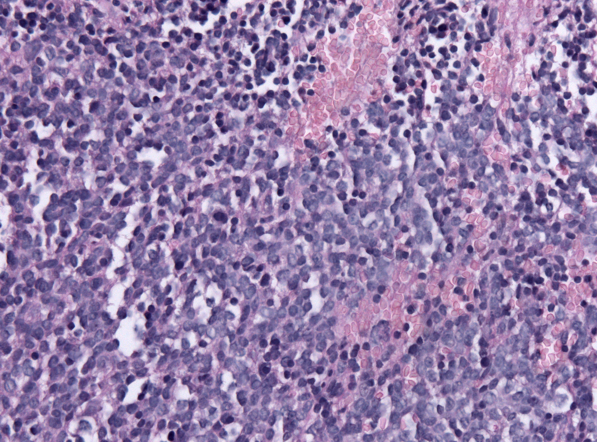
Pineoblastoma HE X200
Pineoblastoma is a malignant tumor of the pineal gland. A pineoblastoma is a supratentorial midline primitive neuroectodermal tumor. Pineoblastoma can present at any age, but is most common in young children. They account for 0.001% of all primary CNS neoplasms. Epidemiology Pineoblastomas typically occur at very young ages. One study found the average age of presentation to be 4.3 years, with peaks at age 3 and 8. Another cites cases to more commonly occur in patients under 2 years of age. Rates of occurrence for males and females are similar, but may be slightly more common in females. One study found incidence of pineoblastoma to be increased in black patients compared to white patients by around 71%. This difference was most apparent in patients aged 5 to 9 years old. Pathophysiology The pineal gland is a small organ in the center of the brain that is responsible for controlling melatonin secretion. Several tumors can occur in the area of the pineal gland, with the most ... [...More Info...] [...Related Items...] OR: [Wikipedia] [Google] [Baidu] |
Primitive Neuroectodermal Tumor
Primitive neuroectodermal tumor is a malignant (cancerous) neural crest tumor. It is a rare tumor, usually occurring in children and young adults under 25 years of age. The overall 5 year survival rate is about 53%. It gets its name because the majority of the cells in the tumor are derived from neuroectoderm, but have not developed and differentiated in the way a normal neuron would, and so the cells appear "primitive".PNET belongs to the Ewing family of tumors. Genetics Using gene transfer of SV40 large T-antigen in neuronal precursor cells of rats, a brain tumor model was established. The PNETs were histologically indistinguishable from the human counterparts and have been used to identify new genes involved in human brain tumor carcinogenesis. The model was used to confirm p53 as one of the genes involved in human medulloblastomas, but since only about 10% of the human tumors showed mutations in that gene, the model can be used to identify the other binding partners of SV40 ... [...More Info...] [...Related Items...] OR: [Wikipedia] [Google] [Baidu] |
Trilateral Retinoblastoma
Trilateral retinoblastoma is a malignant midline primitive neuroectodermal tumor occurring in patients with inherited uni- or bilateral retinoblastoma. In most cases trilateral retinoblastoma presents itself as pineoblastoma (pineal TRb). In about a fourth of the cases the tumor develops in another intracranial region, most commonly supra- or parasellar (non-pineal TRb), but there are reported cases with non-pineal TRb in the 3rd ventricle. In most cases pineal TRb is diagnosed before the age of 5, but after the diagnosis of retinoblastoma. Non-pineal TRb, however, is often diagnosed simultaneous with retinoblastoma. Prognosis of patients with trilateral retinoblastoma is dismal, only a few patients have survived more than 5 years after diagnosis; all survivors were diagnosed with small tumors in a subclinical stage. Recent advances in (high-dose) chemotherapy treatment regimens and early detection have improved survival of patients with trilateral retinoblastoma. See also * Prim ... [...More Info...] [...Related Items...] OR: [Wikipedia] [Google] [Baidu] |
Glia
Glia, also called glial cells (gliocytes) or neuroglia, are non-neuronal cells in the central nervous system (brain and spinal cord) and the peripheral nervous system that do not produce electrical impulses. They maintain homeostasis, form myelin in the peripheral nervous system, and provide support and protection for neurons. In the central nervous system, glial cells include oligodendrocytes, astrocytes, ependymal cells, and microglia, and in the peripheral nervous system they include Schwann cells and satellite cells. Function They have four main functions: *to surround neurons and hold them in place *to supply nutrients and oxygen to neurons *to insulate one neuron from another *to destroy pathogens and remove dead neurons. They also play a role in neurotransmission and synaptic connections, and in physiological processes such as breathing. While glia were thought to outnumber neurons by a ratio of 10:1, recent studies using newer methods and reappraisal of historical qua ... [...More Info...] [...Related Items...] OR: [Wikipedia] [Google] [Baidu] |
Homer Wright Pseudorosettes
In histopathology, a palisade is a single layer of relatively long cells, arranged loosely perpendicular to a surface and parallel to each other. A rosette is a palisade in a halo or spoke-and-wheel arrangement, surrounding a central core or hub. A pseudorosette is a perivascular radial arrangement of neoplastic cells around a small blood vessel. Rosette A ''rosette'' is a cell formation in a halo or spoke-and-wheel arrangement, surrounding a central core or hub. The central hub may consist of an empty-appearing lumen or a space filled with cytoplasmic processes. The cytoplasm of each of the cells in the rosette is often wedge-shaped with the apex directed toward the central core: the nuclei of the cells participating in the rosette are peripherally positioned and form a ring or halo around the hub. Pathogenesis Rosettes may be considered primary or secondary manifestations of tumor architecture. Primary rosettes form as a characteristic growth pattern of a given tumor type wh ... [...More Info...] [...Related Items...] OR: [Wikipedia] [Google] [Baidu] |
Immunohistochemistry
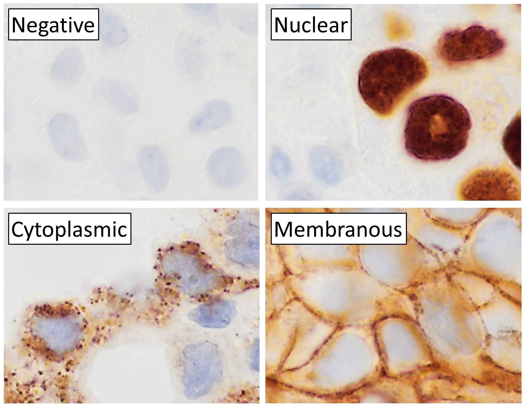
Immunohistochemistry (IHC) is the most common application of immunostaining. It involves the process of selectively identifying antigens (proteins) in cells of a tissue section by exploiting the principle of antibodies binding specifically to antigens in biological tissues. IHC takes its name from the roots "immuno", in reference to antibodies used in the procedure, and "histo", meaning tissue (compare to immunocytochemistry). Albert Coons conceptualized and first implemented the procedure in 1941. Visualising an antibody-antigen interaction can be accomplished in a number of ways, mainly either of the following: * ''Chromogenic immunohistochemistry'' (CIH), wherein an antibody is conjugated to an enzyme, such as peroxidase (the combination being termed immunoperoxidase), that can catalyse a colour-producing reaction. * '' Immunofluorescence'', where the antibody is tagged to a fluorophore, such as fluorescein or rhodamine. Immunohistochemical staining is widely used in t ... [...More Info...] [...Related Items...] OR: [Wikipedia] [Google] [Baidu] |
Neuronal Lineage Marker
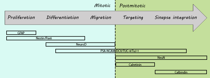
A neuronal lineage marker is an endogenous tag that is expressed in different cells along neurogenesis and differentiated cells such as neurons. It allows detection and identification of cells by using different techniques. A neuronal lineage marker can be either DNA, mRNA or RNA expressed in a cell of interest. It can also be a protein tag, as a partial protein, a protein or an epitope that discriminates between different cell types or different states of a common cell. An ideal marker is specific to a given cell type in normal conditions and/or during injury. Cell markers are very valuable tools for examining the function of cells in normal conditions as well as during disease. The discovery of various proteins specific to certain cells led to the production of cell-type-specific antibodies that have been used to identify cells. The techniques used for its detection can be immunohistochemistry, immunocytochemistry, methods that utilize transcriptional modulators and site-specific ... [...More Info...] [...Related Items...] OR: [Wikipedia] [Google] [Baidu] |
Synaptophysin
Synaptophysin, also known as the major synaptic vesicle protein p38, is a protein that in humans is encoded by the ''SYP'' gene. Genomics The gene is located on the short arm of X chromosome (Xp11.23-p11.22). It is 12,406 bases in length and lies on the minus strand. The encoded protein has 313 amino acids with a predicted molecular weight of 33.845 kDa. Molecular biology The protein is a synaptic vesicle glycoprotein with four transmembrane domains weighing 38kDa. It is present in neuroendocrine cells and in virtually all neurons in the brain and spinal cord that participate in synaptic transmission. It acts as a marker for neuroendocrine tumors, and its ubiquity at the synapse has led to the use of synaptophysin immunostaining for quantification of synapses. The exact function of the protein is unknown: it interacts with the essential synaptic vesicle protein synaptobrevin, but when the synaptophysin gene is experimentally inactivated in animals, they still develop a ... [...More Info...] [...Related Items...] OR: [Wikipedia] [Google] [Baidu] |
Photoreceptor Cell
A photoreceptor cell is a specialized type of neuroepithelial cell found in the retina that is capable of visual phototransduction. The great biological importance of photoreceptors is that they convert light (visible electromagnetic radiation) into signals that can stimulate biological processes. To be more specific, photoreceptor proteins in the cell absorb photons, triggering a change in the cell's membrane potential. There are currently three known types of photoreceptor cells in mammalian eyes: rods, cones, and intrinsically photosensitive retinal ganglion cells. The two classic photoreceptor cells are rods and cones, each contributing information used by the visual system to form an image of the environment, sight. Rods primarily mediate scotopic vision (dim conditions) whereas cones primarily mediate to photopic vision (bright conditions), but the processes in each that supports phototransduction is similar. A third class of mammalian photoreceptor cell was disc ... [...More Info...] [...Related Items...] OR: [Wikipedia] [Google] [Baidu] |
Contrast Enhancement
A contrast agent (or contrast medium) is a substance used to increase the contrast of structures or fluids within the body in medical imaging. Contrast agents absorb or alter external electromagnetism or ultrasound, which is different from radiopharmaceuticals, which emit radiation themselves. In x-ray imaging, contrast agents enhance the radiodensity in a target tissue or structure. In magnetic resonance imaging, contrast agents shorten (or in some instances increase) the relaxation times of nuclei within body tissues in order to alter the contrast in the image. Contrast agents are commonly used to improve the visibility of blood vessels and the gastrointestinal tract. The types of contrast agent are classified according to their intended imaging modalities. Radiocontrast media For radiography, which is based on X-rays, iodine and barium are the most common types of contrast agent. Various sorts of iodinated contrast agents exist, with variations occurring between the osmolari ... [...More Info...] [...Related Items...] OR: [Wikipedia] [Google] [Baidu] |
Neurofilament
Neurofilaments (NF) are classed as type IV intermediate filaments found in the cytoplasm of neurons. They are protein polymers measuring 10 nm in diameter and many micrometers in length. Together with microtubules (~25 nm) and microfilaments (7 nm), they form the neuronal cytoskeleton. They are believed to function primarily to provide structural support for axons and to regulate axon diameter, which influences nerve conduction velocity. The proteins that form neurofilaments are members of the intermediate filament protein family, which is divided into six types based on their gene organization and protein structure. Types I and II are the keratins which are expressed in epithelia. Type III contains the proteins vimentin, desmin, peripherin and glial fibrillary acidic protein (GFAP). Type IV consists of the neurofilament proteins L, M, H and internexin. Type V consists of the nuclear lamins, and type VI consists of the protein nestin. The type IV interm ... [...More Info...] [...Related Items...] OR: [Wikipedia] [Google] [Baidu] |


