|
Phosducin Family
The outer and inner segments of vertebrate retina rod photoreceptor cells contain phosducin, a soluble phosphoprotein that complexes with the beta/gamma-subunits of the guanosine triphosphate-binding protein, transducin. Light-induced changes in cyclic nucleotide levels modulate the phosphorylation of phosducin by protein kinase A. The protein is thought to participate in the regulation of visual phototransduction or in the integration of photoreceptor metabolism. Similar proteins have been isolated from the pineal gland The pineal gland, conarium, or epiphysis cerebri, is a small endocrine gland in the brain of most vertebrates. The pineal gland produces melatonin, a serotonin-derived hormone which modulates sleep, sleep patterns in both circadian rhythm, circ ... and it is believed that the function of the protein is the same in both retina and pineal gland. Human proteins containing this domain References {{DEFAULTSORT:Phosducin Family Protein domains Protein f ... [...More Info...] [...Related Items...] OR: [Wikipedia] [Google] [Baidu] |
Transducin
Transducin (Gt) is a protein naturally expressed in vertebrate retina rods and cones and it is very important in vertebrate phototransduction. It is a type of heterotrimeric G-protein with different α subunits in rod and cone photoreceptors. Light leads to conformational changes in rhodopsin, which in turn leads to the activation of transducin. Transducin activates phosphodiesterase, which results in the breakdown of cyclic guanosine monophosphate (cGMP). The intensity of the flash response is directly proportional to the number of transducin activated. Function in phototransduction Transducin is activated by metarhodopsin II, a conformational change in rhodopsin caused by the absorption of a photon by the rhodopsin moiety retinal. The light causes isomerization of retinal from 11-cis to all-trans. Isomerization causes a change in the opsin to become metarhodopsin II. When metarhodopsin activates transducin, the guanosine diphosphate (GDP) bound to the α subunit (Tα) is ex ... [...More Info...] [...Related Items...] OR: [Wikipedia] [Google] [Baidu] |
Retina
The retina (from la, rete "net") is the innermost, light-sensitive layer of tissue of the eye of most vertebrates and some molluscs. The optics of the eye create a focused two-dimensional image of the visual world on the retina, which then processes that image within the retina and sends nerve impulses along the optic nerve to the visual cortex to create visual perception. The retina serves a function which is in many ways analogous to that of the film or image sensor in a camera. The neural retina consists of several layers of neurons interconnected by synapses and is supported by an outer layer of pigmented epithelial cells. The primary light-sensing cells in the retina are the photoreceptor cells, which are of two types: rods and cones. Rods function mainly in dim light and provide monochromatic vision. Cones function in well-lit conditions and are responsible for the perception of colour through the use of a range of opsins, as well as high-acuity vision used f ... [...More Info...] [...Related Items...] OR: [Wikipedia] [Google] [Baidu] |
Photoreceptor Cell
A photoreceptor cell is a specialized type of neuroepithelial cell found in the retina that is capable of visual phototransduction. The great biological importance of photoreceptors is that they convert light (visible electromagnetic radiation) into signals that can stimulate biological processes. To be more specific, photoreceptor proteins in the cell absorb photons, triggering a change in the cell's membrane potential. There are currently three known types of photoreceptor cells in mammalian eyes: rods, cones, and intrinsically photosensitive retinal ganglion cells. The two classic photoreceptor cells are rods and cones, each contributing information used by the visual system to form an image of the environment, sight. Rods primarily mediate scotopic vision (dim conditions) whereas cones primarily mediate to photopic vision (bright conditions), but the processes in each that supports phototransduction is similar. A third class of mammalian photoreceptor cell was disc ... [...More Info...] [...Related Items...] OR: [Wikipedia] [Google] [Baidu] |
Phosducin
Phosducin, also known as PDC, is a human protein and gene. It belongs to the phosducin family of proteins. This gene encodes a phosphoprotein, which is located in the outer and inner segments of the rod cells in the retina. This protein may participate in the regulation of visual phototransduction or in the integration of photoreceptor metabolism. It modulates the phototransduction cascade by interacting with the beta and gamma subunits of the retinal G-protein transducin. By associating with these subunits only, the Transducin alpha subunit will remain active for longer. This will increase the amount of time of visual excitation. This gene is a potential candidate gene for retinitis pigmentosa and Usher syndrome type II. Alternatively spliced transcript variants encoding different isoforms have been identified. See also *Phosducin family The outer and inner segments of vertebrate retina rod photoreceptor cells contain phosducin, a soluble phosphoprotein that complexes wi ... [...More Info...] [...Related Items...] OR: [Wikipedia] [Google] [Baidu] |
Phosphoprotein
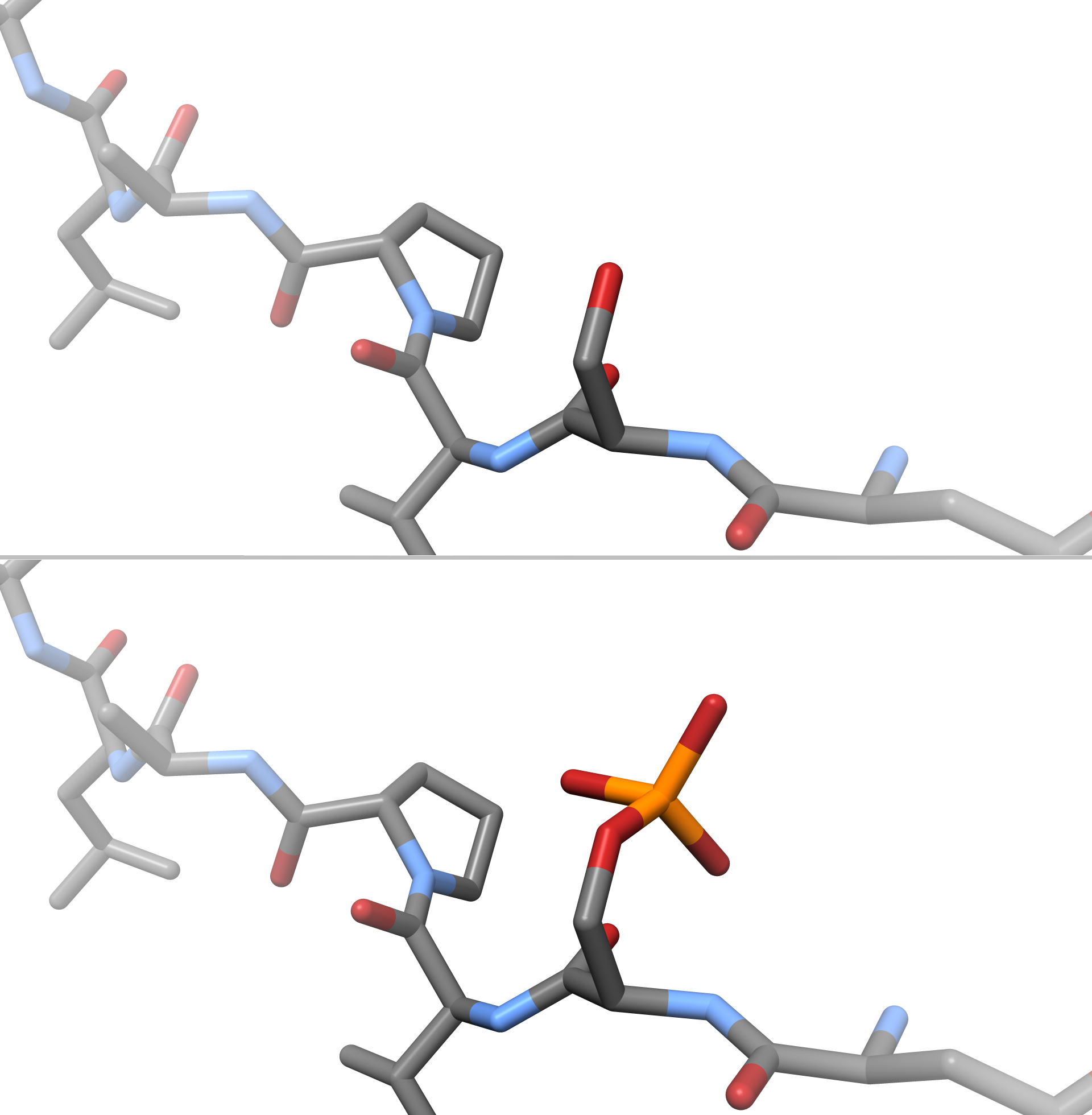
A phosphoprotein is a protein that is posttranslationally modified by the attachment of either a single phosphate group, or a complex molecule such as 5'-phospho-DNA, through a phosphate group. The target amino acid is most often serine, threonine, or tyrosine residues (mostly in eukaryotes), or aspartic acid or histidine residues (mostly in prokaryotes). Biological function The phosphorylation of proteins is a major regulatory mechanism in cells. Clinical significance Phosphoproteins have been proposed as biomarkers for breast cancer. See also *Protein phosphorylation Protein phosphorylation is a reversible post-translational modification of proteins in which an amino acid residue is phosphorylated by a protein kinase by the addition of a covalently bound phosphate group. Phosphorylation alters the structura ... References Phosphoproteins {{protein-stub ... [...More Info...] [...Related Items...] OR: [Wikipedia] [Google] [Baidu] |
Guanosine Triphosphate
Guanosine-5'-triphosphate (GTP) is a purine nucleoside triphosphate. It is one of the building blocks needed for the synthesis of RNA during the transcription process. Its structure is similar to that of the guanosine nucleoside, the only difference being that nucleotides like GTP have phosphates on their ribose sugar. GTP has the guanine nucleobase attached to the 1' carbon of the ribose and it has the triphosphate moiety attached to ribose's 5' carbon. It also has the role of a source of energy or an activator of substrates in metabolic reactions, like that of ATP, but more specific. It is used as a source of energy for protein synthesis and gluconeogenesis. GTP is essential to signal transduction, in particular with G-proteins, in second-messenger mechanisms where it is converted to guanosine diphosphate (GDP) through the action of GTPases. Uses Energy transfer GTP is involved in energy transfer within the cell. For instance, a GTP molecule is generated by o ... [...More Info...] [...Related Items...] OR: [Wikipedia] [Google] [Baidu] |
Transducin
Transducin (Gt) is a protein naturally expressed in vertebrate retina rods and cones and it is very important in vertebrate phototransduction. It is a type of heterotrimeric G-protein with different α subunits in rod and cone photoreceptors. Light leads to conformational changes in rhodopsin, which in turn leads to the activation of transducin. Transducin activates phosphodiesterase, which results in the breakdown of cyclic guanosine monophosphate (cGMP). The intensity of the flash response is directly proportional to the number of transducin activated. Function in phototransduction Transducin is activated by metarhodopsin II, a conformational change in rhodopsin caused by the absorption of a photon by the rhodopsin moiety retinal. The light causes isomerization of retinal from 11-cis to all-trans. Isomerization causes a change in the opsin to become metarhodopsin II. When metarhodopsin activates transducin, the guanosine diphosphate (GDP) bound to the α subunit (Tα) is ex ... [...More Info...] [...Related Items...] OR: [Wikipedia] [Google] [Baidu] |
Phosphorylation
In chemistry, phosphorylation is the attachment of a phosphate group to a molecule or an ion. This process and its inverse, dephosphorylation, are common in biology and could be driven by natural selection. Text was copied from this source, which is available under a Creative Commons Attribution 4.0 International License. Protein phosphorylation often activates (or deactivates) many enzymes. Glucose Phosphorylation of sugars is often the first stage in their catabolism. Phosphorylation allows cells to accumulate sugars because the phosphate group prevents the molecules from diffusing back across their transporter. Phosphorylation of glucose is a key reaction in sugar metabolism. The chemical equation for the conversion of D-glucose to D-glucose-6-phosphate in the first step of glycolysis is given by :D-glucose + ATP → D-glucose-6-phosphate + ADP :ΔG° = −16.7 kJ/mol (° indicates measurement at standard condition) Hepatic cells are freely permeable to glucose, an ... [...More Info...] [...Related Items...] OR: [Wikipedia] [Google] [Baidu] |
Protein Kinase A
In cell biology, protein kinase A (PKA) is a family of enzymes whose activity is dependent on cellular levels of cyclic AMP (cAMP). PKA is also known as cAMP-dependent protein kinase (). PKA has several functions in the cell, including regulation of glycogen, sugar, and lipid metabolism. It should not be confused with 5'-AMP-activated protein kinase ( AMP-activated protein kinase). History Protein kinase A, more precisely known as adenosine 3',5'-monophosphate (cyclic AMP)-dependent protein kinase, abbreviated to PKA, was discovered by chemists Edmond H. Fischer and Edwin G. Krebs in 1968. They won the Nobel Prize in Physiology or Medicine in 1992 for their work on phosphorylation and dephosphorylation and how it relates to PKA activity. PKA is one of the most widely researched protein kinases, in part because of its uniqueness; out of 540 different protein kinase genes that make up the human kinome, only one other protein kinase, casein kinase 2, is known to exist in a p ... [...More Info...] [...Related Items...] OR: [Wikipedia] [Google] [Baidu] |
Pineal Gland
The pineal gland, conarium, or epiphysis cerebri, is a small endocrine gland in the brain of most vertebrates. The pineal gland produces melatonin, a serotonin-derived hormone which modulates sleep patterns in both circadian and seasonal cycles. The shape of the gland resembles a pine cone, which gives it its name. The pineal gland is located in the epithalamus, near the center of the brain, between the two hemispheres, tucked in a groove where the two halves of the thalamus join. The pineal gland is one of the neuroendocrine secretory circumventricular organs in which capillaries are mostly permeable to solutes in the blood. Nearly all vertebrate species possess a pineal gland. The most important exception is a primitive vertebrate, the hagfish. Even in the hagfish, however, there may be a "pineal equivalent" structure in the dorsal diencephalon. The lancelet '' Branchiostoma lanceolatum'', an early chordate which is a close relative to vertebrates, also lacks a ... [...More Info...] [...Related Items...] OR: [Wikipedia] [Google] [Baidu] |
Phosducin-like
Phosducin-like protein is a protein that in humans is encoded by the ''PDCL'' gene. Phosducin-like protein is a putative modulator of heterotrimeric G proteins. The protein shares extensive amino acid sequence homology with phosducin, a phosphoprotein expressed in the retina and pineal gland The pineal gland, conarium, or epiphysis cerebri, is a small endocrine gland in the brain of most vertebrates. The pineal gland produces melatonin, a serotonin-derived hormone which modulates sleep, sleep patterns in both circadian rhythm, circ .... Both phosducin-like protein and phosphoducin have been shown to regulate G-protein signaling by binding to the beta-gamma subunits of G proteins. References Further reading * * * * * * * * * * * * * * * * * * * {{gene-9-stub ... [...More Info...] [...Related Items...] OR: [Wikipedia] [Google] [Baidu] |
PDCL2
Palladium(II) chloride, also known as palladium dichloride and palladous chloride, are the chemical compounds with the formula PdCl2. PdCl2 is a common starting material in palladium chemistry – palladium-based catalysts are of particular value in organic synthesis. It is prepared by the reaction of chlorine with palladium metal at high temperatures. Structure Two forms of PdCl2 are known, denoted α and β. In both forms, the palladium centres adopt a square-planar coordination geometry that is characteristic of Pd(II). Furthermore, in both forms, the Pd(II) centers are linked by μ2-chloride bridges. The α-form of PdCl2 is a polymer, consisting of "infinite" slabs or chains. The β-form of PdCl2 is molecular, consisting of an octahedral cluster of six Pd atoms. Each of the twelve edges of this octahedron is spanned by Cl−. PtCl2 adopts similar structures, whereas NiCl2 adopts the CdCl2 motif, featuring hexacoordinated Ni(II). Two further polymorphs, γ-PdCl2 ... [...More Info...] [...Related Items...] OR: [Wikipedia] [Google] [Baidu] |


-chloride-xtal-3D-balls.png)