|
Persistent Tunica Vasculosa Lentis
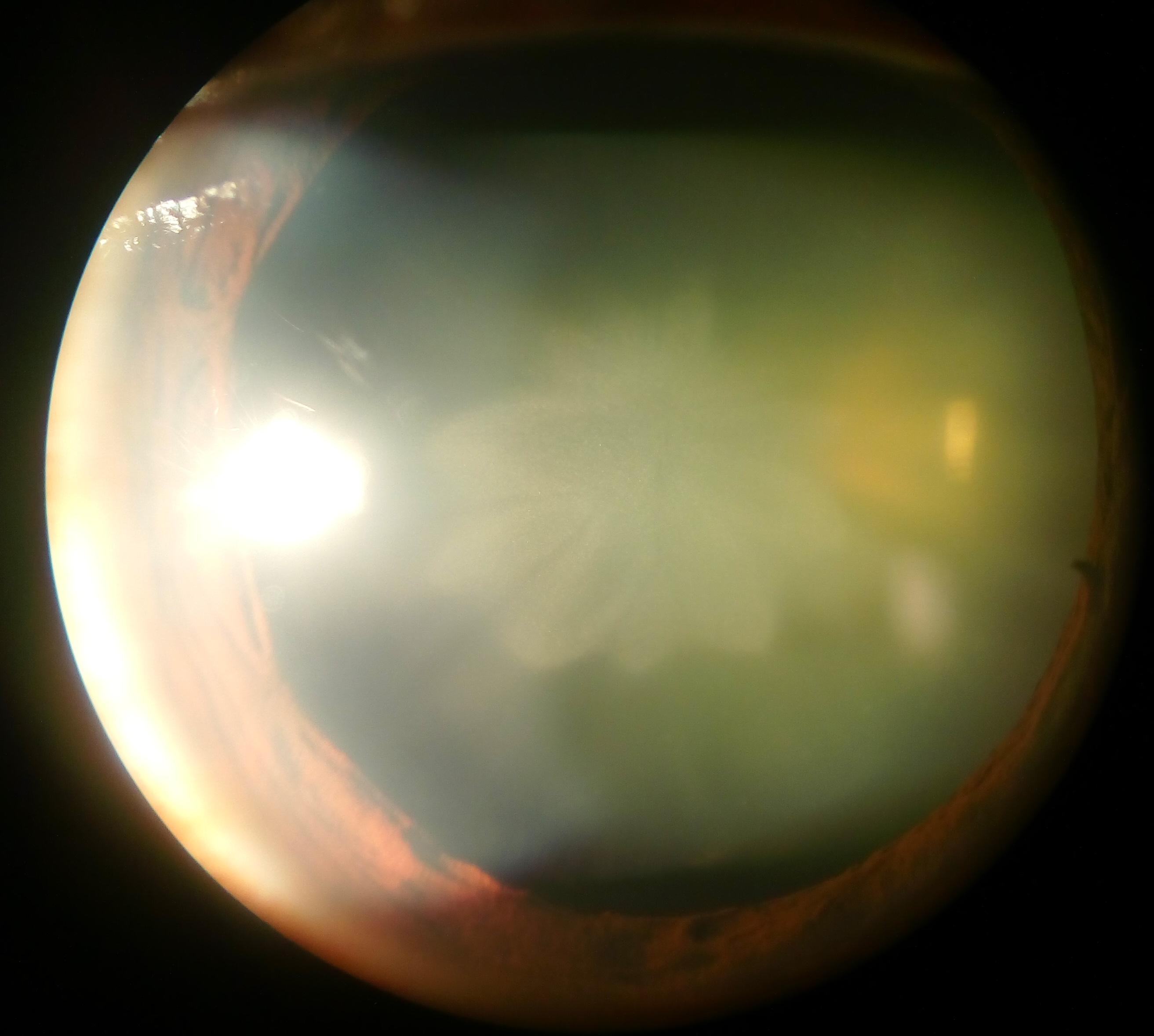
Persistent tunica vasculosa lentis is a congenital ocular anomaly. It is a form of persistent fetal vasculature (PFV). It is a developmental disorder of the vitreous. It is usually unilateral and first noticed in the neonatal period. It may be associated with microphthalmos, cataracts, and increased intraocular pressure. Elongated ciliary processes In the anatomy of the eye, the ciliary processes are formed by the inward folding of the various layers of the choroid, viz. the choroid proper and the lamina basalis, and are received between corresponding foldings of the suspensory ligam ... are visible through the dilated pupil. A USG B-scan confirms diagnosis in the presence of a cataract. See also * Persistent fetal vasculature * Tunica vasculosa lentis References * http://www.djo.harvard.edu/files/1568.pdf * Wright KW, Spiegel PH, Buckley EG. Pediatric Ophthalmology and Strabismus. Springer; US, New York: 2003. p. 17. Eye diseases {{eye-disease-stu ... [...More Info...] [...Related Items...] OR: [Wikipedia] [Google] [Baidu] |
Congenital
A birth defect is an abnormal condition that is present at childbirth, birth, regardless of its cause. Birth defects may result in disability, disabilities that may be physical disability, physical, intellectual disability, intellectual, or developmental disability, developmental. The disabilities can range from mild to severe. Birth defects are divided into two main types: structural disorders in which problems are seen with the shape of a body part and functional disorders in which problems exist with how a body part works. Functional disorders include metabolic disorder, metabolic and degenerative disease, degenerative disorders. Some birth defects include both structural and functional disorders. Birth defects may result from genetic disorder, genetic or chromosome abnormality, chromosomal disorders, exposure to certain medications or chemicals, or certain vertically transmitted infection, infections during pregnancy. Risk factors include folate deficiency, alcohol drink, d ... [...More Info...] [...Related Items...] OR: [Wikipedia] [Google] [Baidu] |
Ocular
An eye is a sensory organ that allows an organism to perceive visual information. It detects light and converts it into electro-chemical impulses in neurons (neurones). It is part of an organism's visual system. In higher organisms, the eye is a complex optical system that collects light from the surrounding environment, regulates its intensity through a diaphragm, focuses it through an adjustable assembly of lenses to form an image, converts this image into a set of electrical signals, and transmits these signals to the brain through neural pathways that connect the eye via the optic nerve to the visual cortex and other areas of the brain. Eyes with resolving power have come in ten fundamentally different forms, classified into compound eyes and non-compound eyes. Compound eyes are made up of multiple small visual units, and are common on insects and crustaceans. Non-compound eyes have a single lens and focus light onto the retina to form a single image. This type of eye i ... [...More Info...] [...Related Items...] OR: [Wikipedia] [Google] [Baidu] |
Persistent Fetal Vasculature
Persistent fetal vasculature (PFV), also known as persistent fetal vasculature syndrome (PFVS), and until 1997 known primarily as persistent hyperplastic primary vitreous (PHPV), is a rare congenital anomaly which occurs when blood vessels within the developing eye, known as the embryonic hyaloid vasculature network, fail to regress as they normally would in-utero after the eye is fully developed. Defects which arise from this lack of vascular regression are diverse; as a result, the presentation, symptoms, and prognosis of affected patients vary widely, ranging from clinical insignificance to irreversible blindness. The underlying structural causes of PFV are considered to be relatively common, and the vast majority of cases do not warrant additional intervention. When symptoms do manifest, however, they are often significant, causing detrimental and irreversible visual impairment. Persistent fetal vasculature heightens the lifelong risk of glaucoma, cataracts, intraocular hemorr ... [...More Info...] [...Related Items...] OR: [Wikipedia] [Google] [Baidu] |
Vitreous Humour
The vitreous body (''vitreous'' meaning "glass-like"; , ) is the clear gel that fills the space between the lens and the retina of the eyeball (the vitreous chamber) in humans and other vertebrates. It is often referred to as the vitreous humor (also spelled humour), from Latin meaning liquid, or simply "the vitreous". Vitreous fluid or "liquid vitreous" is the liquid component of the vitreous gel, found after a vitreous detachment. It is not to be confused with the aqueous humor, the other fluid in the eye that is found between the cornea and lens. Structure The vitreous humor is a transparent, colorless, gelatinous mass that fills the space in the eye between the lens and the retina. It is surrounded by a layer of collagen called the vitreous membrane (or hyaloid membrane or vitreous cortex) separating it from the rest of the eye. It makes up four-fifths of the volume of the eyeball. The vitreous humour is fluid-like near the centre, and gel-like near the edges. The vit ... [...More Info...] [...Related Items...] OR: [Wikipedia] [Google] [Baidu] |
Neonatal
In common terminology, a baby is the very young offspring of adult human beings, while infant (from the Latin word ''infans'', meaning 'baby' or 'child') is a formal or specialised synonym. The terms may also be used to refer to Juvenile (organism), juveniles of other organisms. A newborn is, in colloquial use, a baby who is only hours, days, or weeks old; while in medical contexts, a newborn or neonate (from Latin, ''neonatus'', newborn) is an infant in the first 28 days after Human birth, birth (the term applies to Preterm birth, premature, Pregnancy#Term, full term, and Postterm pregnancy, postmature infants). Infants born prior to 37 weeks of gestation are called "premature", those born between 39 and 40 weeks are "full term", those born through 41 weeks are "late term", and anything beyond 42 weeks is considered "post term". Before birth, the offspring is called a fetus. The term ''infant'' is typically applied to very young children under one year of age; however, defini ... [...More Info...] [...Related Items...] OR: [Wikipedia] [Google] [Baidu] |
Microphthalmos
Microphthalmia (Greek: , ), also referred as microphthalmos, is a developmental disorder of the eye in which one (unilateral microphthalmia) or both (bilateral microphthalmia) eyes are abnormally small and have anatomic malformations. Microphthalmia is a distinct condition from anophthalmia and nanophthalmia. Although sometimes referred to as 'simple microphthalmia', nanophthalmia is a condition in which the size of the eye is small but no anatomical alterations are present. Presentation Microphthalmia is a congenital disorder in which the globe of the eye is unusually small and structurally disorganized. While the axis of an adult human eye has an average length of about , a diagnosis of microphthalmia generally corresponds to an axial length below in adults. Additionally, the diameter of the cornea is about in affected newborns and in adults with the condition. The presence of a small eye within the orbit can be a normal incidental finding but in many cases it is atypical an ... [...More Info...] [...Related Items...] OR: [Wikipedia] [Google] [Baidu] |
Cataracts
A cataract is a cloudy area in the lens of the eye that leads to a decrease in vision of the eye. Cataracts often develop slowly and can affect one or both eyes. Symptoms may include faded colours, blurry or double vision, halos around light, trouble with bright lights, and difficulty seeing at night. This may result in trouble driving, reading, or recognizing faces. Poor vision caused by cataracts may also result in an increased risk of falling and depression. Cataracts cause 51% of all cases of blindness and 33% of visual impairment worldwide. Cataracts are most commonly due to aging but may also occur due to trauma or radiation exposure, be present from birth, or occur following eye surgery for other problems. Risk factors include diabetes, longstanding use of corticosteroid medication, smoking tobacco, prolonged exposure to sunlight, and alcohol. In addition to these, poor nutrition, obesity, chronic kidney disease, and autoimmune diseases have been recognized in ... [...More Info...] [...Related Items...] OR: [Wikipedia] [Google] [Baidu] |
Ciliary Processes
In the anatomy of the eye, the ciliary processes are formed by the inward folding of the various layers of the choroid, viz. the choroid proper and the lamina basalis, and are received between corresponding foldings of the suspensory ligament of the lens. Anatomy They are arranged in a circle, and form a sort of frill behind the iris, around the margin of the lens A lens is a transmissive optical device that focuses or disperses a light beam by means of refraction. A simple lens consists of a single piece of transparent material, while a compound lens consists of several simple lenses (''elements'') .... They vary from sixty to eighty in number, lie side by side, and may be divided into large and small; the former are about 2.5 mm. in length, and the latter, consisting of about one-third of the entire number, are situated in spaces between them, but without regular arrangement. They are attached by their periphery to three or four of the ridges of the orb ... [...More Info...] [...Related Items...] OR: [Wikipedia] [Google] [Baidu] |
Persistent Fetal Vasculature
Persistent fetal vasculature (PFV), also known as persistent fetal vasculature syndrome (PFVS), and until 1997 known primarily as persistent hyperplastic primary vitreous (PHPV), is a rare congenital anomaly which occurs when blood vessels within the developing eye, known as the embryonic hyaloid vasculature network, fail to regress as they normally would in-utero after the eye is fully developed. Defects which arise from this lack of vascular regression are diverse; as a result, the presentation, symptoms, and prognosis of affected patients vary widely, ranging from clinical insignificance to irreversible blindness. The underlying structural causes of PFV are considered to be relatively common, and the vast majority of cases do not warrant additional intervention. When symptoms do manifest, however, they are often significant, causing detrimental and irreversible visual impairment. Persistent fetal vasculature heightens the lifelong risk of glaucoma, cataracts, intraocular hemorr ... [...More Info...] [...Related Items...] OR: [Wikipedia] [Google] [Baidu] |
Tunica Vasculosa Lentis
The tunica vasculosa lentis is an extensive capillary network, spreading over the posterior and lateral surfaces of the lens of the eye. It disappears normally shortly after birth, through apoptosis. The structure was not studied properly and in detail until the 1960s, when new technologies developed to allow the preservation of the networks in fetuses. The scanning electron microscope finally enabled researchers to see the network even in very small laboratory animals such as the mouse embryo. See also * Comparative vertebrate anatomy * Ophthalmology * Persistent tunica vasculosa lentis Persistent tunica vasculosa lentis is a congenital ocular anomaly. It is a form of persistent fetal vasculature (PFV). It is a developmental disorder of the vitreous. It is usually unilateral and first noticed in the neonatal period. It may be ... References Human eye anatomy {{Eye-stub ... [...More Info...] [...Related Items...] OR: [Wikipedia] [Google] [Baidu] |


