|
Peroneal Nerves
The common fibular nerve (also known as the common peroneal nerve, external popliteal nerve, or lateral popliteal nerve) is a nerve in the lower leg that provides sensation over the wikt:posterolateral, posterolateral part of the leg and the knee joint. It divides at the knee into two terminal branches: the superficial fibular nerve and deep fibular nerve, which innervate the muscles of the lateral and anterior compartments of the leg respectively. When the common fibular nerve is damaged or Nerve compression syndrome, compressed, foot drop can ensue. Structure The common fibular nerve is the smaller terminal branch of the sciatic nerve. The common fibular nerve has root values of L4, L5, S1, and S2. It arises from the superior angle of the popliteal fossa and extends to the lateral angle of the popliteal fossa, along the medial border of the biceps femoris. It then winds around the neck of the fibula to pierce the fibularis longus and divides into terminal branches of the superfi ... [...More Info...] [...Related Items...] OR: [Wikipedia] [Google] [Baidu] |
Anterior Compartment Of Leg
The anterior compartment of the leg is a fascial compartment of the lower leg. It contains muscles that produce dorsiflexion and participate in inversion and eversion of the foot, as well as vascular and nervous elements, including the anterior tibial artery and veins and the deep fibular nerve. Muscles The muscles of the compartment are: * tibialis anterior * extensor hallucis longus * extensor digitorum longus * fibularis (peroneus) tertius Function The compartment contains muscles that are dorsiflexors and participate in inversion and eversion of the foot. Innervation and blood supply The anterior compartment of the leg is supplied by the deep fibular nerve (deep peroneal nerve), a branch of the common fibular nerve. The nerve contains axons from the L4, L5, and S1 spinal nerves. Blood for the compartment is supplied by the anterior tibial artery, which runs between the tibialis anterior and extensor digitorum longus muscles. When the artery crosses the exte ... [...More Info...] [...Related Items...] OR: [Wikipedia] [Google] [Baidu] |
Tibialis Anterior
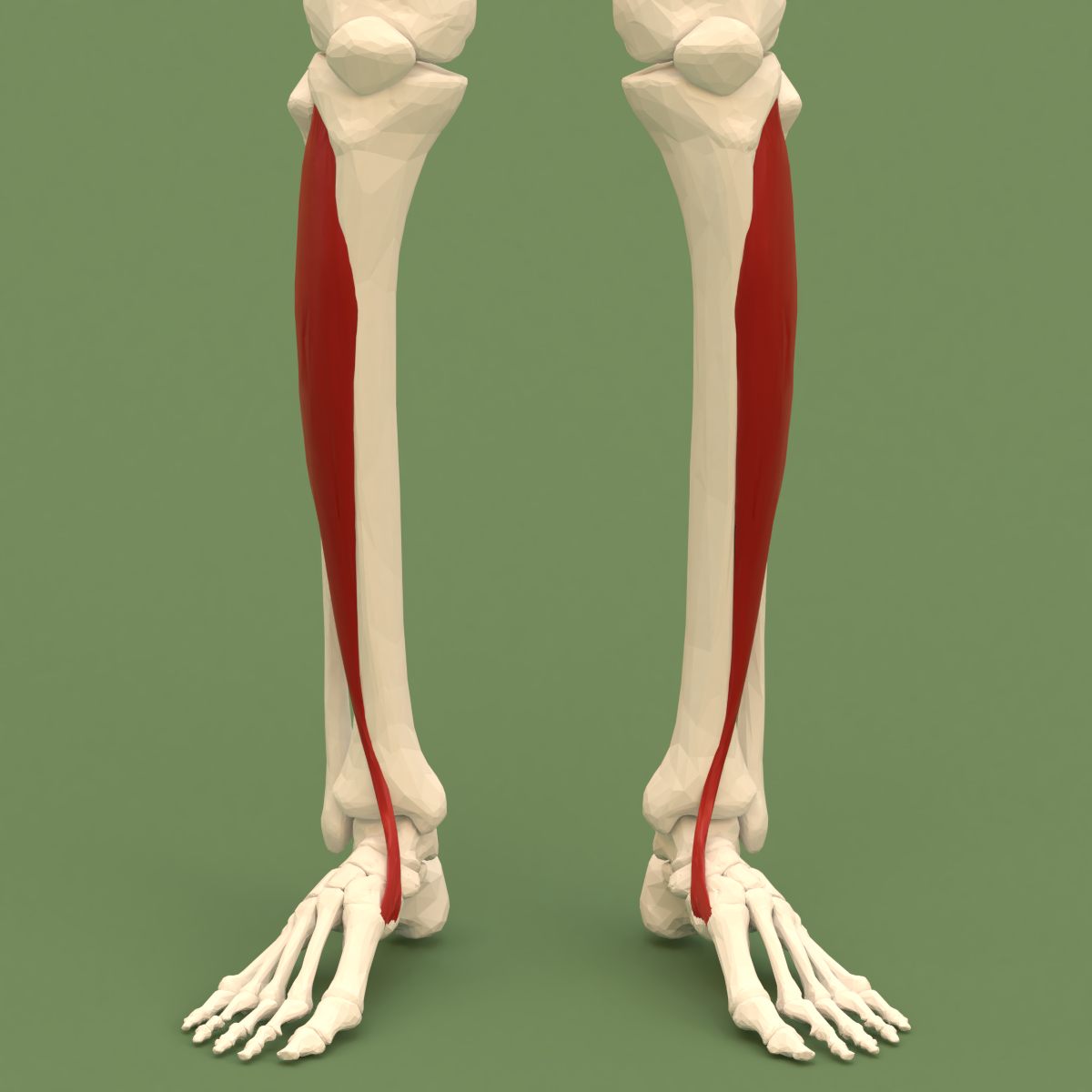
The tibialis anterior muscle is a muscle of the anterior compartment of the lower leg. It originates from the upper portion of the tibia; it inserts into the medial cuneiform and first metatarsal bones of the foot. It acts to dorsiflex and invert the foot. This muscle is mostly located near the shin. It is situated on the lateral side of the tibia; it is thick and fleshy above, tendinous below. The tibialis anterior overlaps the anterior tibial vessels and deep peroneal nerve in the upper part of the leg. Structure The tibialis anterior muscle is the most medial muscle of the anterior compartment of the leg. The muscle ends in a tendon which is apparent on the anteriomedial dorsal aspect of the foot close to the ankle. Its tendon is ensheathed in a synovial sheath. The tendon passes through the medial compartment superior and inferior extensor retinacula of the foot. Origin The tibialis anterior muscle arises from the upper 2/3 of the lateral surface of the tibia and the ... [...More Info...] [...Related Items...] OR: [Wikipedia] [Google] [Baidu] |
Sciatic Nerve
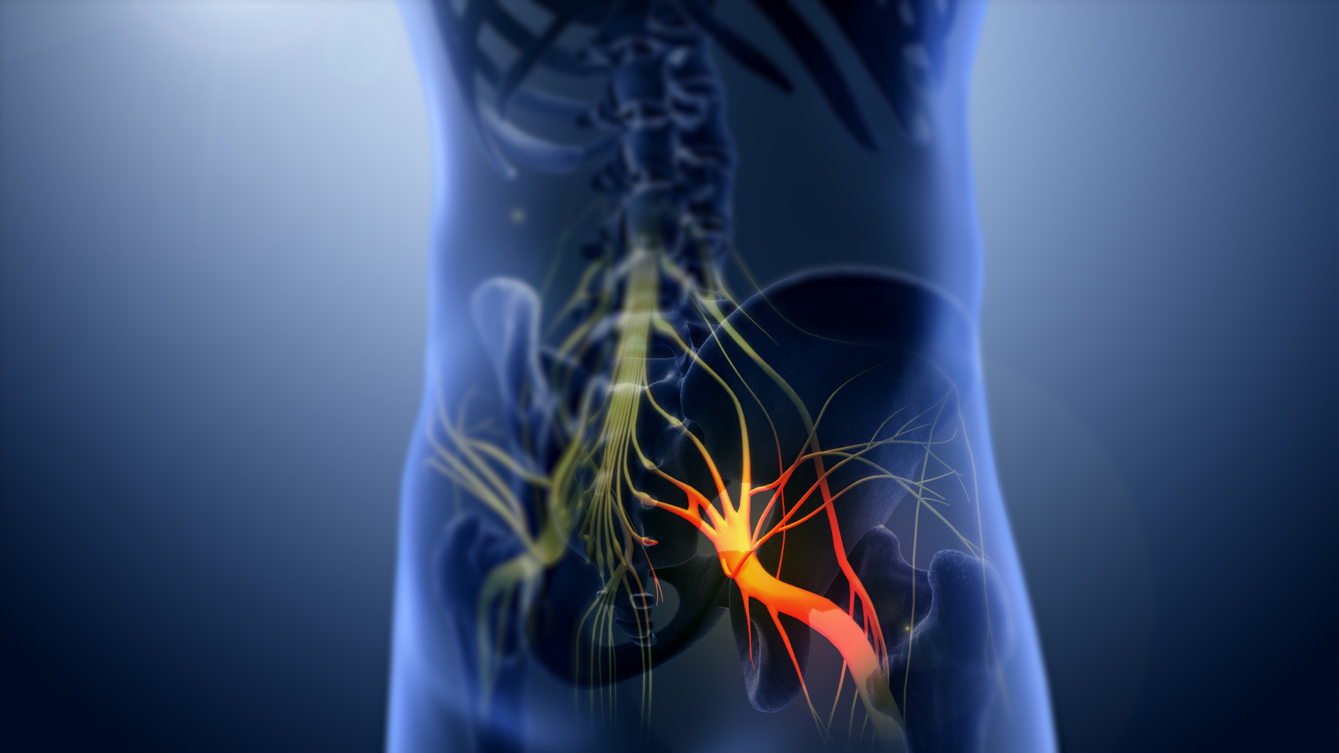
The sciatic nerve, also called the ischiadic nerve, is a large nerve in humans and other vertebrate animals. It is the largest branch of the sacral plexus and runs alongside the hip joint and down the right lower limb. It is the longest and widest single nerve in the human body, going from the top of the leg to the foot on the posterior aspect. The sciatic nerve has no cutaneous branches for the thigh. This nerve provides the connection to the nervous system for the skin of the lateral leg and the whole foot, the muscles of the back of the thigh, and those of the leg and foot. It is derived from Spinal nerve, spinal nerves Lumbar spinal nerve 4, L4 to Sacral spinal nerve 3, S3. It contains Axon, fibres from both the anterior and posterior divisions of the lumbosacral plexus. Structure In humans, the sciatic nerve is formed from the L4 to S3 segments of the sacral plexus, a collection of nerve fibres that emerge from the Sacrum, sacral part of the spinal cord. The lumbosacral trunk ... [...More Info...] [...Related Items...] OR: [Wikipedia] [Google] [Baidu] |
Fibular Muscles
The fibula (: fibulae or fibulas) or calf bone is a leg bone on the lateral side of the tibia, to which it is connected above and below. It is the smaller of the two bones and, in proportion to its length, the most slender of all the long bones. Its upper extremity is small, placed toward the back of the head of the tibia, below the knee joint and excluded from the formation of this joint. Its lower extremity inclines a little forward, so as to be on a plane anterior to that of the upper end; it projects below the tibia and forms the lateral part of the ankle joint. Structure The bone has the following components: * Lateral malleolus * Interosseous membrane connecting the fibula to the tibia, forming a syndesmosis joint * The superior tibiofibular articulation is an arthrodial joint between the lateral condyle of the tibia and the head of the fibula. * The inferior tibiofibular articulation (tibiofibular syndesmosis) is formed by the rough, convex surface of the medial side of ... [...More Info...] [...Related Items...] OR: [Wikipedia] [Google] [Baidu] |
Fibular Veins
In anatomy, the fibular veins (also known as peroneal veins) are accompanying veins (venae comitantes) of the fibular artery. Structure The fibular veins are deep veins that help carry blood from the lateral compartment of the leg. They drain into the posterior tibial veins, which in turn drain into the popliteal vein. The fibular veins accompany the fibular artery. See also * Fibular artery * Common fibular nerve * Venae comitantes Additional images File:Gray440_color.png, Cross-section through middle of leg. File:Ultrasonography of thrombosis of the fibular veins, coronal plane, annotated.jpg, Coronal plane The dorsal plane (also known as the coronal plane or frontal plane, especially in human anatomy) is an anatomical plane that divides the body into Anatomical terms of location#Dorsal and ventral, dorsal and ventral sections. It is perpendicular t ... (seen from medial side of lower leg) ultrasonography of deep vein thrombosis of the fibular veins, seen as hypere ... [...More Info...] [...Related Items...] OR: [Wikipedia] [Google] [Baidu] |
Peroneal Strike
A peroneal strike is a temporarily disabling blow to the common fibular (peroneal) nerve of the leg, just above the knee. The attacker aims roughly a hand span above the exterior side of the knee, towards the back of the leg. This causes a temporary loss of motor control of the leg, accompanied by numbness and a painful tingling sensation from the point of impact all the way down the leg, usually lasting anywhere from 30 seconds to 5 hours in duration. The strike is commonly made with the knee, a baton, or shin kick, but can be done by anything forcefully impacting the nerve. The technique is a part of the pressure point control tactics used in martial arts and by law enforcement agents. The peroneal strike was used against detainees during the 2002 Bagram torture and prisoner abuse scandal.Rashid, Ahmed. ''Descent into Chaos: The U.S. and the Disaster in Pakistan, Afghanistan and Central Asia''. 2008. New York: Viking Penguin, 2009 See also *Charley horse *Pain compliance *Ku ... [...More Info...] [...Related Items...] OR: [Wikipedia] [Google] [Baidu] |
Superficial Fibular Nerve
The superficial fibular nerve (also known as superficial peroneal nerve) is a mixed (motor and sensory) nerve that provides motor innervation to the fibularis longus and fibularis brevis muscles, and sensory innervation to skin over the antero-lateral aspect of the leg along with the greater part of the dorsum of the foot (with the exception of the first web space, which is innervated by the deep fibular nerve). Structure Lateral side of the leg The superficial fibular nerve is the main nerve of the lateral compartment of the leg. It begins at the lateral side of the neck of fibula, and runs through the fibularis longus and fibularis brevis muscles. In the middle third of the leg, it descends between the fibularis longus and fibularis brevis, and then reaches the anterior border of the fibularis brevis to enter the groove between the fibularis brevis and the extensor digitorum longus under the deep fascia of leg. It becomes superficial at the junction of upper two-thirds and ... [...More Info...] [...Related Items...] OR: [Wikipedia] [Google] [Baidu] |
Extensor Digitorum Brevis Muscle
The extensor digitorum brevis muscle (sometimes EDB) is a muscle Muscle is a soft tissue, one of the four basic types of animal tissue. There are three types of muscle tissue in vertebrates: skeletal muscle, cardiac muscle, and smooth muscle. Muscle tissue gives skeletal muscles the ability to muscle contra ... on the upper surface of the foot that helps extend digits 2 through 4. Structure The muscle originates from the forepart of the upper and lateral surface of the calcaneus (in front of the groove for the peroneus brevis tendon), from the interosseous talocalcaneal ligament and the stem of the inferior extensor retinaculum. The fibres pass obliquely forwards and medially across the dorsum of the foot and end in four tendons. The medial part of the muscle, also known as extensor hallucis brevis, ends in a tendon which crosses the dorsalis pedis artery and inserts into the dorsal surface of the base of the proximal phalanx of the great toe. The other three tendons ins ... [...More Info...] [...Related Items...] OR: [Wikipedia] [Google] [Baidu] |
NYU Langone Medical Center
NYU Langone Health is an integrated Health system, academic health system located in New York City, New York, United States. The health system consists of the New York University Grossman School of Medicine, NYU Grossman School of Medicine and NYU Grossman Long Island School of Medicine, both part of New York University (NYU), and more than 320 locations throughout the New York City Region and Florida, including seven inpatient facilities: #Tisch Hospital, Tisch Hospital; #Kimmel Pavilion, Kimmel Pavilion; #NYU Langone Orthopedic Hospital, NYU Langone Orthopedic Hospital; Hassenfeld Children's Hospital; NYU Langone Hospital – Brooklyn, NYU Langone Hospital–Brooklyn; NYU Langone Hospital – Long Island, NYU Langone Hospital–Long Island; and NYU Langone Hospital — Suffolk. It is also home to #Rusk Rehabilitation, Rusk Rehabilitation. NYU Langone Health is one of the largest Hospital network, healthcare systems in the Northeastern United States, Northeast, with more than 53,0 ... [...More Info...] [...Related Items...] OR: [Wikipedia] [Google] [Baidu] |
Johns Hopkins School Of Medicine
The Johns Hopkins University School of Medicine (JHUSOM) is the medical school of Johns Hopkins University, a private research university in Baltimore, Maryland. Established in 1893 following the construction of the Johns Hopkins Hospital, the school is known for being a center of biomedical research and innovation, and is routinely ranked as one of the top medical schools in the United States. The School of Medicine is located in Baltimore, Maryland, where it shares a campus in East Baltimore with the School of Nursing, Bloomberg School of Public Health, and Johns Hopkins Hospital, the school's primary teaching hospital. As part of the larger Johns Hopkins Medicine health system, the school is also affiliated with numerous regional hospitals and medical centers including the Johns Hopkins Bayview Medical Center, Johns Hopkins Howard County Medical Center, Suburban Hospital in Montgomery County, Sibley Memorial Hospital in Washington, D.C. History Before his death in ... [...More Info...] [...Related Items...] OR: [Wikipedia] [Google] [Baidu] |
Nerve Decompression
A nerve decompression is a neurosurgical procedure to relieve chronic, direct pressure on a nerve to treat nerve entrapment, a pain syndrome characterized by severe chronic pain and muscle weakness. In this way a nerve decompression targets the underlying pathophysiology of the syndrome and is considered a first-line surgical treatment option for peripheral nerve pain. Despite treating the underlying cause of the disease, the symptoms may not be fully reversible as delays in diagnosis can allow permanent damage to occur to the nerve and surrounding microvasculature. Traditionally only nerves accessible with open surgery have been good candidates, however innovations in laparoscopy and nerve-sparing techniques made nearly all nerves in the body good candidates, as surgical access is no longer a barrier. Surgical planning Surgical planning is distinct from diagnosis of entrapment. Diagnosis will focus on a binary decision: does the patient have entrapment or not? A diagnosis may ... [...More Info...] [...Related Items...] OR: [Wikipedia] [Google] [Baidu] |
Yoga Foot Drop
Vajrasana (), Thunderbolt Pose, or Diamond Pose, is a kneeling asana in hatha yoga and modern yoga as exercise. Ancient texts describe a variety of poses under this name. Etymology and origins The name comes from the Sanskrit words ''vajra'', a weapon whose name means "thunderbolt" or "diamond", and ''asana'' (, āsana) meaning "posture" or "seat". The name Vajrasana denotes a medieval meditation seat, but its usage varied. The 15th century ''Hatha Yoga Pradipika'' called it a synonym of Siddhasana, where one of the heels presses the root of the penis; according to ''Yoga-Mimamsa'' III.2 p. 135, this explains the reference to the vajra weapon. The 17th century ''Gheranda Samhita'' 2.12 describes what ''Light on Yoga'' calls Virasana, with the feet beside the buttocks, while in other texts Vajrasana appears to mean the modern kneeling-down position, with the buttocks resting on the feet. The yoga scholar Norman Sjoman notes that ''Light on Yoga'' is unclear here, as it ... [...More Info...] [...Related Items...] OR: [Wikipedia] [Google] [Baidu] |


