|
Nerve Decompression
A nerve decompression is a neurosurgical procedure to relieve chronic, direct pressure on a nerve to treat Nerve compression syndrome, nerve entrapment, a pain syndrome characterized by severe chronic pain and muscle weakness. In this way a nerve decompression targets the underlying pathophysiology of the syndrome and is considered a first-line surgical treatment option for peripheral nerve pain. Despite treating the underlying cause of the disease, the symptoms may not be fully reversible as delays in diagnosis can allow permanent damage to occur to the nerve and surrounding microvasculature. Traditionally only nerves accessible with open surgery have been good candidates, however innovations in laparoscopy and nerve-sparing techniques made nearly all nerves in the body good candidates, as surgical access is no longer a barrier. Surgical planning Surgical planning is distinct from diagnosis of entrapment. Diagnosis will focus on a binary decision: does the patient have entrapmen ... [...More Info...] [...Related Items...] OR: [Wikipedia] [Google] [Baidu] |
Neurosurgical
Neurosurgery or neurological surgery, known in common parlance as brain surgery, is the medical specialty concerned with the surgical treatment of disorders which affect any portion of the nervous system including the brain, spinal cord and peripheral nervous system. Education and context In different countries, there are different requirements for an individual to legally practice neurosurgery, and there are varying methods through which they must be educated. In most countries, neurosurgeon training requires a minimum period of seven years after graduating from medical school. United States In the United States, a neurosurgeon must generally complete four years of undergraduate education, four years of medical school, and seven years of Residency (medicine), residency (PGY-1-7). Most, but not all, residency programs have some component of basic science or clinical research. Neurosurgeons may pursue additional training in the form of a Fellowship (medicine), fellowship after re ... [...More Info...] [...Related Items...] OR: [Wikipedia] [Google] [Baidu] |
Diffusion MRI
Diffusion-weighted magnetic resonance imaging (DWI or DW-MRI) is the use of specific MRI sequences as well as software that generates images from the resulting data that uses the diffusion of water molecules to generate contrast in MR images. It allows the mapping of the diffusion process of molecules, mainly water, in biological tissues, in vivo and non-invasively. Molecular diffusion in tissues is not random, but reflects interactions with many obstacles, such as macromolecules, fibers, and membranes. Water molecule diffusion patterns can therefore reveal microscopic details about tissue architecture, either normal or in a diseased state. A special kind of DWI, diffusion tensor imaging (DTI), has been used extensively to map white matter tractography in the brain. Introduction In diffusion weighted imaging (DWI), the intensity of each image element ( voxel) reflects the best estimate of the rate of water diffusion at that location. Because the mobility of water is driven by ... [...More Info...] [...Related Items...] OR: [Wikipedia] [Google] [Baidu] |
Sciatic Nerve
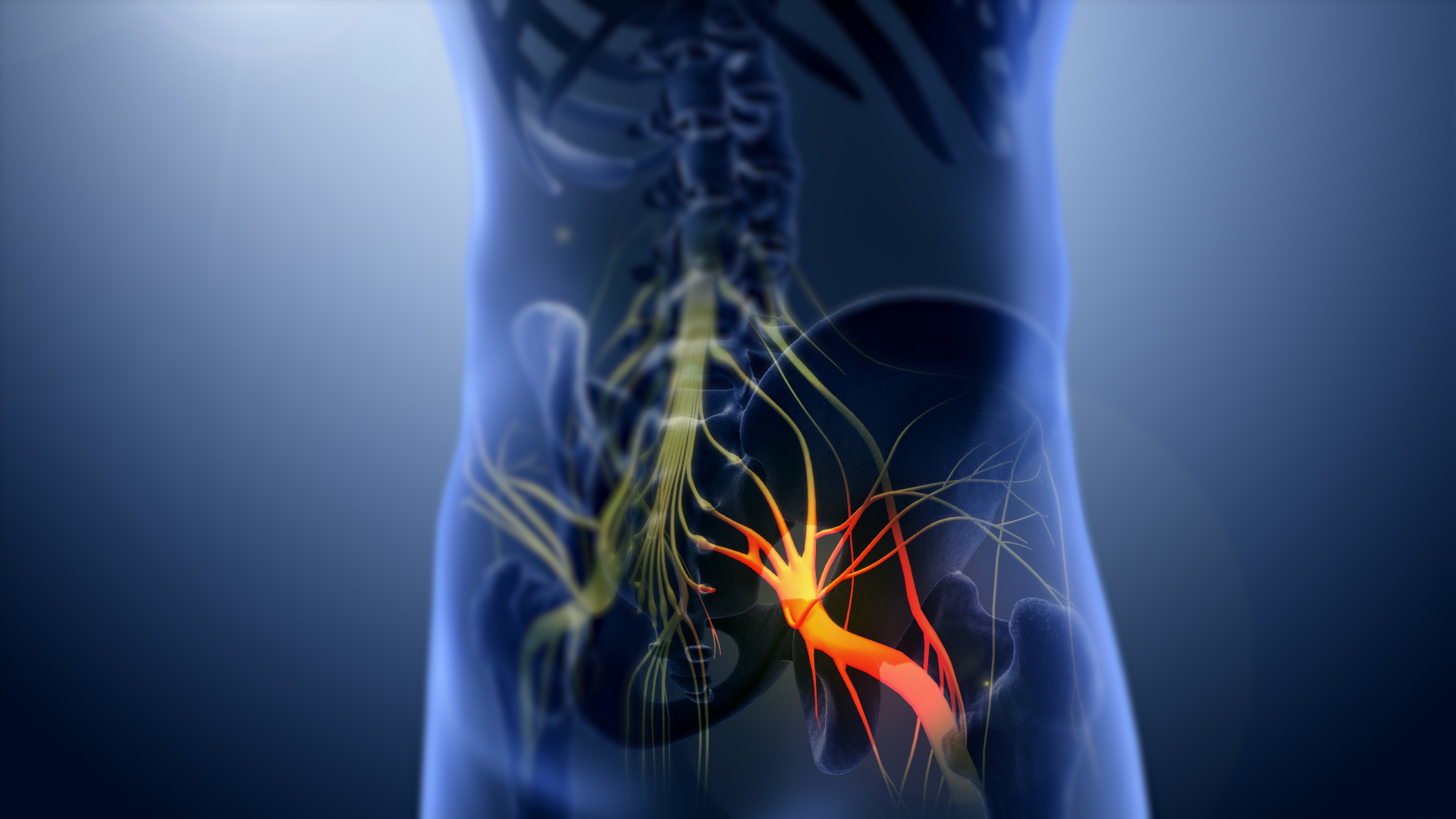
The sciatic nerve, also called the ischiadic nerve, is a large nerve in humans and other vertebrate animals which is the largest branch of the sacral plexus and runs alongside the hip joint and down the lower limb. It is the longest and widest single nerve in the human body, going from the top of the leg to the foot on the posterior aspect. The sciatic nerve has no cutaneous branches for the thigh. This nerve provides the connection to the nervous system for the skin of the lateral leg and the whole foot, the muscles of the back of the thigh, and those of the leg and foot. It is derived from spinal nerves L4 to S3. It contains fibers from both the anterior and posterior divisions of the lumbosacral plexus. Structure In humans, the sciatic nerve is formed from the L4 to S3 segments of the sacral plexus, a collection of nerve fibres that emerge from the sacral part of the spinal cord. The lumbosacral trunk from the L4 and L5 roots descends between the sacral promontory and ala ... [...More Info...] [...Related Items...] OR: [Wikipedia] [Google] [Baidu] |
Piriformis Syndrome
Piriformis syndrome is a condition which is believed to result from compression of the sciatic nerve by the piriformis muscle. Symptoms may include pain and numbness in the buttocks and down the leg. Often symptoms are worsened with sitting or running. Causes may include trauma to the gluteal muscle, spasms of the piriformis muscle, anatomical variation, or an overuse injury. Few cases in athletics, however, have been described. Diagnosis is difficult as there is no definitive test. A number of physical exam maneuvers can be supportive. Medical imaging is typically normal. Other conditions that may present similarly include a herniated disc. Treatment may include avoiding activities that cause symptoms, stretching, physiotherapy, and medication such as NSAIDs. Steroid or botulinum toxin injections may be used in those who do not improve. Surgery is not typically recommended. The frequency of the condition is unknown, with different groups arguing it is more or less common ... [...More Info...] [...Related Items...] OR: [Wikipedia] [Google] [Baidu] |
Piriformis Muscle
The piriformis muscle () is a flat, pyramidally-shaped muscle in the gluteal region of the lower limbs. It is one of the six muscles in the lateral rotator group. The piriformis muscle has its origin upon the front surface of the sacrum, and inserts onto the greater trochanter of the femur. Depending upon the given position of the leg, it acts either as external (lateral) rotator of the thigh or as abductor of the thigh. It is innervated by the piriformis nerve. Structure The piriformis is a flat muscle, and is pyramidal in shape. Origin The piriformis muscle originates from the anterior (front) surface of the sacrum by three fleshy digitations attached to the second, third, and fourth sacral vertebra. It also arises from the superior margin of the greater sciatic notch, the gluteal surface of the ilium (near the posterior inferior iliac spine), the sacroiliac joint capsule, and (sometimes) the sacrotuberous ligament (more specifically, the superior part of the ... [...More Info...] [...Related Items...] OR: [Wikipedia] [Google] [Baidu] |
Migraine
Migraine (, ) is a common neurological disorder characterized by recurrent headaches. Typically, the associated headache affects one side of the head, is pulsating in nature, may be moderate to severe in intensity, and could last from a few hours to three days. Non-headache symptoms may include nausea, vomiting, and sensitivity to light, sound, or smell. The pain is generally made worse by physical activity during an attack,as PDF although regular may prevent future attacks. Up to one-third of people affected have |
Migraine Surgery
Migraine surgery is a surgical operation undertaken with the goal of reducing or preventing migraines. Migraine surgery most often refers to surgical decompression of one or several nerves in the head and neck which have been shown to trigger migraine symptoms in many migraine sufferers. Following the development of nerve decompression techniques for the relief of migraine pain in the year 2000, these procedures have been extensively studied and shown to be effective in appropriate candidates. The nerves that are most often addressed in migraine surgery are found outside of the skull, in the face and neck, and include the supra-orbital and supra-trochlear nerves in the forehead, the zygomaticotemporal nerve and auriculotemporal nerves in the temple region, and the greater occipital, lesser occipital, and third occipital nerves in the back of the neck. Nerve impingement in the nasal cavity has additionally been shown to be a trigger of migraine symptoms. Indications and pat ... [...More Info...] [...Related Items...] OR: [Wikipedia] [Google] [Baidu] |
Diabetic Neuropathy
Diabetic neuropathy is various types of nerve damage associated with diabetes mellitus. Symptoms depend on the site of nerve damage and can include motor changes such as weakness; sensory symptoms such as numbness, tingling, or pain; or autonomic changes such as urinary symptoms. These changes are thought to result from microvascular injury involving small blood vessels that supply nerves ( vasa nervorum). Relatively common conditions which may be associated with diabetic neuropathy include distal symmetric polyneuropathy; third, fourth, or sixth cranial nerve palsy; mononeuropathy; mononeuropathy multiplex; diabetic amyotrophy; and autonomic neuropathy. Signs and symptoms Diabetic neuropathy can affect any peripheral nerves including sensory neurons, motor neurons, and the autonomic nervous system. Therefore, diabetic neuropathy has the potential to affect essentially any organ system and can cause a range of symptoms. There are several distinct syndromes based on the orga ... [...More Info...] [...Related Items...] OR: [Wikipedia] [Google] [Baidu] |
Trigeminal Neuralgia
Trigeminal neuralgia (TN or TGN), also called Fothergill disease, tic douloureux, or trifacial neuralgia is a long-term pain disorder that affects the trigeminal nerve, the nerve responsible for sensation in the face and motor functions such as biting and chewing. It is a form of neuropathic pain. There are two main types: typical and atypical trigeminal neuralgia. The typical form results in episodes of severe, sudden, shock-like pain in one side of the face that lasts for seconds to a few minutes. Groups of these episodes can occur over a few hours. The atypical form results in a constant burning pain that is less severe. Episodes may be triggered by any touch to the face. Both forms may occur in the same person. It is regarded as one of the most painful disorders known to medicine, and often results in depression. The exact cause is unknown, but believed to involve loss of the myelin of the trigeminal nerve. This might occur due to compression from a blood vessel as the ner ... [...More Info...] [...Related Items...] OR: [Wikipedia] [Google] [Baidu] |
Microvascular Decompression
Microvascular decompression (MVD), also known as the Jannetta procedure, is a neurosurgical procedure used to treat trigeminal neuralgia (along with other cranial nerve neuralgias) a pain syndrome characterized by severe episodes of intense facial pain, and hemifacial spasm. The procedure is also used experimentally to treat tinnitus and vertigo caused by vascular compression on the vestibulocochlear nerve. History Nicholas Andre first described trigeminal neuralgia in 1756. In 1891 Sir Victor Horsley proposed the first open surgical procedure for the disorder involving the sectioning of preganglionic rootlets of the trigeminal nerve. Walter Dandy in 1925 was an advocate of partial sectioning of the nerve in the posterior cranial fossa. During this procedure he noted compression of the nerve by vascular loops, and in 1932 proposed the theory that trigeminal neuralgia was caused by compression of the nerve by blood vessels, typically the superior cerebellar artery. With the adve ... [...More Info...] [...Related Items...] OR: [Wikipedia] [Google] [Baidu] |
Cauda Equina Syndrome
Cauda equina syndrome (CES) is a condition that occurs when the bundle of nerves below the end of the spinal cord known as the cauda equina is damaged. Signs and symptoms include low back pain, pain that radiates down the leg, numbness around the anus, and loss of bowel or bladder control. Onset may be rapid or gradual. The cause is usually a disc herniation in the lower region of the back. Other causes include spinal stenosis, cancer, trauma, epidural abscess, and epidural hematoma. The diagnosis is suspected based on symptoms and confirmed by medical imaging such as MRI or CT scan. CES is generally treated surgically via laminectomy. Sudden onset is regarded as a medical emergency requiring prompt surgical decompression, with delay causing permanent loss of function. Permanent bladder problems, sexual dysfunction or numbness may occur despite surgery. A poor outcome occurs in about 20% of people despite treatment. About 1 in 70,000 people is affected every year. It was ... [...More Info...] [...Related Items...] OR: [Wikipedia] [Google] [Baidu] |
Laminectomy
A laminectomy is a surgical procedure that removes a portion of a vertebra called the lamina, which is the roof of the spinal canal. It is a major spine operation with residual scar tissue and may result in postlaminectomy syndrome. Depending on the problem, more conservative treatments (e.g., small endoscopic procedures, without bone removal) may be viable. Method The lamina is a posterior arch of the vertebral bone lying between the spinous process (which juts out in the middle) and the more lateral pedicles and the transverse processes of each vertebra. The pair of laminae, along with the spinous process, make up the posterior wall of the bony spinal canal. Although the literal meaning of laminectomy is 'excision of the lamina', a conventional laminectomy in neurosurgery and orthopedics involves excision of the supraspinous ligament and some or all of the spinous process. Removal of these structures with an open technique requires disconnecting the many muscles of th ... [...More Info...] [...Related Items...] OR: [Wikipedia] [Google] [Baidu] |


_1.jpg)


