|
Oncocytes
An oncocyte is an epithelial cell characterized by an excessive number of mitochondria, resulting in an abundant acidophilic, granular cytoplasm. Oncocytes can be benign or malignant. Other names Also known as: *''Hürthle cell'' (thyroid gland only) *'' Oxyphilic cell'' *''Askanazy cell'' *''Apocrine-type metaplasia'' (breast gland only). *''Oncocytic cell'' Etymology Derived from the Greek root onco-, which means mass, bulk. See also * Hurthle cell carcinoma, a variant of follicular thyroid carcinoma. *Oncocytoma, a tumour composed of oncocytes, may be found as a less common salivary gland neoplasm also known as oxyphilic adenoma. *Renal oncocytoma A renal oncocytoma is a tumour of the kidney made up of oncocytes, epithelial cells with an excess amount of mitochondria. Signs and symptoms Renal oncocytomas are often asymptomatic and are frequently discovered by chance on a CT or ultrasoun ..., a kidney tumour composed of oncocytes. References {{Reflist External lin ... [...More Info...] [...Related Items...] OR: [Wikipedia] [Google] [Baidu] |
Oncocytoma
An oncocytoma is a tumor made up of oncocytes, epithelial cell (biology), cells characterized by an excessive amount of mitochondria, resulting in an abundant acidophilic, granular cytoplasm. The cells and the tumor that they compose are often benign but sometimes may be premalignant or malignant. Presentation An oncocytoma is an epithelial tumor composed of oncocytes, large eosinophilic cells having small, round, benign-appearing Cell nucleus, nuclei with large nucleoli. Oncocytoma can arise in a number of organs. Renal oncocytoma Renal oncocytoma is thought to arise from the intercalated cells of collecting ducts of the kidney. It represents 5% to 15% of surgically resected renal neoplasms. Salivary gland oncocytoma An salivary gland oncocytoma (also known as an oxyphilic adenoma) is a well-circumscribed, benign neoplasm, neoplastic growth comprising about one percent of all salivary gland tumors. The histopathology is marked by sheets of large, swollen polyhedron, polyhedr ... [...More Info...] [...Related Items...] OR: [Wikipedia] [Google] [Baidu] |
Hürthle Cell
A Hürthle cell is a transformed (metaplasia) thyroid follicular cell with "enlarged mitochondria and enlarged round nuclei with prominent nucleoli", resulting in eosinophilia in the cytoplasm. Oncocytes in the thyroid are often called Hürthle cells. Although the terms oncocyte, oxyphil cell, and Hürthle cell are used interchangeably, "Hürthle cell" is used only to indicate cells of thyroid follicular origin.Cannon, J. (2011). The Significance of Hurthle Cells in Thyroid Disease. The Oncologist. doi:10.1634/theoncologist.2010-0253 Diseases While Hurthle cells can occur in healthy thyroid glands, they are often associated with Hashimoto's thyroiditis and Graves' disease. Hürthle cell neoplasms can be separated into Hürthle cell adenomas (benign tumours) and carcinomas (malignant tumours) arising from the follicular epithelium of the thyroid gland. The latter is a relatively rare form of differentiated thyroid cancer, accounting for only 3-10% of all differentiated thyr ... [...More Info...] [...Related Items...] OR: [Wikipedia] [Google] [Baidu] |
Renal Oncocytoma
A renal oncocytoma is a tumour of the kidney made up of oncocytes, epithelial cells with an excess amount of mitochondria. Signs and symptoms Renal oncocytomas are often asymptomatic and are frequently discovered by chance on a CT or ultrasound of the abdomen. Possible signs and symptoms of a renal oncocytoma include blood in the urine, flank pain, and an abdominal mass. Pathophysiology Renal oncocytoma is thought to arise from the intercalated cells of collecting ducts of the kidney. It represent 5% to 15% of surgically resected renal neoplasms. Ultrastructurally, the eosinophilic cells have numerous mitochondria. Histologic appearance An oncocytoma is an epithelial tumor composed of oncocytes, large eosinophilic cells having small, round, benign-appearing nuclei with large nucleoli and excessive amounts of mitochondria. Diagnosis In gross appearance, the tumors are tan or mahogany brown, well circumscribed and contain a central scar. They may achieve a large size (u ... [...More Info...] [...Related Items...] OR: [Wikipedia] [Google] [Baidu] |
Apocrine
Apocrine () is a term used to classify the mode of secretion of exocrine glands. In apocrine secretion, secretory cells accumulate material at their apical ends, often forming blebs or "snouts", and this material then buds off from the cells, forming extracellular vesicles. The secretory cells therefore lose part of their cytoplasm in the process of secretion. An example of true apocrine glands is the mammary glands, responsible for secreting breast milk. Apocrine glands are also found in the anogenital region and axilla The axilla (: axillae or axillas; also known as the armpit, underarm or oxter) is the area on the human body directly under the shoulder joint. It includes the axillary space, an anatomical space within the shoulder girdle between the arm a ...e. Apocrine secretion is less damaging to the gland than holocrine secretion (which destroys a cell) but more damaging than merocrine secretion ( exocytosis). File:405 Modes of Secretion by Glands Apocrine.p ... [...More Info...] [...Related Items...] OR: [Wikipedia] [Google] [Baidu] |
Thyroid Gland
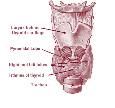
The thyroid, or thyroid gland, is an endocrine gland in vertebrates. In humans, it is a butterfly-shaped gland located in the neck below the Adam's apple. It consists of two connected lobes. The lower two thirds of the lobes are connected by a thin band of tissue called the isthmus (: isthmi). Microscopically, the functional unit of the thyroid gland is the spherical thyroid follicle, lined with follicular cells (thyrocytes), and occasional parafollicular cells that surround a lumen containing colloid. The thyroid gland secretes three hormones: the two thyroid hormones triiodothyronine (T3) and thyroxine (T4)and a peptide hormone, calcitonin. The thyroid hormones influence the metabolic rate and protein synthesis and growth and development in children. Calcitonin plays a role in calcium homeostasis. Secretion of the two thyroid hormones is regulated by thyroid-stimulating hormone (TSH), which is secreted from the anterior pituitary gland. TSH is regulated by th ... [...More Info...] [...Related Items...] OR: [Wikipedia] [Google] [Baidu] |
Epithelial Cells
Epithelium or epithelial tissue is a thin, continuous, protective layer of cells with little extracellular matrix. An example is the epidermis, the outermost layer of the skin. Epithelial ( mesothelial) tissues line the outer surfaces of many internal organs, the corresponding inner surfaces of body cavities, and the inner surfaces of blood vessels. Epithelial tissue is one of the four basic types of animal tissue, along with connective tissue, muscle tissue and nervous tissue. These tissues also lack blood or lymph supply. The tissue is supplied by nerves. There are three principal shapes of epithelial cell: squamous (scaly), columnar, and cuboidal. These can be arranged in a singular layer of cells as simple epithelium, either simple squamous, simple columnar, or simple cuboidal, or in layers of two or more cells deep as stratified (layered), or ''compound'', either squamous, columnar or cuboidal. In some tissues, a layer of columnar cells may appear to be stratified due ... [...More Info...] [...Related Items...] OR: [Wikipedia] [Google] [Baidu] |
Follicular Thyroid Carcinoma
Follicular thyroid cancer accounts for 15% of thyroid cancer and occurs more commonly in women over 50 years of age. Thyroglobulin (Tg) can be used as a tumor marker for well-differentiated follicular thyroid cancer. Thyroid follicular cells are the thyroid cells responsible for the production and secretion of thyroid hormones. Cause Associated mutations Approximately one-half of follicular thyroid carcinomas have mutations in the Ras subfamily of oncogenes, most notably HRAS, NRAS, and KRAS. Mutations in MINPP1 have likewise been observed, as well as germline PTEN gene mutations responsible for Cowden syndrome of which follicular thyroid cancer is a feature. Also, a chromosomal translocation specific for follicular thyroid carcinomas is one between paired box gene 8 (PAX-8), a gene important in thyroid development, and the gene encoding peroxisome proliferator-activated receptor γ 1 (PPARγ1), a nuclear hormone receptor contributing to terminal differentiation of cells. The ... [...More Info...] [...Related Items...] OR: [Wikipedia] [Google] [Baidu] |
Metaplasia
Metaplasia () is the transformation of a cell type to another cell type. The change from one type of cell to another may be part of a normal maturation process, or caused by some sort of abnormal stimulus. In simplistic terms, it is as if the original cells are not robust enough to withstand their environment, so they transform into another cell type better suited to their environment. If the stimulus causing metaplasia is removed or ceases, tissues return to their normal pattern of differentiation. Metaplasia is not synonymous with dysplasia, and is not considered to be an actual cancer. It is also contrasted with heteroplasia, which is the spontaneous abnormal growth of cytologic and histologic elements. Today, metaplastic changes are usually considered to be an early phase of carcinogenesis, specifically for those with a history of cancers or who are known to be susceptible to carcinogenic changes. Metaplastic change is thus often viewed as a premalignant condition th ... [...More Info...] [...Related Items...] OR: [Wikipedia] [Google] [Baidu] |
Oxyphil Cell (pathology)
Oxyphil cells are found in oncocytomas of the kidney, endocrine glands, and salivary gland The salivary glands in many vertebrates including mammals are exocrine glands that produce saliva through a system of ducts. Humans have three paired major salivary glands ( parotid, submandibular, and sublingual), as well as hundreds of min ...s. References External links * Cell biology {{pathology-stub ... [...More Info...] [...Related Items...] OR: [Wikipedia] [Google] [Baidu] |
Cytopathology Of Warthin's Tumor
Cytopathology (from Greek , ''kytos'', "a hollow"; , ''pathos'', "fate, harm"; and , ''-logia'') is a branch of pathology that studies and diagnoses diseases on the cellular level. The discipline was founded by George Nicolas Papanicolaou in 1928. Cytopathology is generally used on samples of free cells or tissue fragments, in contrast to histopathology, which studies whole tissues. Cytopathology is frequently, less precisely, called "cytology", which means "the study of cells". Cytopathology is commonly used to investigate diseases involving a wide range of body sites, often to aid in the diagnosis of cancer but also in the diagnosis of some infectious diseases and other inflammatory conditions. For example, a common application of cytopathology is the Pap smear, a screening tool used to detect precancerous cervical lesions that may lead to cervical cancer. Cytopathologic tests are sometimes called smear tests because the samples may be smeared across a glass microscope slid ... [...More Info...] [...Related Items...] OR: [Wikipedia] [Google] [Baidu] |
Histopathology Of Apocrine Metaplasia Of Breast, Annotated
Histopathology (compound of three Greek words: 'tissue', 'suffering', and ''-logia'' 'study of') is the microscopic examination of tissue in order to study the manifestations of disease. Specifically, in clinical medicine, histopathology refers to the examination of a biopsy or surgical specimen by a pathologist, after the specimen has been processed and histological sections have been placed onto glass slides. In contrast, cytopathology examines free cells or tissue micro-fragments (as "cell blocks "). Collection of tissues Histopathological examination of tissues starts with surgery, biopsy, or autopsy. The tissue is removed from the body or plant, and then, often following expert dissection in the fresh state, placed in a fixative which stabilizes the tissues to prevent decay. The most common fixative is 10% neutral buffered formalin (corresponding to 3.7% w/v formaldehyde in neutral buffered water, such as phosphate buffered saline). Preparation for histology Th ... [...More Info...] [...Related Items...] OR: [Wikipedia] [Google] [Baidu] |


