|
Oncocytoma
An oncocytoma is a tumor made up of oncocytes, epithelial cell (biology), cells characterized by an excessive amount of mitochondria, resulting in an abundant acidophilic, granular cytoplasm. The cells and the tumor that they compose are often benign but sometimes may be premalignant or malignant. Presentation An oncocytoma is an epithelial tumor composed of oncocytes, large eosinophilic cells having small, round, benign-appearing Cell nucleus, nuclei with large nucleoli. Oncocytoma can arise in a number of organs. Renal oncocytoma Renal oncocytoma is thought to arise from the intercalated cells of collecting ducts of the kidney. It represents 5% to 15% of surgically resected renal neoplasms. Salivary gland oncocytoma An salivary gland oncocytoma (also known as an oxyphilic adenoma) is a well-circumscribed, benign neoplasm, neoplastic growth comprising about one percent of all salivary gland tumors. The histopathology is marked by sheets of large, swollen polyhedron, polyhedr ... [...More Info...] [...Related Items...] OR: [Wikipedia] [Google] [Baidu] |
Renal Oncocytoma
A renal oncocytoma is a tumour of the kidney made up of oncocytes, epithelial cells with an excess amount of mitochondria. Signs and symptoms Renal oncocytomas are often asymptomatic and are frequently discovered by chance on a CT or ultrasound of the abdomen. Possible signs and symptoms of a renal oncocytoma include blood in the urine, flank pain, and an abdominal mass. Pathophysiology Renal oncocytoma is thought to arise from the intercalated cells of collecting ducts of the kidney. It represent 5% to 15% of surgically resected renal neoplasms. Ultrastructurally, the eosinophilic cells have numerous mitochondria. Histologic appearance An oncocytoma is an epithelial tumor composed of oncocytes, large eosinophilic cells having small, round, benign-appearing nuclei with large nucleoli and excessive amounts of mitochondria. Diagnosis In gross appearance, the tumors are tan or mahogany brown, well circumscribed and contain a central scar. They may achieve a large size (u ... [...More Info...] [...Related Items...] OR: [Wikipedia] [Google] [Baidu] |
Oncocyte
An oncocyte is an epithelial cell characterized by an excessive number of mitochondria, resulting in an abundant acidophilic, granular cytoplasm. Oncocytes can be benign or malignant. Other names Also known as: *'' Hürthle cell'' (thyroid gland only) *'' Oxyphilic cell'' *''Askanazy cell'' *'' Apocrine-type metaplasia'' (breast gland only). *''Oncocytic cell'' Etymology Derived from the Greek root onco-, which means mass, bulk. See also * Hurthle cell carcinoma, a variant of follicular thyroid carcinoma. *Oncocytoma, a tumour composed of oncocytes, may be found as a less common salivary gland neoplasm also known as oxyphilic adenoma. *Renal oncocytoma A renal oncocytoma is a tumour of the kidney made up of oncocytes, epithelial cells with an excess amount of mitochondria. Signs and symptoms Renal oncocytomas are often asymptomatic and are frequently discovered by chance on a CT or ultrasoun ..., a kidney tumour composed of oncocytes. References {{Reflist External ... [...More Info...] [...Related Items...] OR: [Wikipedia] [Google] [Baidu] |
Mitochondria
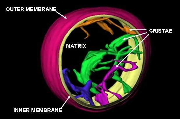
A mitochondrion () is an organelle found in the cells of most eukaryotes, such as animals, plants and fungi. Mitochondria have a double membrane structure and use aerobic respiration to generate adenosine triphosphate (ATP), which is used throughout the cell as a source of chemical energy. They were discovered by Albert von Kölliker in 1857 in the voluntary muscles of insects. The term ''mitochondrion'', meaning a thread-like granule, was coined by Carl Benda in 1898. The mitochondrion is popularly nicknamed the "powerhouse of the cell", a phrase popularized by Philip Siekevitz in a 1957 ''Scientific American'' article of the same name. Some cells in some multicellular organisms lack mitochondria (for example, mature mammalian red blood cells). The multicellular animal '' Henneguya salminicola'' is known to have retained mitochondrion-related organelles despite a complete loss of their mitochondrial genome. A large number of unicellular organisms, such as microspo ... [...More Info...] [...Related Items...] OR: [Wikipedia] [Google] [Baidu] |
Hürthle Cell
A Hürthle cell is a transformed (metaplasia) thyroid follicular cell with "enlarged mitochondria and enlarged round nuclei with prominent nucleoli", resulting in eosinophilia in the cytoplasm. Oncocytes in the thyroid are often called Hürthle cells. Although the terms oncocyte, oxyphil cell, and Hürthle cell are used interchangeably, "Hürthle cell" is used only to indicate cells of thyroid follicular origin.Cannon, J. (2011). The Significance of Hurthle Cells in Thyroid Disease. The Oncologist. doi:10.1634/theoncologist.2010-0253 Diseases While Hurthle cells can occur in healthy thyroid glands, they are often associated with Hashimoto's thyroiditis and Graves' disease. Hürthle cell neoplasms can be separated into Hürthle cell adenomas (benign tumours) and carcinomas (malignant tumours) arising from the follicular epithelium of the thyroid gland. The latter is a relatively rare form of differentiated thyroid cancer, accounting for only 3-10% of all differentiated thyr ... [...More Info...] [...Related Items...] OR: [Wikipedia] [Google] [Baidu] |
Micrograph
A micrograph is an image, captured photographically or digitally, taken through a microscope or similar device to show a magnify, magnified image of an object. This is opposed to a macrograph or photomacrograph, an image which is also taken on a microscope but is only slightly magnified, usually less than 10 times. Micrography is the practice or art of using microscopes to make photographs. A photographic micrograph is a photomicrograph, and one taken with an electron microscope is an electron micrograph. A micrograph contains extensive details of microstructure. A wealth of information can be obtained from a simple micrograph like behavior of the material under different conditions, the phases found in the system, failure analysis, grain size estimation, elemental analysis and so on. Micrographs are widely used in all fields of microscopy. Types Photomicrograph A light micrograph or photomicrograph is a micrograph prepared using an optical microscope, a process referred to ... [...More Info...] [...Related Items...] OR: [Wikipedia] [Google] [Baidu] |
Neoplasm
A neoplasm () is a type of abnormal and excessive growth of tissue. The process that occurs to form or produce a neoplasm is called neoplasia. The growth of a neoplasm is uncoordinated with that of the normal surrounding tissue, and persists in growing abnormally, even if the original trigger is removed. This abnormal growth usually forms a mass, which may be called a tumour or tumor.'' ICD-10 classifies neoplasms into four main groups: benign neoplasms, in situ neoplasms, malignant neoplasms, and neoplasms of uncertain or unknown behavior. Malignant neoplasms are also simply known as cancers and are the focus of oncology. Prior to the abnormal growth of tissue, such as neoplasia, cells often undergo an abnormal pattern of growth, such as metaplasia or dysplasia. However, metaplasia or dysplasia does not always progress to neoplasia and can occur in other conditions as well. The word neoplasm is from Ancient Greek 'new' and 'formation, creation'. Types A neoplasm ... [...More Info...] [...Related Items...] OR: [Wikipedia] [Google] [Baidu] |
Nephrectomy
A nephrectomy is the surgical removal of a kidney, performed to treat a number of kidney diseases including kidney cancer. It is also done to remove a normal healthy kidney from a living or deceased donor, which is part of a kidney transplant procedure. History The first recorded nephrectomy was performed in 1861 by Erastus B. Wolcott in Wisconsin. The patient had had a large tumor and the operation was initially successful, but the patient died fifteen days later. The first planned nephrectomy was performed by the German surgeon Gustav Simon (surgeon), Gustav Simon on August 2, 1869, in Heidelberg. Simon practiced the operation beforehand in animal experiments. He proved that one healthy kidney can be sufficient for urine excretion in humans. Indications There are various indications for this procedure, including renal cell carcinoma, a non-functioning kidney (which may cause arterial hypertension, high blood pressure) and a congenitally small kidney (in which the kidney is sw ... [...More Info...] [...Related Items...] OR: [Wikipedia] [Google] [Baidu] |
Gross Examination
Gross processing, "grossing" or "gross pathology" is the process by which pathology specimens undergo examination with the bare eye to obtain diagnosis, diagnostic information, as well as cutting and tissue sampling in order to prepare material for subsequent histopathology, microscopic ''examination.'' Responsibility Gross examination of surgical pathology, surgical specimens is typically performed by a pathology, pathologist, or by a pathologists' assistant working within a pathology practice. Individuals trained in these fields are often able to gather diagnostically critical information in this stage of processing, including the stage and margin status of surgically removed tumors. Steps The initial step in any examination of a clinical specimen is confirmation of the identity of the patient and the anatomy, anatomical site from which the specimen was obtained. Sufficient clinical data should be communicated by the clinical team to the pathology team in order to guide the app ... [...More Info...] [...Related Items...] OR: [Wikipedia] [Google] [Baidu] |
Carcinomas
Carcinoma is a malignancy that develops from epithelial cells. Specifically, a carcinoma is a cancer that begins in a tissue that lines the inner or outer surfaces of the body, and that arises from cells originating in the endodermal, mesodermal or ectodermal germ layer during embryogenesis. Carcinomas occur when the DNA of a cell is damaged or altered and the cell begins to grow uncontrollably and becomes malignant. It is from the (itself derived from meaning ''crab''). Classification As of 2004, no simple and comprehensive classification system has been devised and accepted within the scientific community. Traditionally, however, malignancies have generally been classified into various types using a combination of criteria, including: The cell type from which they start; specifically: * Epithelial cells ⇨ carcinoma * Non-hematopoietic mesenchymal cells ⇨ sarcoma * Hematopoietic cells ** Bone marrow–derived cells that normally mature in the bloodstream ⇨ leukemi ... [...More Info...] [...Related Items...] OR: [Wikipedia] [Google] [Baidu] |
Adenomas
An adenoma is a benign tumor of epithelial tissue with glandular origin, glandular characteristics, or both. Adenomas can grow from many glandular organs, including the adrenal glands, pituitary gland, thyroid, prostate, and others. Some adenomas grow from epithelial tissue in nonglandular areas but express glandular tissue structure (as can happen in familial polyposis coli). Although adenomas are benign, they should be treated as pre-cancerous. Over time adenomas may transform to become malignant, at which point they are called adenocarcinomas. Most adenomas do not transform. However, even though benign, they have the potential to cause serious health complications by compressing other structures (mass effect) and by producing large amounts of hormones in an unregulated, non-feedback-dependent manner (causing paraneoplastic syndromes). Some adenomas are too small to be seen macroscopically but can still cause clinical symptoms. Histopathology Adenoma is a benign tumor of g ... [...More Info...] [...Related Items...] OR: [Wikipedia] [Google] [Baidu] |
Acidophilic
Acidophiles or acidophilic organisms are those that thrive under highly acidic conditions (usually at pH 5.0 or below). These organisms can be found in different branches of the tree of life, including Archaea, Bacteria,Becker, A.Types of Bacteria Living in Acidic pH" Retrieved 10 May 2017. and Eukarya. Examples A list of these organisms includes: Archaea :* Sulfolobales, an order in the Thermoproteota branch of Archaea :* Thermoplasmatales, an order in the Euryarchaeota branch of Archaea :* ARMAN, in the Euryarchaeota branch of Archaea :* '' Acidianus brierleyi, A. infernus'', facultatively anaerobic thermoacidophilic archaebacteria :* ''Halarchaeum acidiphilum'', acidophilic member of the Halobacteriacaeae :* ''Metallosphaera sedula'', thermoacidophilic Bacteria :* Acidobacteriota, a phylum of Bacteria :* Acidithiobacillales, an order of Pseudomonadota e.g. ''A. ferrooxidans, A. thiooxidans'' :*''Thiobacillus prosperus, T. acidophilus, T. organovorus, T. cuprinus'' :*'' Ace ... [...More Info...] [...Related Items...] OR: [Wikipedia] [Google] [Baidu] |


