|
Nucleolus (including Pre-rRNA Components)
The nucleolus (; : nucleoli ) is the largest structure in the nucleus of eukaryotic cells. It is best known as the site of ribosome biogenesis. The nucleolus also participates in the formation of signal recognition particles and plays a role in the cell's response to stress. Nucleoli are made of proteins, DNA and RNA, and form around specific chromosomal regions called nucleolar organizing regions. Malfunction of the nucleolus is the cause of several human conditions called "nucleolopathies" and the nucleolus is being investigated as a target for cancer chemotherapy. History The nucleolus was identified by bright-field microscopy during the 1830s. Theodor Schwann in his 1839 treatise described that Schleiden had identified small corpuscles in nuclei, and named the structures "Kernkörperchen". In a 1947 translation of the work to English, the structure was named "nucleolus". Little was known about the function of the nucleolus until 1964, when a study of nucleoli by ... [...More Info...] [...Related Items...] OR: [Wikipedia] [Google] [Baidu] |
Diagram Human Cell Nucleus
A diagram is a symbolic representation of information using visualization techniques. Diagrams have been used since prehistoric times on walls of caves, but became more prevalent during the Enlightenment. Sometimes, the technique uses a three-dimensional visualization which is then projected onto a two-dimensional surface. The word ''graph'' is sometimes used as a synonym for diagram. Overview The term "diagram" in its commonly used sense can have a general or specific meaning: * ''visual information device'' : Like the term "illustration", "diagram" is used as a collective term standing for the whole class of technical genres, including graphs, technical drawings and tables. * ''specific kind of visual display'' : This is the genre that shows qualitative data with shapes that are connected by lines, arrows, or other visual links. In science the term is used in both ways. For example, Anderson (1997) stated more generally: "diagrams are pictorial, yet abstract, represent ... [...More Info...] [...Related Items...] OR: [Wikipedia] [Google] [Baidu] |
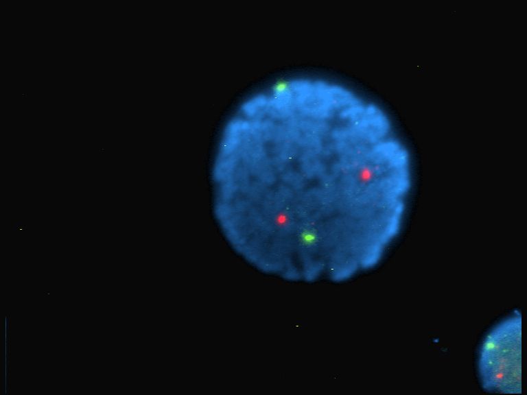
Nucleic Acid Hybridization
In molecular biology, hybridization (or hybridisation) is a phenomenon in which single-stranded deoxyribonucleic acid (DNA) or ribonucleic acid (RNA) molecules anneal to complementary DNA or RNA. Though a double-stranded DNA sequence is generally stable under physiological conditions, changing these conditions in the laboratory (generally by raising the surrounding temperature) will cause the molecules to separate into single strands. These strands are complementary to each other but may also be complementary to other sequences present in their surroundings. Lowering the surrounding temperature allows the single-stranded molecules to anneal or “hybridize” to each other. DNA replication and transcription of DNA into RNA both rely upon nucleotide hybridization, as do molecular biology techniques including Southern blots and Northern blots, the polymerase chain reaction (PCR), and most approaches to DNA sequencing. Applications Hybridization is a basic property of nucleotide ... [...More Info...] [...Related Items...] OR: [Wikipedia] [Google] [Baidu] |
Fluorescence Recovery After Photobleaching
Fluorescence recovery after photobleaching (FRAP) is a method for determining the kinetics of diffusion through tissue or cells. It is capable of quantifying the two-dimensional lateral diffusion of a molecularly thin film containing fluorescently labeled probes, or to examine single cells. This technique is very useful in biological studies of cell membrane diffusion and protein binding. In addition, surface deposition of a fluorescing phospholipid bilayer (or monolayer) allows the characterization of hydrophilic (or hydrophobic) surfaces in terms of surface structure and free energy. Similar, though less well known, techniques have been developed to investigate the 3-dimensional diffusion and binding of molecules inside the cell; they are also referred to as FRAP. Experimental setup The basic apparatus comprises an optical microscope, a light source and some fluorescent probe. Fluorescent emission is contingent upon absorption of a specific optical wavelength or color which ... [...More Info...] [...Related Items...] OR: [Wikipedia] [Google] [Baidu] |
Photobleaching
In optics, photobleaching (sometimes termed fading) is the photochemical alteration of a dye or a fluorophore molecule such that it is permanently unable to fluoresce. This is caused by cleaving of covalent bonds or non-specific reactions between the fluorophore and surrounding molecules. Such irreversible modifications in covalent bonds are caused by transition from a singlet state to the triplet state of the fluorophores. The number of excitation cycles to achieve full bleaching varies. In microscopy, photobleaching may complicate the observation of fluorescent molecules, since they will eventually be destroyed by the light exposure necessary to stimulate them into fluorescing. This is especially problematic in time-lapse microscopy. However, photobleaching may also be used prior to applying the (primarily antibody-linked) fluorescent molecules, in an attempt to quench autofluorescence. This can help improve the signal-to-noise ratio. Photobleaching may also be exploited to s ... [...More Info...] [...Related Items...] OR: [Wikipedia] [Google] [Baidu] |
Fluorophore
A fluorophore (or fluorochrome, similarly to a chromophore) is a fluorescent chemical compound that can re-emit light upon light excitation. Fluorophores typically contain several combined aromatic groups, or planar or cyclic molecules with several π bonds. Fluorophores are sometimes used alone, as a tracer in fluids, as a dye for staining of certain structures, as a substrate of enzymes, or as a probe or indicator (when its fluorescence is affected by environmental aspects such as polarity or ions). More generally they are covalently bonded to macromolecules, serving as a markers (or dyes, or tags, or reporters) for affine or bioactive reagents (antibodies, peptides, nucleic acids). Fluorophores are notably used to stain tissues, cells, or materials in a variety of analytical methods, such as fluorescent imaging and spectroscopy. Fluorescein, via its amine-reactive isothiocyanate derivative fluorescein isothiocyanate (FITC), has been one of the most popular fluorophores ... [...More Info...] [...Related Items...] OR: [Wikipedia] [Google] [Baidu] |
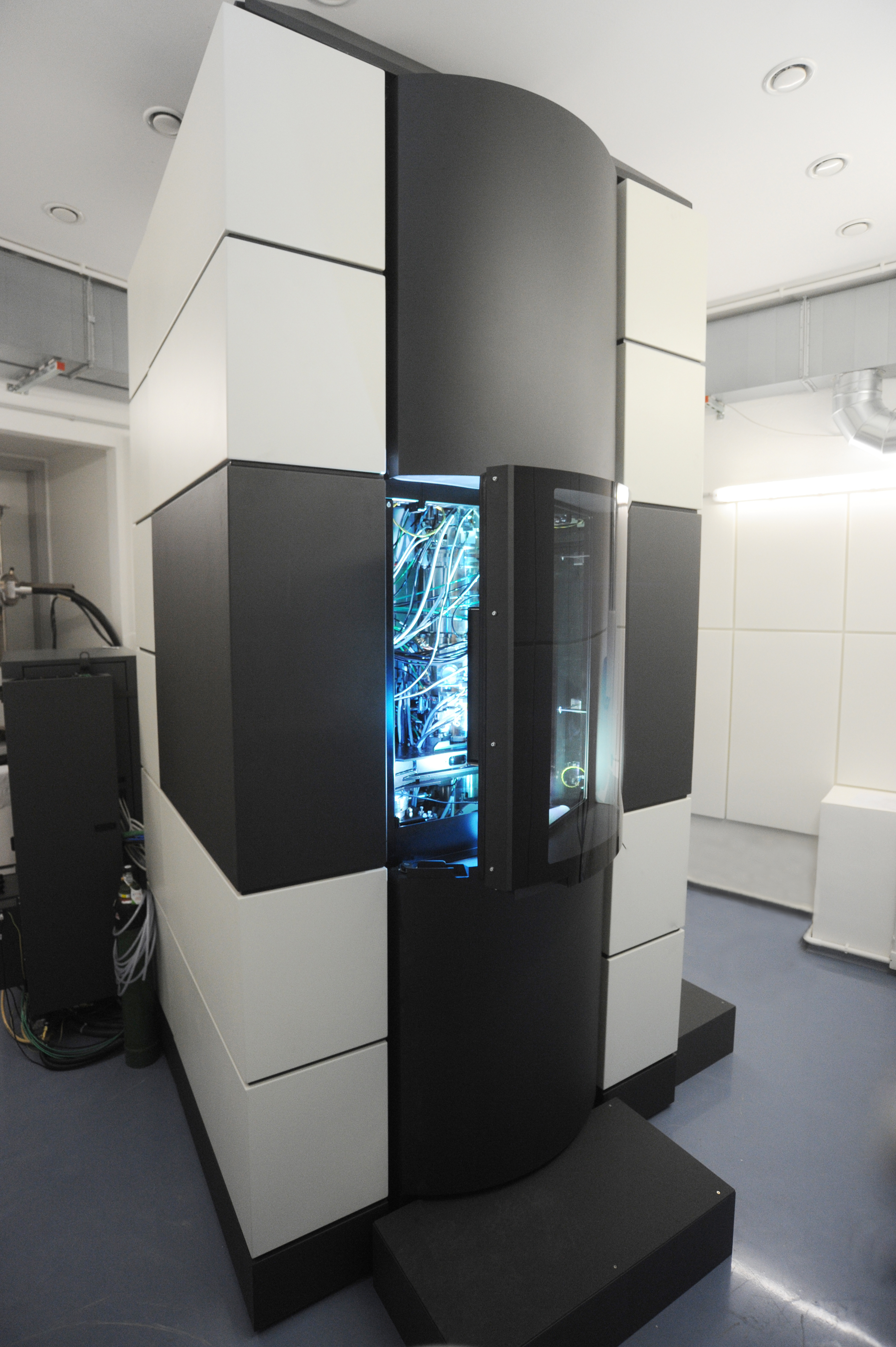
Electron Microscope
An electron microscope is a microscope that uses a beam of electrons as a source of illumination. It uses electron optics that are analogous to the glass lenses of an optical light microscope to control the electron beam, for instance focusing it to produce magnified images or electron diffraction patterns. As the wavelength of an electron can be up to 100,000 times smaller than that of visible light, electron microscopes have a much higher Angular resolution, resolution of about 0.1 nm, which compares to about 200 nm for optical microscope, light microscopes. ''Electron microscope'' may refer to: * Transmission electron microscopy, Transmission electron microscope (TEM) where swift electrons go through a thin sample * Scanning transmission electron microscopy, Scanning transmission electron microscope (STEM) which is similar to TEM with a scanned electron probe * Scanning electron microscope (SEM) which is similar to STEM, but with thick samples * Electron microprobe sim ... [...More Info...] [...Related Items...] OR: [Wikipedia] [Google] [Baidu] |
Ultrastructure
Ultrastructure (or ultra-structure) is the architecture of cells and biomaterials that is visible at higher magnifications than found on a standard optical light microscope. This traditionally meant the resolution and magnification range of a conventional transmission electron microscope (TEM) when viewing biological specimens such as cells, tissue, or organs. Ultrastructure can also be viewed with scanning electron microscopy and super-resolution microscopy, although TEM is a standard histology technique for viewing ultrastructure. Such cellular structures as organelles, which allow the cell to function properly within its specified environment, can be examined at the ultrastructural level. Ultrastructure, along with molecular phylogeny, is a reliable phylogenetic way of classifying organisms. Features of ultrastructure are used industrially to control material properties and promote biocompatibility. History In 1931, German engineers Max Knoll and Ernst Ruska invente ... [...More Info...] [...Related Items...] OR: [Wikipedia] [Google] [Baidu] |
Intergenic Region
An intergenic region is a stretch of DNA sequences located between genes. Intergenic regions may contain functional elements and junk DNA. Properties and functions Intergenic regions may contain a number of functional DNA sequences such as promoters and regulatory elements, enhancers, spacers, and (in eukaryotes) centromeres. They may also contain origins of replication, scaffold attachment regions, and transposons and viruses. Non-functional DNA elements such as pseudogenes and repetitive DNA, both of which are types of junk DNA, can also be found in intergenic regions—although they may also be located within genes in introns. It is possible that these regions contain as of yet unidentified functional elements, such as non-coding genes or regulatory sequences. This indeed occurs occasionally, but the amount of functional DNA discovered usually constitute only a tiny fraction of the overall amount of intergenic or intronic DNA. Intergenic regions in differ ... [...More Info...] [...Related Items...] OR: [Wikipedia] [Google] [Baidu] |
Amniote
Amniotes are tetrapod vertebrate animals belonging to the clade Amniota, a large group that comprises the vast majority of living terrestrial animal, terrestrial and semiaquatic vertebrates. Amniotes evolution, evolved from amphibious Stem tetrapoda, stem tetrapod ancestors during the Carboniferous geologic period, period. Amniota is defined as the smallest crown clade containing humans, the Greek tortoise, and the Nile crocodile. Amniotes are distinguished from the other living tetrapod clade — the anamniote, non-amniote lissamphibians (frogs/toads, salamanders/newts and caecilians) — by: the development of three fetal membranes, extraembryonic membranes (amnion for embryonic protection, chorion for gas exchange, and allantois for metabolic waste disposal or storage); thicker and keratinized skin; rib, costal respiration (breathing by expanding/constricting the rib cage); the presence of adrenal cortex, adrenocortical and chromaffin cell, chromaffin tissues as adrenal g ... [...More Info...] [...Related Items...] OR: [Wikipedia] [Google] [Baidu] |
Anamniotes
The anamniotes are an informal group of craniates comprising all fish and amphibians, which lay their eggs in aquatic environments. They are distinguished from the amniotes (reptiles, birds and mammals), which can reproduce on dry land either by laying shelled eggs or by carrying fertilized eggs within the female. Older sources, particularly before the 20th century, may refer to anamniotes as "lower vertebrates" and amniotes as "higher vertebrates", based on the antiquated idea of the evolutionary great chain of being. The name "anamniote" is a back-formation word created by adding the prefix '' an-'' to the word ''amniote'', which in turn refers to the amnion, an extraembryonic membrane present during the amniotes' embryonic development which serves as a biochemical barrier that shields the embryo from environmental fluctuations by regulating the oxygen, carbon dioxide and metabolic waste exchanges and secreting a cushioning fluid. As the name suggests, anamniote embr ... [...More Info...] [...Related Items...] OR: [Wikipedia] [Google] [Baidu] |
NPM1
Nucleophosmin (NPM), also known as nucleolar phosphoprotein B23 or numatrin, is a protein that in humans is encoded by the ''NPM1'' gene. Gene In humans, the ''NPM1'' gene is located on the long arm of chromosome 5 (5q35). The gene spans 23 kb and contains 12 exons. Three transcript variants have been described. The longest isoform (294 amino acids long), encoded by transcript variant 1, is the major and the most well studied isoform of Nucleophosmin. Transcript variant 2 is produced by skipping an in-frame exon (exon 8) to produce an isoform that is 265 amino acids long. However, this isoform is not well characterized and its functions and expression pattern is not well understood. Transcript variant 3 is produced by using an alternate exon (exon 10) which results in an isoform 259 amino acids long with a different C-terminal sequence. The isoforms 1 and 3 of human NPM1 are B23.1 and B23.2 respectively in rat. The isoform 1 is localized to the nucleolus as is reported for rat ... [...More Info...] [...Related Items...] OR: [Wikipedia] [Google] [Baidu] |


