|
Nuchal Lines
The nuchal lines are four curved lines on the external surface of the occipital bone: * The upper, often faintly marked, is named the highest nuchal line, but is sometimes referred to as the Mempin line or linea suprema, and it attaches to the epicranial aponeurosis. * Below the highest nuchal line is the superior nuchal line. To it is attached, the splenius capitis muscle, the trapezius muscle, and the occipitalis. * From the external occipital protuberance a ridge or crest, the external occipital crest also called the median nuchal line, often faintly marked, descends to the foramen magnum, and affords attachment to the nuchal ligament. * Running from the middle of this line is the inferior nuchal line. Attached are the obliquus capitis superior muscle, rectus capitis posterior major muscle, and rectus capitis posterior minor muscle The rectus capitis posterior minor (or rectus capitis posticus minor) is a muscle in the upper back part of the neck. It is one of the suboccipita ... [...More Info...] [...Related Items...] OR: [Wikipedia] [Google] [Baidu] |
Occipital Bone
The occipital bone () is a neurocranium, cranial dermal bone and the main bone of the occiput (back and lower part of the skull). It is trapezoidal in shape and curved on itself like a shallow dish. The occipital bone lies over the occipital lobes of the cerebrum. At the base of the skull in the occipital bone, there is a large oval opening called the foramen magnum, which allows the passage of the spinal cord. Like the other cranial bones, it is classed as a flat bone. Due to its many attachments and features, the occipital bone is described in terms of separate parts. From its front to the back is the basilar part of occipital bone, basilar part, also called the basioccipital, at the sides of the foramen magnum are the lateral parts of occipital bone, lateral parts, also called the exoccipitals, and the back is named as the squamous part of occipital bone, squamous part. The basilar part is a thick, somewhat quadrilateral piece in front of the foramen magnum and directed toward ... [...More Info...] [...Related Items...] OR: [Wikipedia] [Google] [Baidu] |
Epicranial Aponeurosis
The epicranial aponeurosis (aponeurosis epicranialis, galea aponeurotica) is an aponeurosis (a tough layer of dense fibrous tissue). It covers the upper part of the skull in humans and many other animals. Structure In humans, the epicranial aponeurosis originates from the external occipital protuberance and highest nuchal lines of the occipital bone. It merges with the occipitofrontalis muscle. In front, it forms a short and narrow prolongation between its union with the frontalis muscle (the frontal part of the occipitofrontalis muscle). On either side, the epicranial aponeurosis attaches to the anterior auricular muscles and the superior auricular muscles. Here it is less aponeurotic, and is continued over the temporal fascia to the zygomatic arch as a layer of laminated areolar tissue. It is closely connected to the integument by the firm, dense, fibro-fatty layer which forms the superficial fascia of the scalp. It is attached to the pericranium by loose cellular tis ... [...More Info...] [...Related Items...] OR: [Wikipedia] [Google] [Baidu] |
Splenius Capitis Muscle
The splenius capitis () () is a broad, straplike muscle in the back of the neck. It pulls on the base of the skull from the vertebrae in the neck and upper thorax. It is involved in movements such as shaking the head. Structure It arises from the lower half of the nuchal ligament, from the spinous process of the seventh cervical vertebra, and from the spinous processes of the upper three or four thoracic vertebrae. The fibers of the muscle are directed upward and laterally and are inserted, under cover of the sternocleidomastoideus, into the mastoid process of the temporal bone, and into the rough surface on the occipital bone The occipital bone () is a neurocranium, cranial dermal bone and the main bone of the occiput (back and lower part of the skull). It is trapezoidal in shape and curved on itself like a shallow dish. The occipital bone lies over the occipital lob ... just below the lateral third of the superior nuchal line. The splenius capitis is deep to ster ... [...More Info...] [...Related Items...] OR: [Wikipedia] [Google] [Baidu] |
Trapezius Muscle
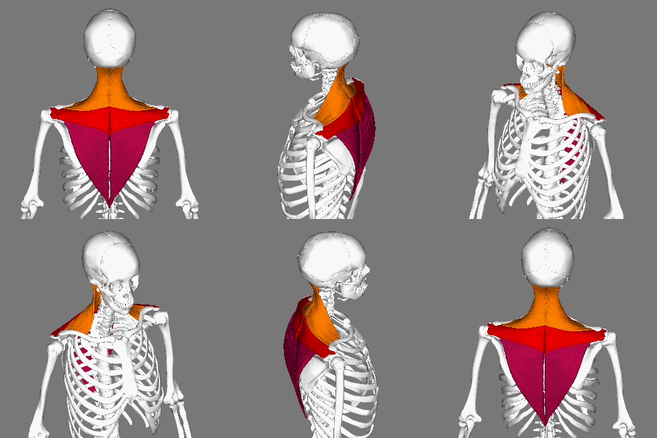
The trapezius is a large paired trapezoid-shaped surface muscle that extends longitudinally from the occipital bone to the lower thoracic vertebrae of the human spine, spine and laterally to the spine of the scapula. It moves the scapula and supports the arm. The trapezius has three functional parts: * an upper (descending) part which supports the weight of the arm; * a middle region (transverse), which retracts the scapula; and * a lower (ascending) part which medially rotates and depresses the scapula. Name and history The trapezius muscle resembles a trapezoid, trapezium, also known as a trapezoid, or diamond-shaped quadrilateral. The word "spinotrapezius" refers to the human trapezius, although it is not commonly used in modern texts. In other mammals, it refers to a portion of the analogous muscle. Structure The ''superior'' or ''upper'' (or descending) fibers of the trapezius originate from the spinous process of C7, the external occipital protuberance, the me ... [...More Info...] [...Related Items...] OR: [Wikipedia] [Google] [Baidu] |
Occipitalis
The occipitalis muscle (occipital belly) is a muscle which covers parts of the skull. Some sources consider the occipital muscle to be a distinct muscle. However, Terminologia Anatomica currently classifies it as part of the occipitofrontalis muscle along with the frontalis muscle. The occipitalis muscle is thin and quadrilateral in form. It arises from tendinous fibers from the lateral two-thirds of the superior nuchal line of the occipital bone and from the mastoid process of the temporal and ends in the epicranial aponeurosis. The occipitalis muscle is innervated by the posterior auricular nerve (a branch of the facial nerve) and its function is to move the scalp back. The muscles receives blood from the occipital artery. Additional image File:Occipitalis muscle animation small.gif, Position of occipitalis muscle (shown in red). See also * Occipitofrontalis muscle The occipitofrontalis muscle (epicranius muscle) is a muscle which covers parts of the skull. It consists o ... [...More Info...] [...Related Items...] OR: [Wikipedia] [Google] [Baidu] |
External Occipital Protuberance
External may refer to: * Externality, in economics, the cost or benefit that affects a party who did not choose to incur that cost or benefit * Externals, a fictional group of X-Men antagonists See also * * Internal (other) {{disambig ... [...More Info...] [...Related Items...] OR: [Wikipedia] [Google] [Baidu] |
External Occipital Crest
The external occipital crest is part of the external surface of the squamous part of the occipital bone. It is a ridge along the midline, beginning at the external occipital protuberance and descending to the foramen magnum, that gives attachment to the nuchal ligament The nuchal ligament is a ligament at the back of the neck that is continuous with the supraspinous ligament. Structure The nuchal ligament extends from the external occipital protuberance on the skull and median nuchal line to the spinous p .... It is also called the median nuchal line. References External links Bones of the head and neck {{musculoskeletal-stub ... [...More Info...] [...Related Items...] OR: [Wikipedia] [Google] [Baidu] |
Foramen Magnum
The foramen magnum () is a large, oval-shaped opening in the occipital bone of the skull. It is one of the several oval or circular openings (foramina) in the base of the skull. The spinal cord, an extension of the medulla oblongata, passes through the foramen magnum as it exits the cranial cavity. Apart from the transmission of the medulla oblongata and its membranes, the foramen magnum transmits the vertebral arteries, the anterior and posterior spinal arteries, the tectorial membranes and alar ligaments. It also transmits the accessory nerve into the skull. The foramen magnum is a very important feature in bipedal mammals. One of the attributes of a biped's foramen magnum is a forward shift of the anterior border of the cerebellar tentorium; this is caused by the shortening of the cranial base. Studies on the foramen magnum position have shown a connection to the functional influences of both posture and locomotion. The forward shift of the foramen magnum is apparent in b ... [...More Info...] [...Related Items...] OR: [Wikipedia] [Google] [Baidu] |
Nuchal Ligament
The nuchal ligament is a ligament at the back of the neck that is continuous with the supraspinous ligament. Structure The nuchal ligament extends from the external occipital protuberance on the skull and median nuchal line to the spinous process of the seventh cervical vertebra in the lower part of the neck. From the anterior border of the nuchal ligament, a fibrous lamina is given off. This is attached to the posterior tubercle of the atlas, and to the spinous processes of the cervical vertebrae, and forms a septum between the muscles on either side of the neck. The trapezius and splenius capitis muscle attach to the nuchal ligament. Function It is a tendon-like structure that has developed independently in humans and other animals well adapted for running. In some four-legged animals, particularly ungulates and canids, the nuchal ligament serves to sustain the weight of the head. Clinical significance In Chiari malformation treatment, decompression and duraplas ... [...More Info...] [...Related Items...] OR: [Wikipedia] [Google] [Baidu] |
Obliquus Capitis Superior Muscle
The obliquus capitis superior muscle () is a small muscle in the upper back part of the neck. It is one of the suboccipital muscles. It attaches inferiorly at the transverse process of the atlas (first cervical vertebra); it attaches superiorly at the external surface of the occipital bone. The muscle is innervated by the suboccipital nerve (the posterior ramus of the first cervical spinal nerve). It acts at the atlanto-occipital joint to extend the head and bend the head to the same side. Anatomy The obliquus capitis superior muscle is one of the suboccipital muscles. It forms the superolateral boundary of the suboccipital triangle. It extends superoposteriorly from its inferior attachment to its superior attachment, becoming wider superiorly. Attachments The muscle's inferior attachment is at the superior surface of the transverse process of the atlas (C1). Its superior attachment is onto the lateral portion of the external surface of the occipital bone between the ... [...More Info...] [...Related Items...] OR: [Wikipedia] [Google] [Baidu] |
Rectus Capitis Posterior Major Muscle
The rectus capitis posterior major (or rectus capitis posticus major) is a muscle in the upper back part of the neck. It is one of the suboccipital muscles. Its inferior attachment is at the spinous process of the axis (Second cervical vertebra); its superior attachment is onto the outer surface of the occipital bone on and around the side part of the inferior nuchal line. The muscle is innervated by the suboccipital nerve (the posterior ramus of cervical spinal nerve C1). The muscle acts to extend the head and rotate the head to its side. Anatomy The rectus capitis posterior major muscle is one of the suboccipital muscles. It forms the superomedial boundary of the suboccipital triangle. The muscle extends obliquely superiolaterally from its inferior attachment to its superior attachment. It becomes broader superiorly. Attachments Its inferior attachment is (via a pointed tendon) at (the external aspect of) the (bifid) spinous process of the axis (cervical vertebra C2) ... [...More Info...] [...Related Items...] OR: [Wikipedia] [Google] [Baidu] |

