|
Obliquus Capitis Superior Muscle
The obliquus capitis superior muscle () is a small muscle in the upper back part of the neck. It is one of the suboccipital muscles. It attaches inferiorly at the transverse process of the atlas (first cervical vertebra); it attaches superiorly at the external surface of the occipital bone. The muscle is innervated by the suboccipital nerve (the posterior ramus of the first cervical spinal nerve). It acts at the atlanto-occipital joint to extend the head and bend the head to the same side. Anatomy The obliquus capitis superior muscle is one of the suboccipital muscles. It forms the superolateral boundary of the suboccipital triangle. It extends superoposteriorly from its inferior attachment to its superior attachment, becoming wider superiorly. Attachments The muscle's inferior attachment is at the superior surface of the transverse process of the atlas (C1). Its superior attachment is onto the lateral portion of the external surface of the occipital bone between the ... [...More Info...] [...Related Items...] OR: [Wikipedia] [Google] [Baidu] |
Human Skull
The skull, or cranium, is typically a bony enclosure around the brain of a vertebrate. In some fish, and amphibians, the skull is of cartilage. The skull is at the head end of the vertebrate. In the human, the skull comprises two prominent parts: the neurocranium and the facial skeleton, which evolved from the first pharyngeal arch. The skull forms the frontmost portion of the axial skeleton and is a product of cephalization and vesicular enlargement of the brain, with several special senses structures such as the eyes, ears, nose, tongue and, in fish, specialized tactile organs such as barbels near the mouth. The skull is composed of three types of bone: cranial bones, facial bones and ossicles, which is made up of a number of fused flat and irregular bones. The cranial bones are joined at firm fibrous junctions called sutures and contains many foramina, fossae, processes, and sinuses. In zoology, the openings in the skull are called fenestrae, the ... [...More Info...] [...Related Items...] OR: [Wikipedia] [Google] [Baidu] |
Spinal Nerve
A spinal nerve is a mixed nerve, which carries Motor neuron, motor, Sensory neuron, sensory, and Autonomic nervous system, autonomic signals between the spinal cord and the body. In the human body there are 31 pairs of spinal nerves, one on each side of the vertebral column. These are grouped into the corresponding cervical vertebrae, cervical, thoracic vertebrae, thoracic, lumbar vertebrae, lumbar, sacral vertebrae, sacral and coccygeal vertebrae, coccygeal regions of the spine. There are eight pairs of cervical nerves, twelve pairs of thoracic nerves, five pairs of lumbar nerves, five pairs of sacral nerves, and one pair of coccygeal nerves. The spinal nerves are part of the peripheral nervous system. Structure Each spinal nerve is a mixed nerve, formed from the combination of nerve root axon, fibers from its Dorsal root of spinal nerve, dorsal and Ventral root of spinal nerve, ventral roots. The dorsal root is the afferent nerve fiber, afferent sensory root and carries sen ... [...More Info...] [...Related Items...] OR: [Wikipedia] [Google] [Baidu] |
Occipital Bone
The occipital bone () is a neurocranium, cranial dermal bone and the main bone of the occiput (back and lower part of the skull). It is trapezoidal in shape and curved on itself like a shallow dish. The occipital bone lies over the occipital lobes of the cerebrum. At the base of the skull in the occipital bone, there is a large oval opening called the foramen magnum, which allows the passage of the spinal cord. Like the other cranial bones, it is classed as a flat bone. Due to its many attachments and features, the occipital bone is described in terms of separate parts. From its front to the back is the basilar part of occipital bone, basilar part, also called the basioccipital, at the sides of the foramen magnum are the lateral parts of occipital bone, lateral parts, also called the exoccipitals, and the back is named as the squamous part of occipital bone, squamous part. The basilar part is a thick, somewhat quadrilateral piece in front of the foramen magnum and directed toward ... [...More Info...] [...Related Items...] OR: [Wikipedia] [Google] [Baidu] |
Back Muscles
The human back, also called the dorsum (: dorsa), is the large posterior area of the human body, rising from the top of the buttocks to the back of the neck. It is the surface of the body opposite from the chest and the abdomen. The vertebral column runs the length of the back and creates a central area of recession. The breadth of the back is created by the shoulders at the top and the pelvis at the bottom. Back pain is a common medical condition, generally benign in origin. Structure The central feature of the human back is the vertebral column, specifically the length from the top of the thoracic vertebrae to the bottom of the lumbar vertebrae, which houses the spinal cord in its spinal canal, and which generally has some curvature that gives shape to the back. The ribcage extends from the spine at the top of the back (with the top of the ribcage corresponding to the T1 vertebra), more than halfway down the length of the back, leaving an area with less protection between the ... [...More Info...] [...Related Items...] OR: [Wikipedia] [Google] [Baidu] |
Posterior Ramus Of Spinal Nerve
The dorsal ramus of spinal nerve, posterior ramus of spinal nerve, or posterior primary division is the posterior division of a spinal nerve. The dorsal rami provide motor innervation to the deep (a.k.a. intrinsic or true) muscles of the back, and sensory innervation to the skin of the posterior portion of the head, neck and back. A spinal nerve splits within the intervertebral foramen to form a dorsal ramus and a ventral ramus. The dorsal ramus then turns to course posterior-ward before splitting into a medial branch and a lateral branch. Both these branches provide motor innervation to deep back muscles. In the neck and upper back, the medial branch is also responsible for providing sensory innervation of the skin; in the lower back, the lateral branch does so. All medial branches additionally also provide sensory innervation to the zygapophyseal joints and periosteum of the vertebral column. Structure Ventral root axons join with dorsal root ganglia to form mixed spinal nerves ... [...More Info...] [...Related Items...] OR: [Wikipedia] [Google] [Baidu] |
Rectus Capitis Posterior Major Muscle
The rectus capitis posterior major (or rectus capitis posticus major) is a muscle in the upper back part of the neck. It is one of the suboccipital muscles. Its inferior attachment is at the spinous process of the axis (Second cervical vertebra); its superior attachment is onto the outer surface of the occipital bone on and around the side part of the inferior nuchal line. The muscle is innervated by the suboccipital nerve (the posterior ramus of cervical spinal nerve C1). The muscle acts to extend the head and rotate the head to its side. Anatomy The rectus capitis posterior major muscle is one of the suboccipital muscles. It forms the superomedial boundary of the suboccipital triangle. The muscle extends obliquely superiolaterally from its inferior attachment to its superior attachment. It becomes broader superiorly. Attachments Its inferior attachment is (via a pointed tendon) at (the external aspect of) the (bifid) spinous process of the axis (cervical vertebra C2) ... [...More Info...] [...Related Items...] OR: [Wikipedia] [Google] [Baidu] |
Semispinalis Capitis
The semispinalis muscles are a group of three muscles belonging to the transversospinales. These are the semispinalis capitis, the semispinalis cervicis and the semispinalis thoracis. Location The semispinalis capitis (''complexus'') is situated at the upper and back part of the neck, deep to the splenius muscles, and medial to the longissimus cervicis and longissimus capitis. It arises by a series of tendons from the tips of the transverse processes of the upper six or seven thoracic and the seventh cervical vertebrae, and from the articular processes of the three cervical vertebrae above this ( C4-C6). The tendons, uniting, form a broad muscle, which passes upward, and is inserted between the superior and inferior nuchal lines of the occipital bone. It lies deep to the trapezius muscle and can be palpated as a firm round muscle mass just lateral to the cervical spinous processes. The semispinalis cervicis (or ''semispinalis colli''), arises by a series of tendinous and ... [...More Info...] [...Related Items...] OR: [Wikipedia] [Google] [Baidu] |
Superior Nuchal Line
The nuchal lines are four curved lines on the external surface of the occipital bone: * The upper, often faintly marked, is named the highest nuchal line, but is sometimes referred to as the Mempin line or linea suprema, and it attaches to the epicranial aponeurosis. * Below the highest nuchal line is the superior nuchal line. To it is attached, the splenius capitis muscle, the trapezius muscle, and the occipitalis. * From the external occipital protuberance a ridge or crest, the external occipital crest also called the median nuchal line, often faintly marked, descends to the foramen magnum, and affords attachment to the nuchal ligament The nuchal ligament is a ligament at the back of the neck that is continuous with the supraspinous ligament. Structure The nuchal ligament extends from the external occipital protuberance on the skull and median nuchal line to the spinous p .... * Running from the middle of this line is the inferior nuchal line. Attached are the obliq ... [...More Info...] [...Related Items...] OR: [Wikipedia] [Google] [Baidu] |
Atlas (vertebra)
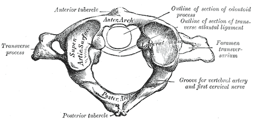
In anatomy, the atlas (C1) is the most superior (first) cervical vertebra of the spine and is located in the neck. The bone is named for Atlas of Greek mythology, just as Atlas bore the weight of the heavens, the first cervical vertebra supports the head. However, the term atlas was first used by the ancient Romans for the seventh cervical vertebra (C7) due to its suitability for supporting burdens. In Greek mythology, Atlas was condemned to bear the weight of the heavens as punishment for rebelling against Zeus. Ancient depictions of Atlas show the globe of the heavens resting at the base of his neck, on C7. Sometime around 1522, anatomists decided to call the first cervical vertebra the atlas. Scholars believe that by switching the designation atlas from the seventh to the first cervical vertebra Renaissance anatomists were commenting that the point of man's burden had shifted from his shoulders to his head—that man's true burden was not a physical load, but rather, his min ... [...More Info...] [...Related Items...] OR: [Wikipedia] [Google] [Baidu] |
Transverse Process
Each vertebra (: vertebrae) is an irregular bone with a complex structure composed of bone and some hyaline cartilage, that make up the vertebral column or spine, of vertebrates. The proportions of the vertebrae differ according to their spinal segment and the particular species. The basic configuration of a vertebra varies; the vertebral body (also ''centrum'') is of bone and bears the load of the vertebral column. The upper and lower surfaces of the vertebra body give attachment to the intervertebral discs. The posterior part of a vertebra forms a vertebral arch, in eleven parts, consisting of two pedicles (pedicle of vertebral arch), two laminae, and seven processes. The laminae give attachment to the ligamenta flava (ligaments of the spine). There are vertebral notches formed from the shape of the pedicles, which form the intervertebral foramina when the vertebrae articulate. These foramina are the entry and exit conduits for the spinal nerves. The body of the vertebr ... [...More Info...] [...Related Items...] OR: [Wikipedia] [Google] [Baidu] |
Suboccipital Triangle
The suboccipital triangle is a region of the neck bounded by the following three muscles of the suboccipital group of muscles: * Rectus capitis posterior major - above and medially * Obliquus capitis superior - above and laterally * Obliquus capitis inferior - below and laterally (Rectus capitis posterior minor is also in this region but does not form part of the triangle) It is covered by a layer of dense fibro-fatty tissue, situated beneath the semispinalis capitis. The floor is formed by the posterior atlantooccipital membrane, and the posterior arch of the atlas. In the deep groove on the upper surface of the posterior arch of the atlas are the vertebral artery and the first cervical or suboccipital nerve. In the past, the vertebral artery was accessed here in order to conduct angiography of the circle of Willis. Presently, formal angiography of the circle of Willis is performed via catheter angiography, with access usually being acquired at the common femoral artery. Alte ... [...More Info...] [...Related Items...] OR: [Wikipedia] [Google] [Baidu] |
Atlanto-occipital Joint
The atlanto-occipital joint (''Articulatio atlantooccipitalis'') is an articulation between the atlas bone and the occipital bone. It consists of a pair of condyloid joints. It is a synovial joint. Structure The atlanto-occipital joint is an articulation between the atlas bone and the occipital bone. It consists of a pair of condyloid joints. It is a synovial joint. Ligaments The ligaments connecting the bones are: * Two articular capsules * Posterior atlanto-occipital membrane * Anterior atlanto-occipital membrane Capsule The capsules of the atlantooccipital articulation surround the condyles of the occipital bone, and connect them with the articular processes of the atlas: they are thin and loose. Variation Atlantooccipital fusion, also known as occipitalization of the atlas, is a congenital or acquired anomaly characterized by the partial or complete fusion of the atlas to the base of the occipital bone. It is found in 0.12% to 0.72% of the population. This fusio ... [...More Info...] [...Related Items...] OR: [Wikipedia] [Google] [Baidu] |


