|
Neurohypophysial Hormone
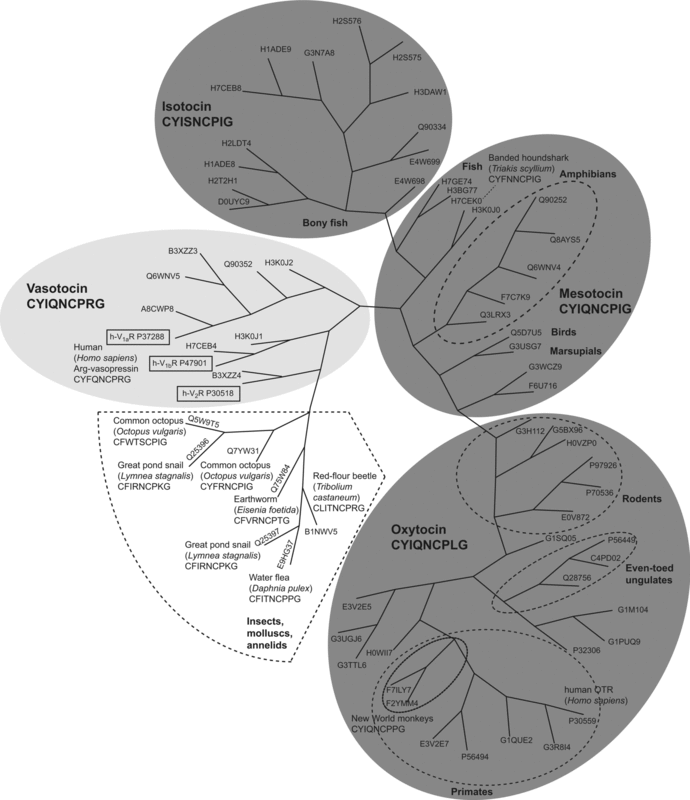
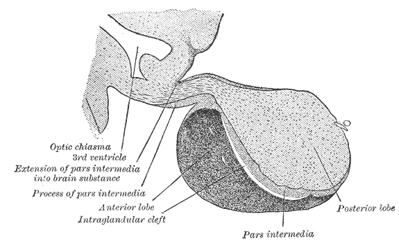
The neurohypophysial hormones form a family of structurally and functionally related peptide hormones. Their representatives in humans are oxytocin and vasopressin. They are named after the location of their release into the blood, the neurohypophysis (another name for the posterior pituitary). Most of the circulating oxytocin and vasopressin hormones are synthesized in magnocellular neurosecretory cells of the supraoptic nucleus and paraventricular nucleus of the hypothalamus. They are then transported in neurosecretory granules along axons within the hypothalamo-neurohypophysial tract by axoplasmic flow to axon terminals forming the pars nervosa of the posterior pituitary. There, they are stored in Herring bodies Herring bodies or neurosecretory bodies are structures found in the posterior pituitary. They represent the terminal end of the axons from the hypothalamus, and hormones are temporarily stored in these locations. They are neurosecretory termin ... and can ... [...More Info...] [...Related Items...] OR: [Wikipedia] [Google] [Baidu] |
Oxytocin
Oxytocin is a peptide hormone and neuropeptide normally produced in the hypothalamus and released by the posterior pituitary. Present in animals since early stages of evolution, in humans it plays roles in behavior that include Human bonding, social bonding, love, reproduction, childbirth, and the Postpartum period, period after childbirth. Oxytocin is released into the bloodstream as a hormone in response to Human sexual activity, sexual activity and during childbirth. It is also available in Oxytocin (medication), pharmaceutical form. In either form, oxytocin stimulates uterine contractions to speed up the process of childbirth. In its natural form, it also plays a role in maternal bonding and lactation, milk production. Production and secretion of oxytocin is controlled by a positive feedback mechanism, where its initial release stimulates production and release of further oxytocin. For example, when oxytocin is released during a contraction of the uterus at the start of c ... [...More Info...] [...Related Items...] OR: [Wikipedia] [Google] [Baidu] |
Axoplasmic Flow
Axonal transport, also called axoplasmic transport or axoplasmic flow, is a cellular process responsible for movement of mitochondrion, mitochondria, lipids, synaptic vesicles, proteins, and other organelles to and from a neuron's cell body, through the cytoplasm of its axon called the axoplasm. Since some axons are on the order of meters long, neurons cannot rely on diffusion to carry products of the nucleus and organelles to the ends of their axons. Axonal transport is also responsible for moving molecules destined for degradation from the axon back to the cell body, where they are broken down by lysosomes. Movement toward the cell body is called retrograde transport and movement toward the synapse is called anterograde transport. Mechanism The vast majority of axonal proteins are synthesized in the neuronal cell body and transported along axons. Some mRNA translation has been demonstrated within axons.Si K, Giustetto Axonal transport occurs throughout the life of a neuron a ... [...More Info...] [...Related Items...] OR: [Wikipedia] [Google] [Baidu] |
Protein Families
A protein family is a group of evolutionarily related proteins. In many cases, a protein family has a corresponding gene family, in which each gene encodes a corresponding protein with a 1:1 relationship. The term "protein family" should not be confused with family as it is used in taxonomy. Proteins in a family descend from a common ancestor and typically have similar three-dimensional structures, functions, and significant sequence similarity. Sequence similarity (usually amino-acid sequence) is one of the most common indicators of homology, or common evolutionary ancestry. Some frameworks for evaluating the significance of similarity between sequences use sequence alignment methods. Proteins that do not share a common ancestor are unlikely to show statistically significant sequence similarity, making sequence alignment a powerful tool for identifying the members of protein families. Families are sometimes grouped together into larger clades called superfamilies based on stru ... [...More Info...] [...Related Items...] OR: [Wikipedia] [Google] [Baidu] |
Vasopressin
Mammalian vasopressin, also called antidiuretic hormone (ADH), arginine vasopressin (AVP) or argipressin, is a hormone synthesized from the ''AVP'' gene as a peptide prohormone in neurons in the hypothalamus, and is converted to AVP. It then travels down the axon terminating in the posterior pituitary, and is released from vesicles into the circulation in response to extracellular fluid hypertonicity ( hyperosmolality). AVP has two primary functions. First, it increases the amount of solute-free water reabsorbed back into the circulation from the filtrate in the kidney tubules of the nephrons. Second, AVP constricts arterioles, which increases peripheral vascular resistance and raises arterial blood pressure. A third function is possible. Some AVP may be released directly into the brain from the hypothalamus, and may play an important role in social behavior, sexual motivation and pair bonding, and maternal responses to stress. Vasopressin induces differentiation o ... [...More Info...] [...Related Items...] OR: [Wikipedia] [Google] [Baidu] |
Antidiuretic
An antidiuretic is a chemical substance, substance that helps to control fluid balance in an animal's body by reducing urination, opposing diuresis. Its effects are opposite that of a diuretic. The major endogeny (biology), endogenous antidiuretics are Vasopressin, antidiuretic hormone (ADH; also called vasopressin) and oxytocin. Both of those are also used exogeny, exogenously as pharmaceutical drug, medications in people whose bodies need extra help with fluid balance via suppression of diuresis. In addition, there are various other antidiuretic drugs, some molecule, molecularly close to ADH or oxytocin and others not. Antidiuretics reduce urine volume, particularly in diabetes insipidus (DI), which is one of their main indication (medicine), indications. The antidiuretic hormone class includes vasopressin (ADH), argipressin, desmopressin, lypressin, ornipressin, oxytocin, and terlipressin. Miscellaneous others include chlorpropamide and carbamazepine. See also *Diuretic *Electro ... [...More Info...] [...Related Items...] OR: [Wikipedia] [Google] [Baidu] |
Smooth Muscle
Smooth muscle is one of the three major types of vertebrate muscle tissue, the others being skeletal and cardiac muscle. It can also be found in invertebrates and is controlled by the autonomic nervous system. It is non- striated, so-called because it has no sarcomeres and therefore no striations (''bands'' or ''stripes''). It can be divided into two subgroups, ''single-unit'' and ''multi-unit'' smooth muscle. Within single-unit muscle, the whole bundle or sheet of smooth muscle cells contracts as a syncytium. Smooth muscle is found in the walls of hollow organs, including the stomach, intestines, bladder and uterus. In the walls of blood vessels, and lymph vessels, (excluding blood and lymph capillaries) it is known as vascular smooth muscle. There is smooth muscle in the tracts of the respiratory, urinary, and reproductive systems. In the eyes, the ciliary muscles, iris dilator muscle, and iris sphincter muscle are types of smooth muscles. The iris dilator and s ... [...More Info...] [...Related Items...] OR: [Wikipedia] [Google] [Baidu] |
Pituicyte
Pituicytes are glial cells of the posterior pituitary. Their main role is to assist in the storage and release of neurohypophysial hormones. Structure Pituicytes are located in the pars nervosa of the posterior pituitary and interspersed with unmyelinated axons and Herring bodies. They generally stain dark purple with an H&E stain and are among the easiest structures to identify in the region. Pituicytes have an irregular and branched shape which resembles that of another type of glial cell: the astrocyte. Like astrocytes, their cytoplasm presents specific intermediate filaments made up of glial fibrillary acidic protein (GFAP). Function Pituicytes are similar to astrocytes, another type of glial cell. Their main role is to assist in the storage and release of hormones of the posterior pituitary. Pituicytes surround axonal endings and regulate hormone secretion by releasing their processes from these endings. Clinical significance Pituicytomas are rare tumors that arise from ... [...More Info...] [...Related Items...] OR: [Wikipedia] [Google] [Baidu] |
Herring Bodies
Herring bodies or neurosecretory bodies are structures found in the posterior pituitary. They represent the terminal end of the axons from the hypothalamus, and hormones are temporarily stored in these locations. They are neurosecretory terminals. Antidiuretic hormone (ADH) and oxytocin are both stored in Herring bodies, but are not stored simultaneously in the same Herring body. In addition, each Herring body also contains ATP and a type of neurophysin Neurophysins are carrier proteins which transport the hormones oxytocin and vasopressin to the posterior pituitary from the paraventricular nucleus, paraventricular and supraoptic nucleus of the hypothalamus, respectively. Inside the Neurosecretory .... Neurophysins are binding proteins, of which there are two types: neurophysin I and neurophysin II, which bind to oxytocin and ADH, respectively. Neurophysin and its hormone become a complex considered a single protein and stored in the neurohypophysis. Upon stimulation by th ... [...More Info...] [...Related Items...] OR: [Wikipedia] [Google] [Baidu] |
Axon Terminal
Axon terminals (also called terminal boutons, synaptic boutons, end-feet, or presynaptic terminals) are distal terminations of the branches of an axon. An axon, also called a nerve fiber, is a long, slender projection of a Neuron, nerve cell that conducts electrical impulses called action potentials away from the neuron's cell body to transmit those impulses to other neurons, muscle cells, or glands. Most presynaptic terminals in the central nervous system are formed along the axons (en passant boutons), not at their ends (terminal boutons). Functionally, the axon terminal converts an electrical signal into a chemical signal. When an action potential arrives at an axon terminal (A), neurotransmitter, the neurotransmitter is released and diffuses across the synaptic cleft. If the postsynaptic cell (B) is also a neuron, Neurotransmitter receptor, neurotransmitter receptors generate a small electrical current that changes the postsynaptic potential. If the postsynaptic cell (B) is ... [...More Info...] [...Related Items...] OR: [Wikipedia] [Google] [Baidu] |
Oxytocin Receptor
The oxytocin receptor, also known as OXTR, is a protein which functions as receptor for the hormone and neurotransmitter oxytocin. In humans, the oxytocin receptor is encoded by the ''OXTR'' gene which has been localized to human chromosome 3p25. Function and location The OXTR protein belongs to the G-protein coupled receptor family, specifically Gq, and acts as a receptor for oxytocin. Its activity is mediated by G proteins that activate several different second messenger systems. Oxytocin receptors are expressed by the myoepithelial cells of the mammary gland, and in both the myometrium and endometrium of the uterus at the end of pregnancy. The oxytocin-oxytocin receptor system plays an important role as an inducer of uterine contractions during parturition and of milk ejection. OXTR is also associated with the central nervous system. The gene is believed to play a major role in social, cognitive, and emotional behavior. A decrease in OXTR expression by methylation of th ... [...More Info...] [...Related Items...] OR: [Wikipedia] [Google] [Baidu] |
Paraventricular Nucleus
The paraventricular nucleus (PVN) is a nucleus in the hypothalamus, located next to the third ventricle. Many of its neurons project to the posterior pituitary where they secrete oxytocin, and a smaller amount of vasopressin. Other secretions are corticotropin-releasing hormone (CRH) and thyrotropin-releasing hormone (TRH). CRH and TRH are secreted into the hypophyseal portal system, and target different neurons in the anterior pituitary. Dysfunctions of the PVN can cause hypersomnia in mice. In humans, the dysfunction of the PVN and the other nuclei around it can lead to drowsiness for up to 20 hours per day. The PVN is thought to mediate many diverse functions through different hormones, including osmoregulation, appetite, wakefulness, and the response of the body to stress. Location The paraventricular nucleus lies adjacent to the third ventricle. It lies within the periventricular zone and is not to be confused with the periventricular nucleus, which occupies a mo ... [...More Info...] [...Related Items...] OR: [Wikipedia] [Google] [Baidu] |


