paraventricular nucleus on:
[Wikipedia]
[Google]
[Amazon]
The paraventricular nucleus (PVN) is a nucleus in the
 The magnocellular cells in the PVN elaborate and secrete two peptide hormones:
hypothalamus
The hypothalamus (: hypothalami; ) is a small part of the vertebrate brain that contains a number of nucleus (neuroanatomy), nuclei with a variety of functions. One of the most important functions is to link the nervous system to the endocrin ...
, located next to the third ventricle. Many of its neuron
A neuron (American English), neurone (British English), or nerve cell, is an membrane potential#Cell excitability, excitable cell (biology), cell that fires electric signals called action potentials across a neural network (biology), neural net ...
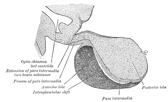
s project to the posterior pituitary
The posterior pituitary (or neurohypophysis) is the posterior lobe of the pituitary gland which is part of the endocrine system. Unlike the anterior pituitary, the posterior pituitary is not glandular, but largely a collection of axonal projec ...
where they secrete oxytocin
Oxytocin is a peptide hormone and neuropeptide normally produced in the hypothalamus and released by the posterior pituitary. Present in animals since early stages of evolution, in humans it plays roles in behavior that include Human bonding, ...
, and a smaller amount of vasopressin
Mammalian vasopressin, also called antidiuretic hormone (ADH), arginine vasopressin (AVP) or argipressin, is a hormone synthesized from the ''AVP'' gene as a peptide prohormone in neurons in the hypothalamus, and is converted to AVP. It ...
. Other secretions are corticotropin-releasing hormone
Corticotropin-releasing hormone (CRH) (also known as corticotropin-releasing factor (CRF) or corticoliberin; corticotropin may also be spelled corticotrophin) is a peptide hormone involved in stress responses. It is a releasing hormone that b ...
(CRH) and thyrotropin-releasing hormone
Thyrotropin-releasing hormone (TRH) is a hypophysiotropic hormone produced by neurons in the hypothalamus that stimulates the release of thyroid-stimulating hormone (TSH) as well as prolactin from the anterior pituitary.
TRH has been used ...
(TRH). CRH and TRH are secreted into the hypophyseal portal system, and target different neurons in the anterior pituitary
The anterior pituitary (also called the adenohypophysis or pars anterior) is a major Organ (anatomy), organ of the endocrine system. The anterior pituitary is the glandular, Anatomical terms of location#Usage in human anatomy, anterior lobe that t ...
. Dysfunctions of the PVN can cause hypersomnia
Hypersomnia is a neurological disorder of excessive time spent sleeping or excessive sleepiness. It can have many possible causes (such as seasonal affective disorder) and can cause distress and problems with functioning. In the fifth edition ...
in mice. In humans, the dysfunction of the PVN and the other nuclei around it can lead to drowsiness for up to 20 hours per day. The PVN is thought to mediate many diverse functions through different hormone
A hormone (from the Ancient Greek, Greek participle , "setting in motion") is a class of cell signaling, signaling molecules in multicellular organisms that are sent to distant organs or tissues by complex biological processes to regulate physio ...
s, including osmoregulation, appetite, wakefulness
Wakefulness is a daily recurring brain state and state of consciousness in which an individual is conscious and engages in coherent cognition, cognitive and behavioral responses to the external world.
Being awake is the opposite of being asleep, ...
, and the response of the body to stress.
Location
The paraventricular nucleus lies adjacent to the third ventricle. It lies within the periventricular zone and is not to be confused with the periventricular nucleus, which occupies a more medial position, beneath the third ventricle. The PVN is highly vascularised and is protected by theblood–brain barrier
The blood–brain barrier (BBB) is a highly selective semipermeable membrane, semipermeable border of endothelium, endothelial cells that regulates the transfer of solutes and chemicals between the circulatory system and the central nervous system ...
, although its neuroendocrine cells extend to sites (in the median eminence and in the posterior pituitary
The posterior pituitary (or neurohypophysis) is the posterior lobe of the pituitary gland which is part of the endocrine system. Unlike the anterior pituitary, the posterior pituitary is not glandular, but largely a collection of axonal projec ...
) beyond the blood–brain barrier. PVN is accounting for only about 1% of the brain volume.In the rat, the PVN consists of approximately 100,000 neurons located in a volume of about 0.5 cubic millimetre.
Neurons
The PVN contains magnocellular neurosecretory cells whoseaxon
An axon (from Greek ἄξων ''áxōn'', axis) or nerve fiber (or nerve fibre: see American and British English spelling differences#-re, -er, spelling differences) is a long, slender cellular extensions, projection of a nerve cell, or neuron, ...
s extend into the posterior pituitary
The posterior pituitary (or neurohypophysis) is the posterior lobe of the pituitary gland which is part of the endocrine system. Unlike the anterior pituitary, the posterior pituitary is not glandular, but largely a collection of axonal projec ...
, parvocellular neurosecretory cells that project to the median eminence, ultimately signalling to the anterior pituitary
The anterior pituitary (also called the adenohypophysis or pars anterior) is a major Organ (anatomy), organ of the endocrine system. The anterior pituitary is the glandular, Anatomical terms of location#Usage in human anatomy, anterior lobe that t ...
, and several populations of other cells that project to many different brain regions including parvocellular preautonomic cells that project to the brainstem
The brainstem (or brain stem) is the posterior stalk-like part of the brain that connects the cerebrum with the spinal cord. In the human brain the brainstem is composed of the midbrain, the pons, and the medulla oblongata. The midbrain is conti ...
and spinal cord
The spinal cord is a long, thin, tubular structure made up of nervous tissue that extends from the medulla oblongata in the lower brainstem to the lumbar region of the vertebral column (backbone) of vertebrate animals. The center of the spinal c ...
.
Magnocellular neurosecretory neurons
 The magnocellular cells in the PVN elaborate and secrete two peptide hormones:
 oxytocin
Oxytocin is a peptide hormone and neuropeptide normally produced in the hypothalamus and released by the posterior pituitary. Present in animals since early stages of evolution, in humans it plays roles in behavior that include Human bonding, ...
and vasopressin
Mammalian vasopressin, also called antidiuretic hormone (ADH), arginine vasopressin (AVP) or argipressin, is a hormone synthesized from the ''AVP'' gene as a peptide prohormone in neurons in the hypothalamus, and is converted to AVP. It ...
.
These hormones are packaged into large vesicles, which are then transported down the unmyelinated
Myelin Sheath ( ) is a lipid-rich material that in most vertebrates surrounds the axons of neurons to Insulator (electricity), insulate them and increase the rate at which electrical impulses (called action potentials) pass along the axon. The my ...
axons of the cells and released from neurosecretory nerve terminals residing in the posterior pituitary gland.
Similar magnocellular neurons are found in the supraoptic nucleus which also secrete vasopressin and a smaller amount of oxytocin.
Parvocellular neurosecretory neurons
The axons of the parvocellular neurosecretory neurons of the PVN project to the median eminence, a neurohemal organ at the base of the brain, where their neurosecretory nerve terminals release their hormones at the primary capillary plexus of the hypophyseal portal system. The median eminence contains fiber terminals from many hypothalamic neuroendocrine neurons, secreting different neurotransmitters or neuropeptides, including vasopressin, corticotropin-releasing hormone (CRH), thyrotropin-releasing hormone (TRH), gonadotropin-releasing hormone (GnRH), growth hormone-releasing hormone (GHRH), dopamine (DA) and somatostatin (growth hormone release inhibiting hormone, GIH) into blood vessels in the hypophyseal portal system. The blood vessels carry the peptides to the anterior pituitary gland, where they regulate the secretion of hormones into the systemic circulation. The parvocellular neurosecretory cells include those that make: *Corticotropin-releasing hormone
Corticotropin-releasing hormone (CRH) (also known as corticotropin-releasing factor (CRF) or corticoliberin; corticotropin may also be spelled corticotrophin) is a peptide hormone involved in stress responses. It is a releasing hormone that b ...
(CRH), which regulates ACTH secretion from the anterior pituitary gland
* Vasopressin
Mammalian vasopressin, also called antidiuretic hormone (ADH), arginine vasopressin (AVP) or argipressin, is a hormone synthesized from the ''AVP'' gene as a peptide prohormone in neurons in the hypothalamus, and is converted to AVP. It ...
, which also regulates ACTH secretion (vasopressin and CRH act synergistically to stimulate ACTH secretion)
* Thyrotropin-releasing hormone
Thyrotropin-releasing hormone (TRH) is a hypophysiotropic hormone produced by neurons in the hypothalamus that stimulates the release of thyroid-stimulating hormone (TSH) as well as prolactin from the anterior pituitary.
TRH has been used ...
(TRH), which regulates TSH and prolactin
Prolactin (PRL), also known as lactotropin and mammotropin, is a protein best known for its role in enabling mammals to produce milk. It is influential in over 300 separate processes in various vertebrates, including humans. Prolactin is secr ...
secretion
Centrally-projecting neurons
As well as neuroendocrine neurons, the PVN containsinterneuron
Interneurons (also called internuncial neurons, association neurons, connector neurons, or intermediate neurons) are neurons that are not specifically motor neurons or sensory neurons. Interneurons are the central nodes of neural circuits, enab ...
s and populations of neurons that project centrally (i.e., to other brain regions). The centrally-projecting neurons include
* Parvocellular oxytocin cells, which project mainly to the brainstem
The brainstem (or brain stem) is the posterior stalk-like part of the brain that connects the cerebrum with the spinal cord. In the human brain the brainstem is composed of the midbrain, the pons, and the medulla oblongata. The midbrain is conti ...
and spinal cord
The spinal cord is a long, thin, tubular structure made up of nervous tissue that extends from the medulla oblongata in the lower brainstem to the lumbar region of the vertebral column (backbone) of vertebrate animals. The center of the spinal c ...
. These neurons are thought to have a role in gastric reflexes and penile erection,
* Parvocellular vasopressin cells, which project to many points in the hypothalamus and limbic system
The limbic system, also known as the paleomammalian cortex, is a set of brain structures located on both sides of the thalamus, immediately beneath the medial temporal lobe of the cerebrum primarily in the forebrain.Schacter, Daniel L. 2012. ''P ...
, as well as to the brainstem and spinal cord (these are involved in blood pressure and temperature regulation), and brown fat thermogenesis.
* Parvocellular CRH neurons, which are thought to be involved in stress-related behaviors.
Afferent inputs
The PVN receives afferent inputs from many brain regions and different parts of the body, by hormonal control. Among these, inputs from neurons in structures adjacent to the anterior wall of the third ventricle (the "AV3V region") carry information about the electrolyte composition of the blood, and about circulating concentrations of such hormones as angiotensin and relaxin, to regulate the magnocellular neurons. Inputs from the brainstem (the nucleus of the solitary tract) and the ventrolateral medulla carry information from the heart andstomach
The stomach is a muscular, hollow organ in the upper gastrointestinal tract of Human, humans and many other animals, including several invertebrates. The Ancient Greek name for the stomach is ''gaster'' which is used as ''gastric'' in medical t ...
. Inputs from the hippocampus
The hippocampus (: hippocampi; via Latin from Ancient Greek, Greek , 'seahorse'), also hippocampus proper, is a major component of the brain of humans and many other vertebrates. In the human brain the hippocampus, the dentate gyrus, and the ...
to the CRH neurones are important regulators of stress responses.
Inputs from neuropeptide Y-containing neurons in the arcuate nucleus coordinate metabolic regulation (via TRH secretion) with regulation of energy intake. Specifically, the projections from the arcuate nucleus seem to exert their effect on appetite via MC4R-expressing oxytocinergic cells of the PVN.
Inputs from suprachiasmatic nucleus
The suprachiasmatic nucleus or nuclei (SCN) is a small region of the brain in the hypothalamus, situated directly above the optic chiasm. It is responsible for regulating sleep cycles in animals. Reception of light inputs from photosensitive r ...
about levels of lighting (circadian rhythms).
Inputs from glucose sensors within the brain stimulate release of vasopressin
Mammalian vasopressin, also called antidiuretic hormone (ADH), arginine vasopressin (AVP) or argipressin, is a hormone synthesized from the ''AVP'' gene as a peptide prohormone in neurons in the hypothalamus, and is converted to AVP. It ...
and corticotropin-releasing hormone
Corticotropin-releasing hormone (CRH) (also known as corticotropin-releasing factor (CRF) or corticoliberin; corticotropin may also be spelled corticotrophin) is a peptide hormone involved in stress responses. It is a releasing hormone that b ...
from parvocellular neurosecretory cells.
References
Further reading
* {{Authority control Endocrine system anatomy Hypothalamus Neuroendocrinology Hormones