|
NPHS1
Nephrin is a protein necessary for the proper functioning of the renal filtration barrier. The renal filtration barrier consists of fenestrated endothelial cells, the glomerular basement membrane, and the podocytes of epithelial cells. Nephrin is a transmembrane protein that is a structural component of the slit diaphragm. They are present on the tips of the podocytes as an intricate mesh and convey strong negative charges which repel protein from crossing into the Bowman's space. A defect in the gene for nephrin, NPHS1, is associated with congenital nephrotic syndrome of the Finnish type and causes massive amounts of protein to be leaked into the urine, or proteinuria. Nephrin is also required for cardiovascular development. Interactions Nephrin has been shown to interact with: * CASK, * CD2AP, * CDH3 and * CTNND1, * FYN, * KIRREL, and * NPHS2. See also * Podocyte Podocytes are cells in Bowman's capsule in the kidneys that wrap around capillaries of the glomerul ... [...More Info...] [...Related Items...] OR: [Wikipedia] [Google] [Baidu] |
Congenital Nephrotic Syndrome
Congenital nephrotic syndrome is a rare kidney disease which manifests in infants during the first 3 months of life, and is characterized by high levels of protein in the urine ( proteinuria), low levels of protein in the blood, and swelling. This disease is primarily caused by genetic mutations which result in damage to components of the glomerular filtration barrier and allow for leakage of plasma proteins into the urinary space. Signs and symptoms Urine protein loss leads to total body swelling (generalized edema) and abdominal distension in the first several weeks to months of life. Fluid retention may lead to cough (from pulmonary edema), ascites, and widened cranial sutures and fontanelles. High urine protein loss can lead to foamy appearance of urine. Infants may be born prematurely with low birth weight, and have meconium stained amniotic fluid or a large placenta. Complications * Frequent, severe infections: urinary loss of immunoglobulins * Malnutrition and poor grow ... [...More Info...] [...Related Items...] OR: [Wikipedia] [Google] [Baidu] |
Finnish-type Nephrosis
Congenital nephrotic syndrome is a rare kidney disease which manifests in infants during the first 3 months of life, and is characterized by high levels of protein in the urine (proteinuria), low levels of protein in the blood, and swelling. This disease is primarily caused by genetic mutations which result in damage to components of the glomerular filtration barrier and allow for leakage of plasma proteins into the urinary space. Signs and symptoms Urine protein loss leads to total body swelling (generalized edema) and abdominal distension in the first several weeks to months of life. Fluid retention may lead to cough (from pulmonary edema), ascites, and widened cranial sutures and fontanelles. High urine protein loss can lead to foamy appearance of urine. Infants may be born prematurely with low birth weight, and have meconium stained amniotic fluid or a large placenta. Complications * Frequent, severe infections: urinary loss of immunoglobulins * Malnutrition and poor growth ... [...More Info...] [...Related Items...] OR: [Wikipedia] [Google] [Baidu] |
NPHS2
Podocin is a protein that in humans is encoded by the ''NPHS2'' gene. Interactions NPHS2 has been shown to interact with Nephrin and CD2AP. See also * Focal segmental glomerulosclerosis Focal segmental glomerulosclerosis (FSGS) is a histopathologic finding of scarring (sclerosis) of glomeruli and damage to renal podocytes.Rosenberg, Avi Z.; Kopp, Jeffrey B. (2017-03-07). "Focal Segmental Glomerulosclerosis". ''Clinical Journal ... References Further reading * * * * * * * * * * * * * * * * * {{gene-1-stub ... [...More Info...] [...Related Items...] OR: [Wikipedia] [Google] [Baidu] |
Protein
Proteins are large biomolecules and macromolecules that comprise one or more long chains of amino acid residues. Proteins perform a vast array of functions within organisms, including catalysing metabolic reactions, DNA replication, responding to stimuli, providing structure to cells and organisms, and transporting molecules from one location to another. Proteins differ from one another primarily in their sequence of amino acids, which is dictated by the nucleotide sequence of their genes, and which usually results in protein folding into a specific 3D structure that determines its activity. A linear chain of amino acid residues is called a polypeptide. A protein contains at least one long polypeptide. Short polypeptides, containing less than 20–30 residues, are rarely considered to be proteins and are commonly called peptides. The individual amino acid residues are bonded together by peptide bonds and adjacent amino acid residues. The sequence of amino acid resid ... [...More Info...] [...Related Items...] OR: [Wikipedia] [Google] [Baidu] |
Podocytes
Podocytes are cells in Bowman's capsule in the kidneys that wrap around capillaries of the glomerulus. Podocytes make up the epithelial lining of Bowman's capsule, the third layer through which filtration of blood takes place. Bowman's capsule filters the blood, retaining large molecules such as proteins while smaller molecules such as water, salts, and sugars are filtered as the first step in the formation of urine. Although various viscera have epithelial layers, the name visceral epithelial cells usually refers specifically to podocytes, which are specialized epithelial cells that reside in the visceral layer of the capsule. One type of specialized epithelial cell is podocalyxin. The podocytes have long foot processes called ''pedicels'', for which the cells are named ('' podo-'' + '' -cyte''). The pedicels wrap around the capillaries and leave slits between them. Blood is filtered through these slits, each known as a filtration slit, slit diaphragm, or slit pore. Sev ... [...More Info...] [...Related Items...] OR: [Wikipedia] [Google] [Baidu] |
Filtration Slits
Podocytes are cells in Bowman's capsule in the kidneys that wrap around capillaries of the glomerulus. Podocytes make up the epithelial lining of Bowman's capsule, the third layer through which filtration of blood takes place. Bowman's capsule filters the blood, retaining large molecules such as proteins while smaller molecules such as water, salts, and sugars are filtered as the first step in the formation of urine. Although various viscera have epithelial layers, the name visceral epithelial cells usually refers specifically to podocytes, which are specialized epithelial cells that reside in the visceral layer of the capsule. One type of specialized epithelial cell is podocalyxin. The podocytes have long foot processes called ''pedicels'', for which the cells are named ('' podo-'' + '' -cyte''). The pedicels wrap around the capillaries and leave slits between them. Blood is filtered through these slits, each known as a filtration slit, slit diaphragm, or slit pore. Several p ... [...More Info...] [...Related Items...] OR: [Wikipedia] [Google] [Baidu] |
Bowman's Capsule
Bowman's capsule (or the Bowman capsule, capsula glomeruli, or glomerular capsule) is a cup-like sac at the beginning of the tubular component of a nephron in the mammalian kidney that performs the first step in the filtration of blood to form urine. A glomerulus is enclosed in the sac. Fluids from blood in the glomerulus are collected in the Bowman's capsule. Structure Outside the capsule, there are two "poles": * The vascular pole is the side with the afferent arteriole and efferent arteriole. * The urinary pole is the side with the proximal convoluted tubule. Inside the capsule, the layers are as follows, from outside to inside: *''Parietal layer''—A single layer of simple squamous epithelium. Does not function in filtration. *''Bowman's space (or "urinary space", or "capsular space")''—Between the visceral and parietal layers, into which the filtrate enters after passing through the filtration slits. *''Visceral layer''—Lies just above the thickened glomerular b ... [...More Info...] [...Related Items...] OR: [Wikipedia] [Google] [Baidu] |
Proteinuria
Proteinuria is the presence of excess proteins in the urine. In healthy persons, urine contains very little protein; an excess is suggestive of illness. Excess protein in the urine often causes the urine to become foamy (although this symptom may also be caused by other conditions). Severe proteinuria can cause nephrotic syndrome in which there is worsening swelling of the body. Signs and symptoms Proteinuria often causes no symptoms and it may only be discovered incidentally. Foamy urine is considered a cardinal sign of proteinuria, but only a third of people with foamy urine have proteinuria as the underlying cause. It may also be caused by bilirubin in the urine ( bilirubinuria), retrograde ejaculation, pneumaturia (air bubbles in the urine) due to a fistula, or drugs such as pyridium. Causes There are three main mechanisms to cause proteinuria: * Due to disease in the glomerulus * Because of increased quantity of proteins in serum (overflow proteinuria) * Due to low ... [...More Info...] [...Related Items...] OR: [Wikipedia] [Google] [Baidu] |
CASK
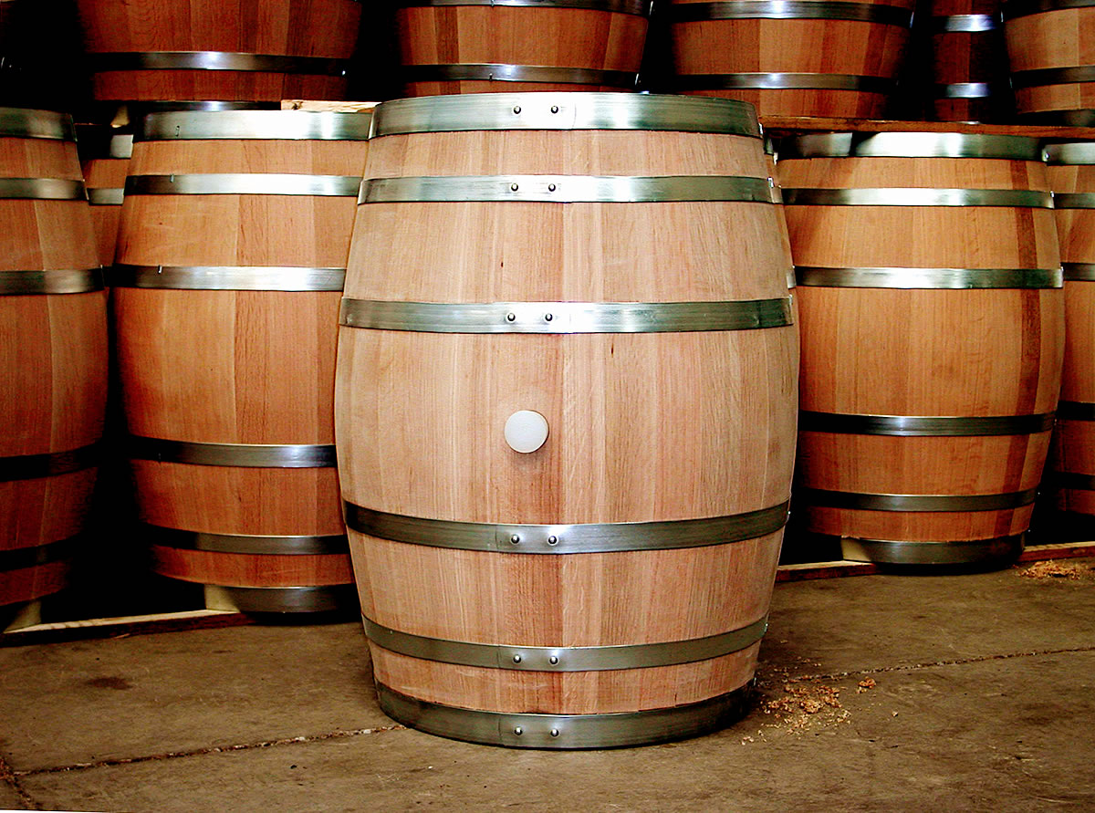
A barrel or cask is a hollow cylindrical container with a bulging center, longer than it is wide. They are traditionally made of wooden staves and bound by wooden or metal hoops. The word vat is often used for large containers for liquids, usually alcoholic beverages; a small barrel or cask is known as a keg. Modern wooden barrels for wine-making are made of French common oak (''Quercus robur''), white oak (''Quercus petraea''), American white oak (''Quercus alba''), more exotic is Mizunara Oak all typically have standard sizes: Recently Oregon Oak (Quercus Garryana) has been used. *"Bordeaux type" , *"Burgundy type" and *"Cognac type" . Modern barrels and casks can also be made of aluminum, stainless steel, and different types of plastic, such as HDPE. Someone who makes barrels is called a "barrel maker" or cooper (coopers also make buckets, vats, tubs, butter churns, hogsheads, firkins, kegs, kilderkins, tierces, rundlets, puncheons, pipes, tuns, butts, pins, tr ... [...More Info...] [...Related Items...] OR: [Wikipedia] [Google] [Baidu] |
CD2AP
CD2-associated protein is a protein that in humans is encoded by the ''CD2AP'' gene. Function This gene encodes a scaffolding molecule that regulates the actin cytoskeleton. The protein directly interacts with filamentous actin and a variety of cell membrane proteins through multiple actin binding sites, SH3 domains, and a proline-rich region containing binding sites for SH3 domains. The cytoplasmic protein localizes to membrane ruffles, lipid rafts, and the leading edges of cells. It is implicated in dynamic actin remodeling and membrane trafficking that occurs during receptor endocytosis and cytokinesis. Haploinsufficiency of this gene is implicated in susceptibility to glomerular disease. Interactions CD2AP has been shown to interact with: * Cbl gene, * NPHS2, * Nephrin, and * RAB4A. See also * Focal segmental glomerulosclerosis Focal segmental glomerulosclerosis (FSGS) is a histopathologic finding of scarring (sclerosis) of glomeruli and damage to renal ... [...More Info...] [...Related Items...] OR: [Wikipedia] [Google] [Baidu] |
CDH3 (gene)
Cadherin-3, also known as P-Cadherin, is a protein that in humans is encoded by the ''CDH3'' gene. Function This gene is a classical cadherin from the cadherin superfamily. The encoded protein is a calcium-dependent cell-cell adhesion glycoprotein composed of five extracellular cadherin repeats, a transmembrane region and a highly conserved cytoplasmic tail. This gene is located in a six-cadherin cluster in a region on the long arm of chromosome 16 that is involved in loss of heterozygosity events in breast and prostate cancer. In addition, aberrant expression of this protein is observed in cervical adenocarcinomas. Clinical significance Mutations in this gene have been associated with congenital hypotrichosis with juvenile macular dystrophy. Interactions CDH3 (gene) has been shown to interact with: * Beta-catenin, * CDH1, * Catenin (cadherin-associated protein), alpha 1, * Nephrin and * Plakoglobin. History Cadherin-3 was first described in 1986 by Masatoshi T ... [...More Info...] [...Related Items...] OR: [Wikipedia] [Google] [Baidu] |


