|
Meroselenidium
''Meroselenidium'' is a genus of parasitic alveolates in the phylum Apicomplexa. Species in this genus infect marine invertebrates. Taxonomy This genus was described by Mackinnon and Ray in 1933. There is one species in this genus – ''Meroselenidium keilini''. Description The trophozoites live within the gut lumen. They measure 200–300 μm × 40–70 μm. There are 30–40 grooves along the body. Four refringent rods are present in the mucron. A vacuole may also be present in the mucron. Schizogony occurs in the intestinal epithelium and gives rise to multiple merozoites. Synergy is caudo-caudal. The gametocysts are 70 μm × 55 μm and give rise to multiple gametes. After fertilization the zygote A zygote (, ) is a eukaryotic cell formed by a fertilization event between two gametes. The zygote's genome is a combination of the DNA in each gamete, and contains all of the genetic information of a new individual organism. In multicellula ... gi ... [...More Info...] [...Related Items...] OR: [Wikipedia] [Google] [Baidu] |
Meroselenidium Keilini
''Meroselenidium'' is a genus of parasitic alveolates in the phylum Apicomplexa. Species in this genus infect marine invertebrates. Taxonomy This genus was described by Doris Mackinnon, Mackinnon and Ray in 1933. There is one species in this genus – ''Meroselenidium keilini''. Description The trophozoites live within the gut lumen. They measure 200–300 μm × 40–70 μm. There are 30–40 grooves along the body. Four refringent rods are present in the mucron. A vacuole may also be present in the mucron. Schizogony occurs in the intestinal epithelium and gives rise to multiple merozoites. Synergy is caudo-caudal. The gametocysts are 70 μm × 55 μm and give rise to multiple gametes. After fertilization the zygote gives rise to ~20 sporocysts. There is no residual body. The sporocysts are bivalved and give rise to multiple sporozoites. The species in this genus, ''Merselenidium keilini'', forms transversely striated folds. Life cycle This species in ... [...More Info...] [...Related Items...] OR: [Wikipedia] [Google] [Baidu] |
Archigregarinorida
The ''Archigregarinorida'' are an order of parasitic alveolates in the phylum Apicomplexa. Species in this order infect marine invertebrates — usually annelids, ascidians, hemichordates and sipunculids. Taxonomy This order was redefined by Levine in 1971. The order currently consists of 76 species in two families — ''Exoschizonidae'' and ''Selenidioididae''. The family ''Exoschizonidae'' contains one genus — ''Exoschizon'' — which has one species. The family ''Selenidioididae'' has six genera: ''Filipodium'' with 3 species, ''Merogregarina'' with one species, ''Meroselenidium'' with one species, '' Platyproteum'' with one species, ''Selenidioides'' with 11 species and ''Veloxidium'' with one species. Phylogenetics DNA studies suggest that the archigregarines are ancestral to the other gregarines. Phylogenetic analysis suggests that this group is paraphyletic and will need division. The Neogregarinorida appear to be derived from the Eugregarinorida. Assuming this is ... [...More Info...] [...Related Items...] OR: [Wikipedia] [Google] [Baidu] |
Selenidioididae
The ''Selenidioididae'' are a family of parasitic alveolates in the phylum Apicomplexa. Species in this order infect marine invertebrates. Taxonomy The order Archigregarinorida was redefined by Levine in 1971Levine N D (1971) Taxonomy of the Archigregarinorida and Selenidiidae (Protozoa, Apicomplexa) J Euk Micro 18 (4) 704-717 DOI: 10.1111/j.1550-7408.1971.tb03401.x and divided into two families: Exoschizonidae The Exoschizonidae are a family in the phylum Apicomplexa. History This family was created by Levine in 1971.Levine ND (1971) Taxonomy of the Archigregarinorida and Selenidiidae (Protozoa, Apicomplexa) J Euk Microbiol 18 (4) 704–717 Taxonom ... and Selenidioididae. There are seven genera and 74 species recognised in this family. Description Species in this family undergo asexual schizogony. Life cycle The species in the family infect the gastrointestinal tract and are presumably transmitted by the orofaecal route but the details of this mechanism are prese ... [...More Info...] [...Related Items...] OR: [Wikipedia] [Google] [Baidu] |
Eukaryota
Eukaryotes () are organisms whose cells have a nucleus. All animals, plants, fungi, and many unicellular organisms, are Eukaryotes. They belong to the group of organisms Eukaryota or Eukarya, which is one of the three domains of life. Bacteria and Archaea (both prokaryotes) make up the other two domains. The eukaryotes are usually now regarded as having emerged in the Archaea or as a sister of the Asgard archaea. This implies that there are only two domains of life, Bacteria and Archaea, with eukaryotes incorporated among archaea. Eukaryotes represent a small minority of the number of organisms, but, due to their generally much larger size, their collective global biomass is estimated to be about equal to that of prokaryotes. Eukaryotes emerged approximately 2.3–1.8 billion years ago, during the Proterozoic eon, likely as Flagellated cell, flagellated phagotrophs. Their name comes from the Greek language, Greek wikt:εὖ, εὖ (''eu'', "well" or "good") and wikt:� ... [...More Info...] [...Related Items...] OR: [Wikipedia] [Google] [Baidu] |
Mucron
A ''mucron'' is an attachment organelle found in archigregarines - an order of epicellular parasitic Conoidasida.Simdyanov TG, Guillou L, Diakin AY, Mikhailov KV, Schrével J, Aleoshin VV. (2017) A new view on the morphology and phylogeny of eugregarines suggested by the evidence from the gregarine ''Ancora sagittata'' (Leuckart, 1860) Labbé, 1899 (Apicomplexa: Eugregarinida) PeerJ 5:e3354 https://peerj.com/articles/3354/?td=wk The mucron is derived from the apical complex, which is found in all members of the phylum Apicomplexa.Adl SM, Simpson AG, Lane CE, Lukeš J, Bass D, Bowser SS, Brown M, Burki F, Dunthorn M, Hampl V, Heiss A, Hoppenrath M, Lara E, leGall L, Lynn DH, McManus H, Mitchell EAD, Mozley-Stanridge SE, Parfrey LW, Pawlowski J, Rueckert S, Shadwick L, Schoch C, Smirnov A, Spiegel FW. (2012) The revised classification of eukaryotes. Journal of Eukaryotic Microbiology 59:429-514. https://doi.org/10.1111/j.1550-7408.2012.00644.x The mucron is located at the anterior ... [...More Info...] [...Related Items...] OR: [Wikipedia] [Google] [Baidu] |
Polychaete
Polychaeta () is a paraphyletic class of generally marine annelid worms, commonly called bristle worms or polychaetes (). Each body segment has a pair of fleshy protrusions called parapodia that bear many bristles, called chaetae, which are made of chitin. More than 10,000 species are described in this class. Common representatives include the lugworm (''Arenicola marina'') and the sandworm or clam worm ''Alitta''. Polychaetes as a class are robust and widespread, with species that live in the coldest ocean temperatures of the abyssal plain, to forms which tolerate the extremely high temperatures near hydrothermal vents. Polychaetes occur throughout the Earth's oceans at all depths, from forms that live as plankton near the surface, to a 2- to 3-cm specimen (still unclassified) observed by the robot ocean probe ''Nereus'' at the bottom of the Challenger Deep, the deepest known spot in the Earth's oceans. Only 168 species (less than 2% of all polychaetes) are known from ... [...More Info...] [...Related Items...] OR: [Wikipedia] [Google] [Baidu] |
Zygote
A zygote (, ) is a eukaryotic cell formed by a fertilization event between two gametes. The zygote's genome is a combination of the DNA in each gamete, and contains all of the genetic information of a new individual organism. In multicellular organisms, the zygote is the earliest developmental stage. In humans and most other anisogamous organisms, a zygote is formed when an egg cell and sperm cell come together to create a new unique organism. In single-celled organisms, the zygote can divide asexually by mitosis to produce identical offspring. German zoologists Oscar and Richard Hertwig made some of the first discoveries on animal zygote formation in the late 19th century. Humans In human fertilization, a released ovum (a haploid secondary oocyte with replicate chromosome copies) and a haploid sperm cell (male gamete) combine to form a single diploid cell called the zygote. Once the single sperm fuses with the oocyte, the latter completes the division of the ... [...More Info...] [...Related Items...] OR: [Wikipedia] [Google] [Baidu] |
Merozoites
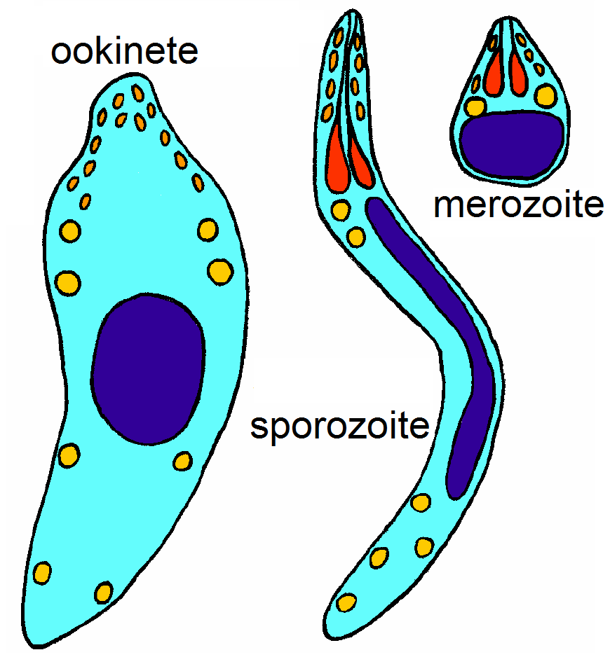
Apicomplexans, a group of intracellular parasites, have life cycle stages that allow them to survive the wide variety of environments they are exposed to during their complex life cycle. Each stage in the life cycle of an apicomplexan organism is typified by a ''cellular variety'' with a distinct morphology and biochemistry. Not all apicomplexa develop all the following cellular varieties and division methods. This presentation is intended as an outline of a hypothetical generalised apicomplexan organism. Methods of asexual replication Apicomplexans (sporozoans) replicate via ways of multiple fission (also known as schizogony). These ways include , and , although the latter is sometimes referred to as schizogony, despite its general meaning. Merogony is an asexually reproductive process of apicomplexa. After infecting a host cell, a trophozoite ( see glossary below) increases in size while repeatedly replicating its nucleus and other organelles. During this process, the orga ... [...More Info...] [...Related Items...] OR: [Wikipedia] [Google] [Baidu] |
Intestinal Epithelium
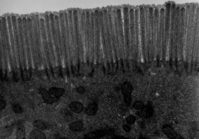
The intestinal epithelium is the single cell layer that form the luminal surface (lining) of both the small and large intestine (colon) of the gastrointestinal tract. Composed of simple columnar epithelial cells, it serves two main functions: absorbing useful substances into the body and restricting the entry of harmful substances. As part of its protective role, the intestinal epithelium forms an important component of the intestinal mucosal barrier. Certain diseases and conditions are caused by functional defects in the intestinal epithelium. On the other hand, various diseases and conditions can lead to its dysfunction which, in turn, can lead to further complications. Structure The intestinal epithelium is part of the intestinal mucosa. The epithelium is composed of a single layer of cells, while the other two layers of the mucosa, the lamina propria and the muscularis mucosae, support and articulate the epithelial layer. To securely contain the contents of the intes ... [...More Info...] [...Related Items...] OR: [Wikipedia] [Google] [Baidu] |
Schizogony
Fission, in biology, is the division of a single entity into two or more parts and the regeneration of those parts to separate entities resembling the original. The object experiencing fission is usually a cell, but the term may also refer to how organisms, bodies, populations, or species split into discrete parts. The fission may be ''binary fission'', in which a single organism produces two parts, or ''multiple fission'', in which a single entity produces multiple parts. Binary fission Organisms in the domains of Archaea and Bacteria reproduce with binary fission. This form of asexual reproduction and cell division is also used by some organelles within eukaryotic organisms (e.g., mitochondria). Binary fission results in the reproduction of a living prokaryotic cell (or organelle) by dividing the cell into two parts, each with the potential to grow to the size of the original. Fission of prokaryotes The single DNA molecule first replicates, then attaches each copy to a d ... [...More Info...] [...Related Items...] OR: [Wikipedia] [Google] [Baidu] |
Vacuole
A vacuole () is a membrane-bound organelle which is present in plant and fungal cells and some protist, animal, and bacterial cells. Vacuoles are essentially enclosed compartments which are filled with water containing inorganic and organic molecules including enzymes in solution, though in certain cases they may contain solids which have been engulfed. Vacuoles are formed by the fusion of multiple membrane vesicles and are effectively just larger forms of these. The organelle has no basic shape or size; its structure varies according to the requirements of the cell. Discovery Contractile vacuoles ("stars") were first observed by Spallanzani (1776) in protozoa, although mistaken for respiratory organs. Dujardin (1841) named these "stars" as ''vacuoles''. In 1842, Schleiden applied the term for plant cells, to distinguish the structure with cell sap from the rest of the protoplasm. In 1885, de Vries named the vacuole membrane as tonoplast. Function The function and ... [...More Info...] [...Related Items...] OR: [Wikipedia] [Google] [Baidu] |


