|
Intestinal Epithelium
The intestinal epithelium is the single cell layer that forms the luminal surface (lining) of both the small and large intestine (colon) of the gastrointestinal tract. Composed of simple columnar epithelium its main functions are absorption, and secretion. Useful substances are absorbed into the body, and the entry of harmful substances is restricted. Secretions include mucins, and peptides. Absorptive cells in the small intestine are known as enterocytes, and in the colon they are known as colonocytes. The other cell types are the secretory cells – goblet cells, Paneth cells, enteroendocrine cells, and Tuft cells. Paneth cells are absent in the colon. As part of its protective role, the intestinal epithelium forms an important component of the intestinal mucosal barrier. Certain diseases and conditions are caused by functional defects in the intestinal epithelium. On the other hand, various diseases and conditions can lead to its dysfunction which, in turn, can lead t ... [...More Info...] [...Related Items...] OR: [Wikipedia] [Google] [Baidu] |
Epithelium
Epithelium or epithelial tissue is a thin, continuous, protective layer of cells with little extracellular matrix. An example is the epidermis, the outermost layer of the skin. Epithelial ( mesothelial) tissues line the outer surfaces of many internal organs, the corresponding inner surfaces of body cavities, and the inner surfaces of blood vessels. Epithelial tissue is one of the four basic types of animal tissue, along with connective tissue, muscle tissue and nervous tissue. These tissues also lack blood or lymph supply. The tissue is supplied by nerves. There are three principal shapes of epithelial cell: squamous (scaly), columnar, and cuboidal. These can be arranged in a singular layer of cells as simple epithelium, either simple squamous, simple columnar, or simple cuboidal, or in layers of two or more cells deep as stratified (layered), or ''compound'', either squamous, columnar or cuboidal. In some tissues, a layer of columnar cells may appear to be stratified d ... [...More Info...] [...Related Items...] OR: [Wikipedia] [Google] [Baidu] |
Microvillus
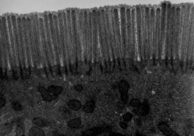
Microvilli (: microvillus) are microscopic cellular membrane protrusions that increase the surface area for diffusion and minimize any increase in volume, and are involved in a wide variety of functions, including absorption, secretion, cellular adhesion, and mechanotransduction. Structure Microvilli are covered in plasma membrane, which encloses cytoplasm and microfilaments. Though these are cellular extensions, there are little or no cellular organelles present in the microvilli. Each microvillus has a dense bundle of cross-linked actin filaments, which serves as its structural core. 20 to 30 tightly bundled actin filaments are cross-linked by bundling proteins fimbrin (or plastin-1), villin and espin to form the core of the microvilli. In the enterocyte microvillus, the structural core is attached to the plasma membrane along its length by lateral arms made of myosin 1a and Ca2+ binding protein calmodulin. Myosin 1a functions through a binding site for filamentous ac ... [...More Info...] [...Related Items...] OR: [Wikipedia] [Google] [Baidu] |
Intestinal Villus
Intestinal villi (: villus) are small, finger-like projections that extend into the lumen of the small intestine. Each villus is approximately 0.5–1.6 mm in length (in humans), and has many microvilli projecting from the enterocytes of its epithelium which collectively form the striated or brush border. Each of these microvilli are about 1 μm in length, around 1000 times shorter than a single villus. The intestinal villi are much smaller than any of the circular folds in the intestine. Villi increase the internal surface area of the intestinal walls making available a greater surface area for absorption. An increased absorptive area is useful because digested nutrients (including monosaccharide and amino acids) pass into the semipermeable villi through diffusion, which is effective only at short distances. In other words, increased surface area (in contact with the fluid in the lumen) decreases the average distance travelled by nutrient molecules, so effectiveness of ... [...More Info...] [...Related Items...] OR: [Wikipedia] [Google] [Baidu] |
Circular Folds
The circular folds (also known as valves of Kerckring, valves of Kerchkring, plicae circulares, ''plicae circulae'', and ''valvulae conniventes'') are large valvular flaps projecting into the lumen of the small intestine. Structure The entire small intestine has circular folds of mucous membrane. The majority extend transversely around the cylinder of the small intestine, for about one-half or two-thirds of its circumference. Some form complete circles. Others have a spiral direction. The latter usually extend a little more than once around the bowel, but occasionally two or three times. While the larger folds are about 1 cm in depth at their broadest part, most folds are smaller. There tends to be an alternating pattern between larger and smaller folds. Distribution They are not found at the commencement of the duodenum, but begin to appear about 2.5 or 5 cm beyond the pylorus. In the lower part of the descending portion, below the point where the bile and pancre ... [...More Info...] [...Related Items...] OR: [Wikipedia] [Google] [Baidu] |
Apoptosis
Apoptosis (from ) is a form of programmed cell death that occurs in multicellular organisms and in some eukaryotic, single-celled microorganisms such as yeast. Biochemistry, Biochemical events lead to characteristic cell changes (Morphology (biology), morphology) and death. These changes include Bleb (cell biology), blebbing, Plasmolysis, cell shrinkage, Karyorrhexis, nuclear fragmentation, Pyknosis, chromatin condensation, Apoptotic DNA fragmentation, DNA fragmentation, and mRNA decay. The average adult human loses 50 to 70 1,000,000,000, billion cells each day due to apoptosis. For the average human child between 8 and 14 years old, each day the approximate loss is 20 to 30 billion cells. In contrast to necrosis, which is a form of traumatic cell death that results from acute cellular injury, apoptosis is a highly regulated and controlled process that confers advantages during an organism's life cycle. For example, the separation of fingers and toes in a developing human embryo ... [...More Info...] [...Related Items...] OR: [Wikipedia] [Google] [Baidu] |
Intestinal Gland
In histology, an intestinal gland (also Crypt (anatomy), crypt of Johann Nathanael Lieberkühn, Lieberkühn and intestinal crypt) is a gland found in between Intestinal villus, villi in the intestinal epithelium, intestinal epithelial lining of the small intestine and large intestine (or colon). The glands and intestinal villi are covered by epithelium, which contains multiple types of Cell (biology), cells: enterocytes (absorbing water and electrolytes), goblet cells (secreting mucus), enteroendocrine cells (secreting hormones), cup cells, myofibroblast, tuft cells, and at the base of the gland, Paneth cells (secreting anti-microbial peptides) and stem cells. Structure Intestinal glands are found in the epithelia of the small intestine, namely the duodenum, jejunum, and ileum, and in the large intestine (colon), where they are sometimes called ''colonic crypts''. Intestinal glands of the small intestine contain a base of replicating stem cells, Paneth cells of the innate immune sy ... [...More Info...] [...Related Items...] OR: [Wikipedia] [Google] [Baidu] |
Crypt (anatomy)
Crypts are anatomical structures that are narrow but deep invagination Invagination is the process of a surface folding in on itself to form a cavity, pouch or tube. In developmental biology, invagination of Epithelium, epithelial sheets occurs in many contexts during Animal embryonic development, embryonic developme ...s into a larger structure. One common type of anatomical crypt is the Crypts of Lieberkühn. However, it is not the only type: some types of tonsils also have crypts. Because these crypts allow external access to the deep portions of the tonsils, these tonsils are more vulnerable to infection. References External links * - "Lymphoid Tissues and Organs: tonsil" Histology of crypt of tonsil at siumed.edu Anatomy {{Anatomy-stub ... [...More Info...] [...Related Items...] OR: [Wikipedia] [Google] [Baidu] |
Stem Cells
In multicellular organisms, stem cells are undifferentiated or partially differentiated cells that can change into various types of cells and proliferate indefinitely to produce more of the same stem cell. They are the earliest type of cell in a cell lineage. They are found in both embryonic and adult organisms, but they have slightly different properties in each. They are usually distinguished from progenitor cells, which cannot divide indefinitely, and precursor or blast cells, which are usually committed to differentiating into one cell type. In mammals, roughly 50 to 150 cells make up the inner cell mass during the blastocyst stage of embryonic development, around days 5–14. These have stem-cell capability. ''In vivo'', they eventually differentiate into all of the body's cell types (making them pluripotent). This process starts with the differentiation into the three germ layers – the ectoderm, mesoderm and endoderm – at the gastrulation stage. However, when t ... [...More Info...] [...Related Items...] OR: [Wikipedia] [Google] [Baidu] |
Villi & Microvilli Of Small Intestine
Villi may refer to: *Plural of Villus (other) *''Le Villi ''Le Villi'' (''The Willis'' or ''The Fairies'') is an opera–ballet in two acts (originally one) composed by Giacomo Puccini to an Italian libretto by Ferdinando Fontana, based on the short story "Les Willis" by Jean-Baptiste Alphonse Karr. Karr ...'', an opera-ballet of 1884 by Giacomo Puccini * Ilkka Villi (born 1975), Finnish actor and writer * Villi Bossi (born 1939), Italian sculptor * Villi Hermann (born 1941), Swiss film director and screenwriter * Villi Baltins, Soviet sprint canoer * Christian Villi (born 1974), Italian association football coach * Olga Villi (1922–1989), Italian model and actress * Villi Boskovsky, an Austrian violinist * Villi Tokarev, a Russian-American singer-songwriter See also * Vili (other) {{disambiguation, given name, surname ... [...More Info...] [...Related Items...] OR: [Wikipedia] [Google] [Baidu] |
Tight Junction
Tight junctions, also known as occluding junctions or ''zonulae occludentes'' (singular, ''zonula occludens''), are multiprotein Cell junction, junctional complexes between epithelial cells, sealing and preventing leakage of solutes and water. They also play a critical role maintaining the structure and permeability of Endothelium, endothelial cells. Tight junctions may also serve as leaky pathways by forming selective channels for small cations, anions, or water. The corresponding junctions that occur in invertebrates are septate junctions. Structure Tight junctions are composed of a branching network of sealing strands, each strand acting independently from the others. Therefore, the efficiency of the junction in preventing ion passage increases exponentially with the number of strands. Each strand is formed from a row of transmembrane proteins embedded in both plasma membranes, with extracellular domains joining one another directly. There are at least 40 different proteins ... [...More Info...] [...Related Items...] OR: [Wikipedia] [Google] [Baidu] |
Muscularis Mucosae
The muscularis mucosae (or lamina muscularis mucosae) is a thin layer ( lamina) of muscle of the gastrointestinal tract, located outside the lamina propria, and separating it from the submucosa. It is present in a continuous fashion from the esophagus to the upper rectum (the exact nomenclature of the rectum's muscle layers is still being debated). A discontinuous muscularis mucosae–like muscle layer is present in the urinary tract, from the renal pelvis to the bladder; as it is discontinuous, it should not be regarded as a true muscularis mucosae. In the gastrointestinal tract, the term ''mucosa'' or ''mucous membrane'' refers to the combination of epithelium, lamina propria, and (where it occurs) muscularis mucosae.H.G. Burkitt et al., ''Wheater's Functional Histology, 3rd ed.'' The etymology suggests this, since the Latin names translate to "the mucosa's own special layer" (''lamina propria mucosae'') and "muscular layer of the mucosa" (''lamina muscularis mucosae''). The ... [...More Info...] [...Related Items...] OR: [Wikipedia] [Google] [Baidu] |


