|
Liquid Biopsy
A liquid biopsy, also known as fluid biopsy or fluid phase biopsy, is the sampling and analysis of non-solid biological tissue, primarily blood. Like traditional biopsy, this type of technique is mainly used as a diagnostic and monitoring tool for diseases such as cancer, with the added benefit of being largely non-invasive. Liquid biopsies may also be used to validate the efficiency of a cancer treatment drug by taking multiple samples in the span of a few weeks. The technology may also prove beneficial for patients after treatment to monitor relapse. The clinical implementation of liquid biopsies is not yet widespread but is becoming standard of care in some areas. Liquid biopsy refers to the molecular analysis in biological fluids of nucleic acids, subcellular structures, especially exosomes, and, in the context of cancer, circulating tumor cells. Types There are several types of liquid biopsy methods; method selection depends on the condition that is being studied. A wid ... [...More Info...] [...Related Items...] OR: [Wikipedia] [Google] [Baidu] |
Blood
Blood is a body fluid in the circulatory system of humans and other vertebrates that delivers necessary substances such as nutrients and oxygen to the cells, and transports metabolic waste products away from those same cells. Blood is composed of blood cells suspended in blood plasma. Plasma, which constitutes 55% of blood fluid, is mostly water (92% by volume), and contains proteins, glucose, mineral ions, and hormones. The blood cells are mainly red blood cells (erythrocytes), white blood cells (leukocytes), and (in mammals) platelets (thrombocytes). The most abundant cells are red blood cells. These contain hemoglobin, which facilitates oxygen transport by reversibly binding to it, increasing its solubility. Jawed vertebrates have an adaptive immune system, based largely on white blood cells. White blood cells help to resist infections and parasites. Platelets are important in the clotting of blood. Blood is circulated around the body through blood vessels by the ... [...More Info...] [...Related Items...] OR: [Wikipedia] [Google] [Baidu] |
Urinalysis
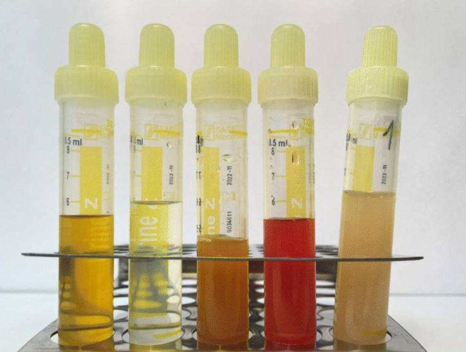
Urinalysis, a portmanteau of the words ''urine'' and ''analysis'', is a Test panel, panel of medical tests that includes physical (macroscopic) examination of the urine, chemical evaluation using urine test strips, and #Microscopic examination, microscopic examination. Macroscopic examination targets parameters such as color, clarity, odor, and specific gravity; urine test strips measure chemical properties such as pH, glucose concentration, and protein levels; and microscopy is performed to identify elements such as Cell (biology), cells, urinary casts, Crystalluria, crystals, and organisms. Background Urine is produced by the filtration of blood in the kidneys. The formation of urine takes place in microscopic structures called nephrons, about one million of which are found in a normal human kidney. Blood enters the kidney though the renal artery and flows through the kidney's vasculature into the Glomerulus (kidney), glomerulus, a tangled knot of capillaries surrounded by Bow ... [...More Info...] [...Related Items...] OR: [Wikipedia] [Google] [Baidu] |
Percutaneous Umbilical Cord Blood Sampling
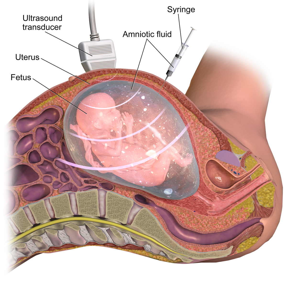
Percutaneous umbilical cord blood sampling (PUBS), also called cordocentesis, fetal blood sampling, or umbilical vein sampling is a diagnostic genetic test that examines blood from the fetal umbilical cord to detect fetal abnormalities. Fetal and maternal blood supply are typically connected in utero with one vein and two arteries to the fetus. The umbilical vein is responsible for delivering oxygen rich blood to the fetus from the mother; the umbilical arteries are responsible for removing oxygen poor blood from the fetus. This allows for the fetus’ tissues to properly perfuse. PUBS provides a means of rapid chromosome analysis and is useful when information cannot be obtained through amniocentesis, chorionic villus sampling, or ultrasound (or if the results of these tests were inconclusive); this test carries a significant risk of complication and is typically reserved for pregnancies determined to be at high risk for genetic defect. It has been used with mothers with immun ... [...More Info...] [...Related Items...] OR: [Wikipedia] [Google] [Baidu] |
Umbilical Cord
In Placentalia, placental mammals, the umbilical cord (also called the navel string, birth cord or ''funiculus umbilicalis'') is a conduit between the developing embryo or fetus and the placenta. During prenatal development, the umbilical cord is physiologically and genetically part of the fetus and (in humans) normally contains two arteries (the umbilical arteries) and one vein (the umbilical vein), buried within Wharton's jelly. The umbilical vein supplies the fetus with oxygenated, nutrient-rich blood from the placenta. Conversely, the fetal heart pumps low-oxygen, nutrient-depleted blood through the umbilical arteries back to the placenta. Structure and development The umbilical cord develops from and contains remnants of the yolk sac and allantois. It forms by the fifth week of human embryogenesis, development, replacing the yolk sac as the source of nutrients for the embryo. The cord is not directly connected to the mother's circulatory system, but instead joins the pla ... [...More Info...] [...Related Items...] OR: [Wikipedia] [Google] [Baidu] |
Fluorescent In Situ Hybridization
Fluorescence ''in situ'' hybridization (FISH) is a cytogenetics, molecular cytogenetic technique that uses hybridization probe, fluorescent probes that bind to only particular parts of a nucleic acid sequence with a high degree of sequence Complementarity (molecular biology), complementarity. It was developed by biomedical researchers in the early 1980s to detect and localize the presence or absence of specific DNA DNA sequence, sequences on chromosomes. Fluorescence microscopy can be used to find out where the fluorescent probe is bound to the chromosomes. FISH is often used for finding specific features in DNA for use in genetic counseling, medicine, and species identification. FISH can also be used to detect and localize specific RNA targets (mRNA, Long non-coding RNA, lncRNA and miRNA) in cells, circulating tumor cells, and tissue samples. In this context, it can help define the spatial-temporal patterns of gene expression within cells and tissues. Probes – RNA and DNA ... [...More Info...] [...Related Items...] OR: [Wikipedia] [Google] [Baidu] |
Karyotyping
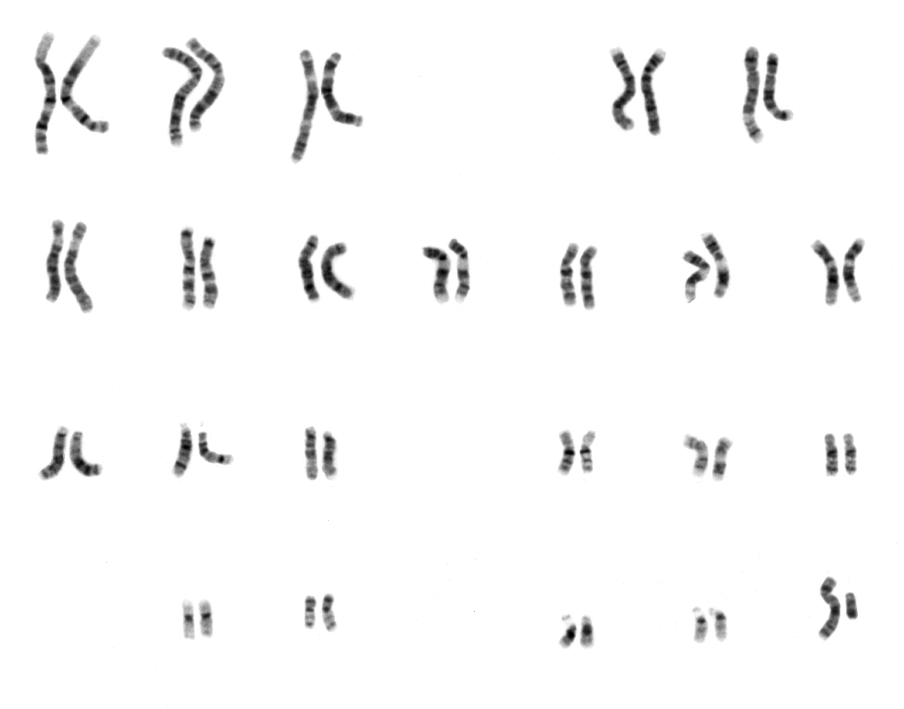
A karyotype is the general appearance of the complete set of chromosomes in the cells of a species or in an individual organism, mainly including their sizes, numbers, and shapes. Karyotyping is the process by which a karyotype is discerned by determining the chromosome complement of an individual, including the number of chromosomes and any abnormalities. A karyogram or idiogram is a graphical depiction of a karyotype, wherein chromosomes are generally organized in pairs, ordered by size and position of centromere for chromosomes of the same size. Karyotyping generally combines light microscopy and photography in the metaphase of the cell cycle, and results in a photomicrographic (or simply micrographic) karyogram. In contrast, a schematic karyogram is a designed graphic representation of a karyotype. In schematic karyograms, just one of the sister chromatids of each chromosome is generally shown for brevity, and in reality they are generally so close together that they look as ... [...More Info...] [...Related Items...] OR: [Wikipedia] [Google] [Baidu] |
Cell-free Fetal DNA
Cell-free fetal DNA (cffDNA) is fetal DNA that circulates freely in the maternal blood. Maternal blood is sampled by venipuncture. Analysis of cffDNA is a method of non-invasive prenatal diagnosis frequently ordered for pregnant women of advanced age. Two hours after delivery, cffDNA is no longer detectable in maternal blood. Background cffDNA originates from placental trophoblasts. Fetal DNA is fragmented when placental microparticles are shed into the maternal blood circulation. cffDNA fragments are approximately 200 base pairs (bp) in length. They are significantly smaller than maternal DNA fragments. The difference in size allows cffDNA to be distinguished from maternal DNA fragments. Approximately 11 to 13.4 percent of the cell-free DNA in maternal blood is of fetal origin. The amount varies widely from one pregnant woman to another. cffDNA is present after five to seven weeks gestation. The amount of cffDNA increases as the pregnancy progresses. The quantity of cffDNA ... [...More Info...] [...Related Items...] OR: [Wikipedia] [Google] [Baidu] |
Prenatal Diagnosis
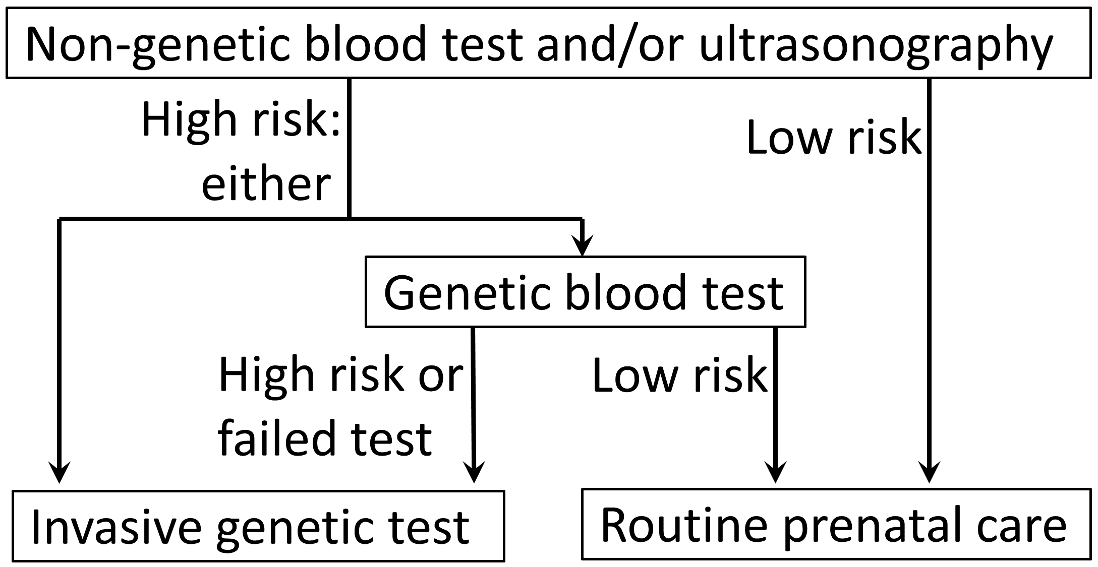
Prenatal testing is a tool that can be used to detect some birth defects at various stages prior to birth. Prenatal testing consists of prenatal screening and prenatal diagnosis, which are aspects of prenatal care that focus on detecting problems with the pregnancy as early as possible. These may be anatomic and physiologic problems with the health of the zygote, embryo, or fetus, either before gestation even starts (as in preimplantation genetic diagnosis) or as early in gestation as practicable. Screening can detect problems such as neural tube defects, chromosome abnormalities, and gene mutations that would lead to genetic disorders and birth defects such as spina bifida, cleft palate, Down syndrome, trisomy 18, Tay–Sachs disease, sickle cell anemia, thalassemia, cystic fibrosis, muscular dystrophy, and fragile X syndrome. Some tests are designed to discover problems which primarily affect the health of the mother, such as PAPP-A to detect pre-eclampsia or glucose tole ... [...More Info...] [...Related Items...] OR: [Wikipedia] [Google] [Baidu] |
Multiplex (assay)
In the biological sciences, a multiplex assay is a type of immunoassay that uses magnetic beads to simultaneously measure multiple analytes in a single experiment. A multiplex assay is a derivative of an ELISA using beads for binding the capture antibody. Multiplex assays are still more common in research than in clinical settings. In a multiplex assay, microspheres of designated colors are coated with antibodies of defined binding specificities. The results can be read by flow cytometry Flow cytometry (FC) is a technique used to detect and measure the physical and chemical characteristics of a population of cells or particles. In this process, a sample containing cells or particles is suspended in a fluid and injected into the ... because the beads are distinguishable by fluorescent signature. The number of analytes measured is determined by the number of different bead colors. Multiplex assays within a given application area or class of technology can be further stratified ... [...More Info...] [...Related Items...] OR: [Wikipedia] [Google] [Baidu] |
ELISA
The enzyme-linked immunosorbent assay (ELISA) (, ) is a commonly used analytical biochemistry assay, first described by Eva Engvall and Peter Perlmann in 1971. The assay is a solid-phase type of enzyme immunoassay (EIA) to detect the presence of a ligand (commonly an amino acid) in a liquid sample using antibodies directed against the ligand to be measured. ELISA has been used as a medical diagnosis, diagnostic tool in medicine, plant pathology, and biotechnology, as well as a quality control check in various industries. In the most simple form of an ELISA, antigens from the sample to be tested are attached to a surface. Then, a matching antibody is applied over the surface so it can bind the antigen. This antibody is linked to an enzyme, and then any unbound antibodies are removed. In the final step, a substance containing the enzyme's Enzyme substrate, substrate is added. If there was binding, the subsequent reaction produces a detectable signal, most commonly a color change. ... [...More Info...] [...Related Items...] OR: [Wikipedia] [Google] [Baidu] |
Cerebrospinal Fluid
Cerebrospinal fluid (CSF) is a clear, colorless Extracellular fluid#Transcellular fluid, transcellular body fluid found within the meninges, meningeal tissue that surrounds the vertebrate brain and spinal cord, and in the ventricular system, ventricles of the brain. CSF is mostly produced by specialized Ependyma, ependymal cells in the choroid plexuses of the ventricles of the brain, and absorbed in the arachnoid granulations. It is also produced by ependymal cells in the lining of the ventricles. In humans, there is about 125 mL of CSF at any one time, and about 500 mL is generated every day. CSF acts as a shock absorber, cushion or buffer, providing basic mechanical and immune system, immunological protection to the brain inside the Human skull, skull. CSF also serves a vital function in the cerebral autoregulation of cerebral blood flow. CSF occupies the subarachnoid space (between the arachnoid mater and the pia mater) and the ventricular system around and inside t ... [...More Info...] [...Related Items...] OR: [Wikipedia] [Google] [Baidu] |


