|
Larsen Syndrome
Larsen syndrome (LS) is a congenital disorder discovered in 1950 by Larsen and associates when they observed dislocation of the large joints and face anomalies in six of their patients.Mitra, N., Kannan, N., Kumar, V.S., Kavita, G. "Larsen Syndrome: A Case Report." Journal of Nepal Paediatric Society. 32.1 (2012): 85-87. Print. Patients with Larsen syndrome normally present with a variety of symptoms, including congenital anterior dislocation of the knees, dislocation of the hips and elbows, flattened facial appearance, prominent foreheads, and depressed nasal bridges.Liang, C.D., Hang, C.L. " Elongation of the Aorta and Multiple Cardiovascular Abnormalities Associated with Larsen Syndrome." Pediatric Cardiology. 22. (2001): 245-246. Print. Larsen syndrome can also cause a variety of cardiovascularKiel, E.A., et al. "Cardiovascular Manifestations in the Larsen Syndrome." Pediatrics. 71.6 (1983): 942-946. Print. and orthopedic abnormalities.Johnston, C.E., Birch, J.G., Daniels, J.L. ... [...More Info...] [...Related Items...] OR: [Wikipedia] [Google] [Baidu] |
Congenital Disorder
A birth defect is an abnormal condition that is present at childbirth, birth, regardless of its cause. Birth defects may result in disability, disabilities that may be physical disability, physical, intellectual disability, intellectual, or developmental disability, developmental. The disabilities can range from mild to severe. Birth defects are divided into two main types: structural disorders in which problems are seen with the shape of a body part and functional disorders in which problems exist with how a body part works. Functional disorders include metabolic disorder, metabolic and degenerative disease, degenerative disorders. Some birth defects include both structural and functional disorders. Birth defects may result from genetic disorder, genetic or chromosome abnormality, chromosomal disorders, exposure to certain medications or chemicals, or certain vertically transmitted infection, infections during pregnancy. Risk factors include folate deficiency, alcohol drink, d ... [...More Info...] [...Related Items...] OR: [Wikipedia] [Google] [Baidu] |
Stenosis
Stenosis () is the abnormal narrowing of a blood vessel or other tubular organ or structure such as foramina and canals. It is also sometimes called a stricture (as in urethral stricture). ''Stricture'' as a term is usually used when narrowing is caused by contraction of smooth muscle (e.g. achalasia, prinzmetal angina); ''stenosis'' is usually used when narrowing is caused by lesion that reduces the space of lumen (e.g. atherosclerosis). The term coarctation is another synonym, but is commonly used only in the context of aortic coarctation. Restenosis is the recurrence of stenosis after a procedure. Examples Examples of vascular stenotic lesions include: * Intermittent claudication (peripheral artery stenosis) * Angina ( coronary artery stenosis) * Carotid artery stenosis which predispose to (strokes and transient ischaemic episodes) * Renal artery stenosis Types In heart valves The types of stenoses in heart valves are: * Pulmonary valve stenosis, which is th ... [...More Info...] [...Related Items...] OR: [Wikipedia] [Google] [Baidu] |
Microdontia
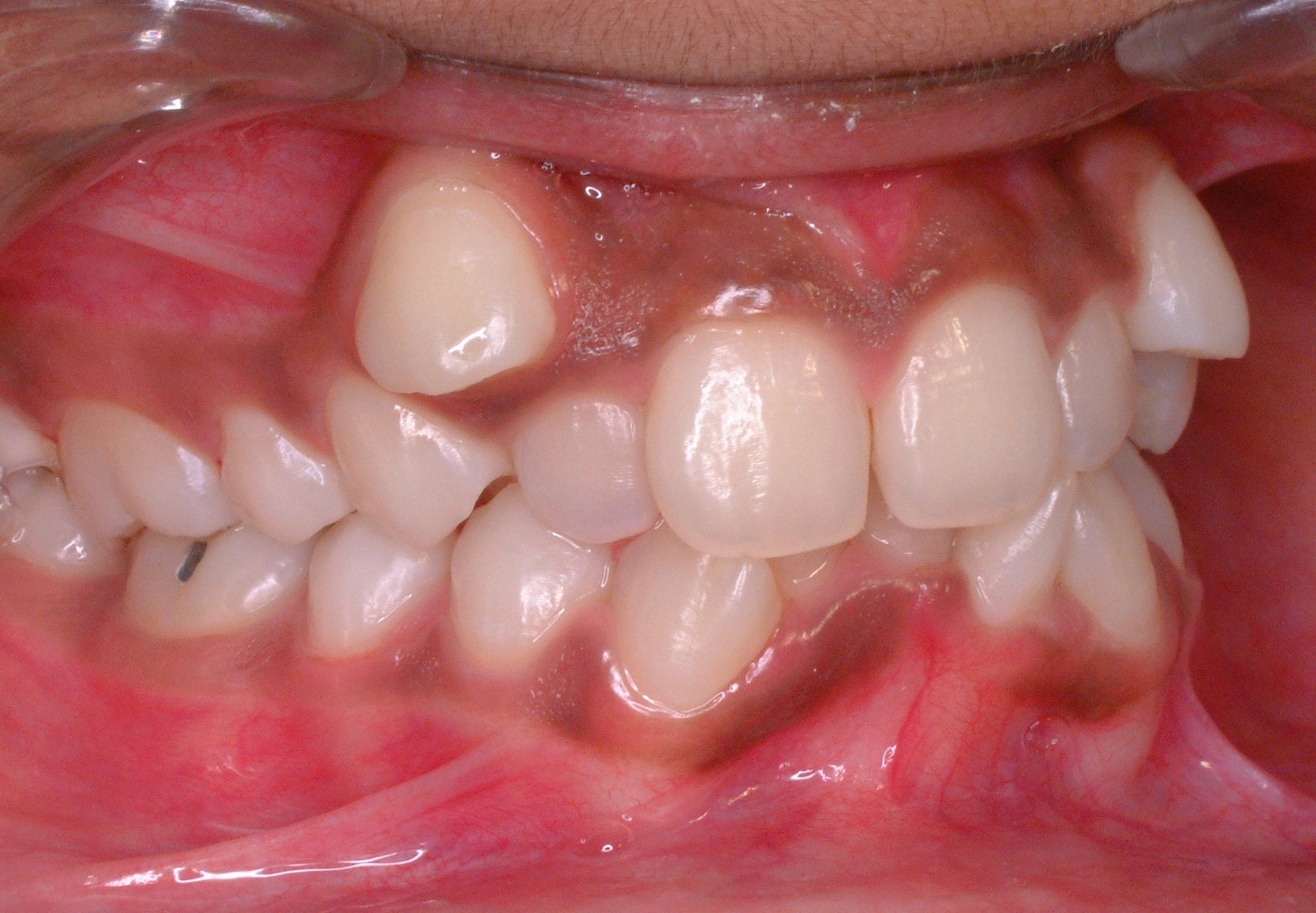
Microdontia is a condition in which one or more teeth appear smaller than normal. In the generalized form, all teeth are involved. In the localized form, only a few teeth are involved. The most common teeth affected are the upper lateral incisors and third molars. Teeth affected by microdontia may also have abnormal shape, and the abnormal size may affect the whole tooth, or only a part of the tooth. Definition Males tend to have larger teeth than females, and tooth size also varies by race. Abnormal tooth size is defined by some as when the dimensions are more than 2 standard deviations from the average. Microdontia is when the teeth are abnormally small, and macrodontia is when the teeth are abnormally large. Classification There are 3 types of microdontia: True generalized All the teeth are smaller than the normal size. True generalized microdontia is very rare, and occurs in pituitary dwarfism. Due to decreased levels of growth hormone the teeth fail to develop to a normal ... [...More Info...] [...Related Items...] OR: [Wikipedia] [Google] [Baidu] |
Malocclusion
In orthodontics, a malocclusion is a misalignment or incorrect relation between the teeth of the upper and lower dental arches when they approach each other as the jaws close. The English-language term dates from 1864; Edward Angle (1855–1930), the "father of modern orthodontics", popularised it. The word derives . The malocclusion classification is based on the relationship of the mesiobuccal cusp of the maxillary first molar and the buccal groove of the mandibular first molar. If this molar relationship exists, then the teeth can align into normal occlusion. According to Angle, malocclusion is any deviation of the occlusion from the ideal. However, assessment for malocclusion should also take into account aesthetics and the impact on functionality. If these aspects are acceptable to the patient despite meeting the formal definition of malocclusion, then treatment may not be necessary. It is estimated that nearly 30% of the population have malocclusions that are cat ... [...More Info...] [...Related Items...] OR: [Wikipedia] [Google] [Baidu] |
Cleft Palate
A cleft lip contains an opening in the upper lip that may extend into the nose. The opening may be on one side, both sides, or in the middle. A cleft palate occurs when the palate (the roof of the mouth) contains an opening into the nose. The term orofacial cleft refers to either condition or to both occurring together. These disorders can result in feeding problems, speech problems, hearing problems, and frequent ear infections. Less than half the time the condition is associated with other disorders. Cleft lip and palate are the result of tissues of the face not joining properly during development. As such, they are a type of birth defect. The cause is unknown in most cases. Risk factors include smoking during pregnancy, diabetes, obesity, an older mother, and certain medications (such as some used to treat seizures). Cleft lip and cleft palate can often be diagnosed during pregnancy with an ultrasound exam. A cleft lip or palate can be successfully treated with surge ... [...More Info...] [...Related Items...] OR: [Wikipedia] [Google] [Baidu] |
Cataracts
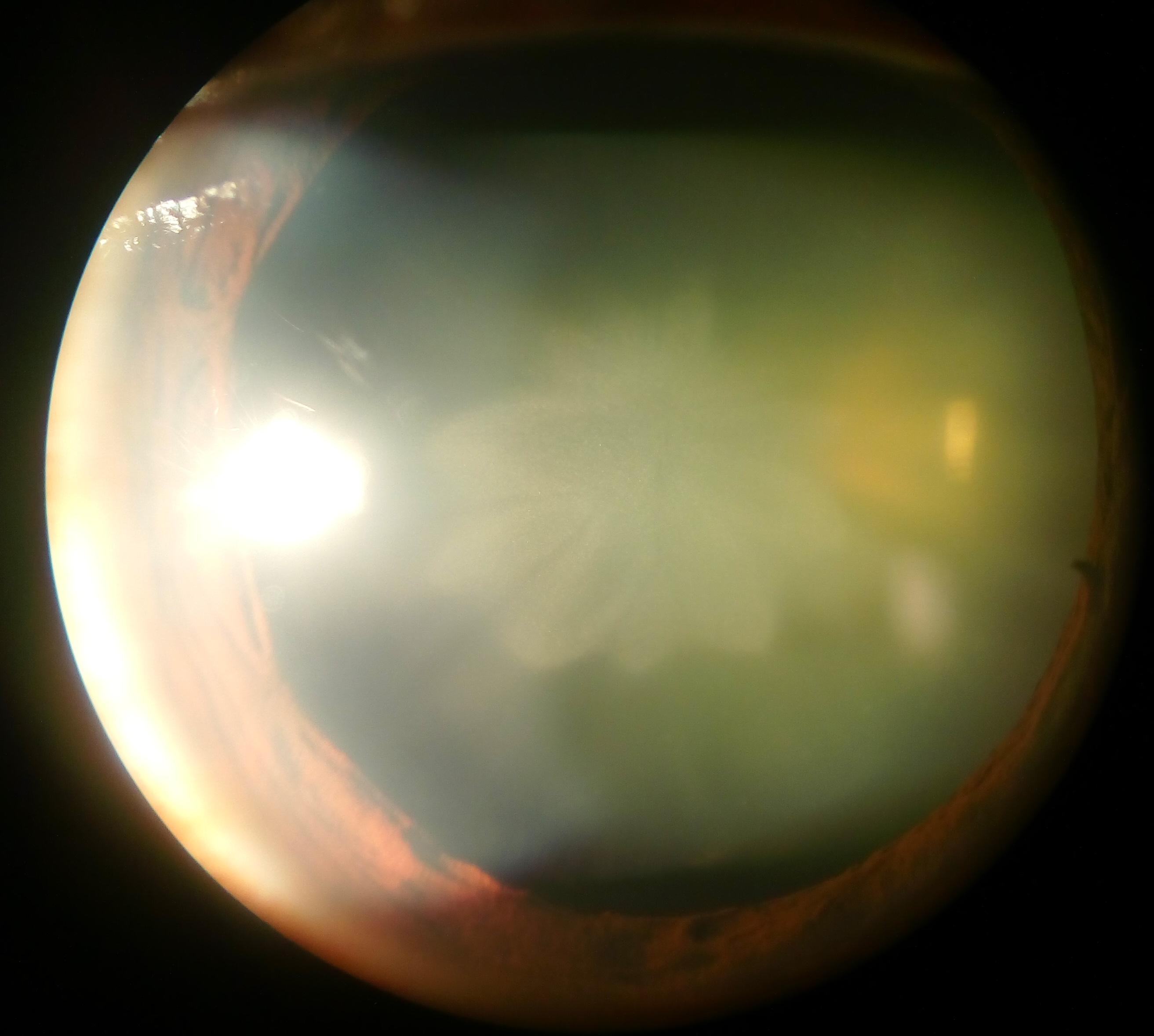
A cataract is a cloudy area in the lens of the eye that leads to a decrease in vision of the eye. Cataracts often develop slowly and can affect one or both eyes. Symptoms may include faded colours, blurry or double vision, halos around light, trouble with bright lights, and difficulty seeing at night. This may result in trouble driving, reading, or recognizing faces. Poor vision caused by cataracts may also result in an increased risk of falling and depression. Cataracts cause 51% of all cases of blindness and 33% of visual impairment worldwide. Cataracts are most commonly due to aging but may also occur due to trauma or radiation exposure, be present from birth, or occur following eye surgery for other problems. Risk factors include diabetes, longstanding use of corticosteroid medication, smoking tobacco, prolonged exposure to sunlight, and alcohol. In addition to these, poor nutrition, obesity, chronic kidney disease, and autoimmune diseases have been recognized in ... [...More Info...] [...Related Items...] OR: [Wikipedia] [Google] [Baidu] |
Ductus Arteriosus
The ductus arteriosus, also called the ductus Botalli, named after the Italian physiologist Leonardo Botallo, is a blood vessel in the developing fetus connecting the trunk of the pulmonary artery to the proximal descending aorta. It allows most of the blood from the right ventricle to bypass the fetus's fluid-filled non-functioning lungs. Upon closure at birth, it becomes the ligamentum arteriosum. Development and structure The ductus arteriosus is formed from the left 6th aortic arches during embryonic development and attaches to the final part of the aortic arch (the isthmus of aorta) and the first part of the pulmonary artery. Disorder: patent ductus arteriosus Consequences Failure of the ductus arteriosus to close after birth results in a condition called patent ductus arteriosus, which results in the abnormal flow of blood from the aorta to the pulmonary artery: a left-to-right shunt. If left uncorrected, this usually leads to pulmonary hypertension followed by r ... [...More Info...] [...Related Items...] OR: [Wikipedia] [Google] [Baidu] |
Aneurysm
An aneurysm is an outward :wikt:bulge, bulging, likened to a bubble or balloon, caused by a localized, abnormal, weak spot on a blood vessel wall. Aneurysms may be a result of a hereditary condition or an acquired disease. Aneurysms can also be a wikt:Special:Search/nidus, nidus (starting point) for clot formation (thrombosis) and Embolism, embolization. As an aneurysm increases in size, the risk of rupture increases, which could lead to uncontrolled bleeding. Although they may occur in any blood vessel, particularly lethal examples include aneurysms of the circle of Willis in the brain, aortic aneurysms affecting the thoracic aorta, and abdominal aortic aneurysms. Aneurysms can arise in the heart itself following a Myocardial infarction, heart attack, including both Ventricular aneurysm, ventricular and atrial septal aneurysms. There are congenital atrial septal defect, atrial septal aneurysms, a rare heart defect. Etymology The word is from Greek language, Greek: ἀνεύρ� ... [...More Info...] [...Related Items...] OR: [Wikipedia] [Google] [Baidu] |
Aortic Dissection
Aortic dissection (AD) occurs when an injury to the innermost layer of the aorta allows blood to flow between the layers of the aortic wall, forcing the layers apart. In most cases, this is associated with a sudden onset of agonizing chest or back pain, often described as "tearing" in character. Vomiting, sweating, and lightheadedness may also occur. Damage to other organs may result from the decreased blood supply, such as stroke, lower extremity ischemia, or mesenteric ischemia. Aortic dissection can quickly lead to death from insufficient blood flow to the heart or complete rupture of the aorta. AD is more common in those with a history of high blood pressure; a number of connective tissue diseases that affect blood vessel wall strength including Marfan syndrome and Ehlers–Danlos syndrome; a bicuspid aortic valve; and previous heart surgery. Major trauma, smoking, cocaine use, pregnancy, a thoracic aortic aneurysm, inflammation of arteries, and abnormal lipid ... [...More Info...] [...Related Items...] OR: [Wikipedia] [Google] [Baidu] |
Tricuspid Valve Prolapse
In medicine, prolapse is a condition in which organs fall down or slip out of place. It is used for organs protruding through the vagina, rectum, or for the misalignment of the valves of the heart. A spinal disc herniation is also sometimes called "disc prolapse". Prolapse means "to fall out of place", from the Latin ' meaning "to fall out". Relating to the uterus, prolapse condition results in an inferior extension of the organ into the vagina, caused by weakened pelvic muscles. Humans Heart valve prolapse The main type of prolapse of heart valves in humans is mitral valve prolapse (MVP), which is a valvular heart disease characterized by the displacement of an abnormally thickened mitral valve leaflet into the left atrium during systole. ''Tricuspid valve prolapse'' can cause tricuspid regurgitation.Page 41 in: Rectal prolapse Rectal prolapse is a condition in which part of the wall or the entire wall of the rectum falls out of place. Rectal prolapse can be a medical e ... [...More Info...] [...Related Items...] OR: [Wikipedia] [Google] [Baidu] |
Patent Ductus Arteriosus
Patent ductus arteriosus (PDA) is a medical condition in which the ''ductus arteriosus'' fails to close after childbirth, birth: this allows a portion of oxygenated blood from the left heart to flow back to the lungs from the aorta, which has a higher blood pressure, to the pulmonary artery, which has a lower blood pressure. Symptoms are uncommon at birth and shortly thereafter, but later in the first year of life there is often the onset of an increased work of breathing and Failure to thrive, failure to gain weight at a normal rate. With time, an uncorrected PDA usually leads to pulmonary hypertension followed by right-sided heart failure. The ''ductus arteriosus'' is a Fetal circulation, fetal blood vessel that normally closes soon after birth. This closure is caused by vessel constriction immediately after birth as circulation changes occur, followed by the occlusion of the vessel’s lumen in the following days. In a PDA, the vessel does not close, but remains ''patent'' (ope ... [...More Info...] [...Related Items...] OR: [Wikipedia] [Google] [Baidu] |


