|
Korfiella
''Korfiella'' is a fungal genus in the family Sarcosomataceae. A monotypic genus, it contains the single species ''Korfiella karnika'', found in India and described as new to science in 1970. Discovery The first scientifically documented collections of ''Korfiella karnika'' were made in November, 1965. Fruitbodies of the fungus were found growing on the rotting stump of a tree in Nainital, India. Specimens were sent to the Royal Botanic Gardens, Kew, where mycologist R.W.G. Dennis noted their similarity to '' Plectania melastoma''. After further consultation with Discomycetes authority Richard Korf, the authors Pant and Tewari erected a new genus, naming it in honour of Korf. The specific epithet ''karnika''—derived from the Sanskrit ''karna'', ("ear")—refers to the fruitbody shape. Pant and Tewari considered the fungus aligned with the tribe ''Urnuleae'' of the family Sarcoscyphaceae, according to the taxonomy of the time. ''Korfiella'' is now classified in the Sarcosoma ... [...More Info...] [...Related Items...] OR: [Wikipedia] [Google] [Baidu] |
Sarcosomataceae
The Sarcosomataceae are a family of fungi in the order Pezizales. According to a 2008 estimate, the family contains 10 genera and 57 species. Most species are found in temperate areas, and are typically saprobic Saprotrophic nutrition or lysotrophic nutrition is a process of chemoheterotrophic extracellular digestion involved in the processing of decayed (dead or waste) organic matter. It occurs in saprotrophs, and is most often associated with fungi ( ... on rotten or buried wood. References Pezizales Ascomycota families {{Pezizomycetes-stub ... [...More Info...] [...Related Items...] OR: [Wikipedia] [Google] [Baidu] |
Richard Korf
Richard Paul "Dick" Korf (May 28, 1925 – August 20, 2016) was an American mycologist and founding co-editor of the journal ''Mycotaxon''. He was a preeminent figure in the study of discomycetes and made significant contributions to the field of fungal nomenclature and taxonomy. Korf was professor emeritus of mycology at Cornell University and director emeritus of Cornell University's Plant Pathology Herbarium. Early life and education Korf was born on May 28, 1925, to an upper-middle-class family with homes in Westchester County, New York, and New Fairfield, Connecticut. While attending the prestigious Riverdale Country School in New York City, Korf was placed in charge of his biology class after their teacher joined the military. "In retrospect," Korf wrote, "I am convinced that this experience had an enormous impact on my future and on my decision to enter the teaching profession." In 1942, shortly after his 17th birthday, Korf enrolled at Cornell University "with the vagu ... [...More Info...] [...Related Items...] OR: [Wikipedia] [Google] [Baidu] |
Fungi
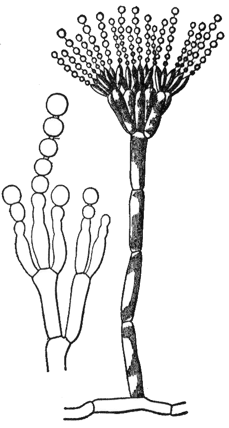
A fungus (plural, : fungi or funguses) is any member of the group of Eukaryote, eukaryotic organisms that includes microorganisms such as yeasts and Mold (fungus), molds, as well as the more familiar mushrooms. These organisms are classified as a Kingdom (biology), kingdom, separately from the other eukaryotic kingdoms, which by one traditional classification include Plantae, Animalia, Protozoa, and Chromista. A characteristic that places fungi in a different kingdom from plants, bacteria, and some protists is chitin in their cell walls. Fungi, like animals, are heterotrophs; they acquire their food by absorbing dissolved molecules, typically by secreting digestive enzymes into their environment. Fungi do not photosynthesize. Growth is their means of motility, mobility, except for spores (a few of which are flagellated), which may travel through the air or water. Fungi are the principal decomposers in ecological systems. These and other differences place fungi in a single gro ... [...More Info...] [...Related Items...] OR: [Wikipedia] [Google] [Baidu] |
Sarcoscyphaceae
The ''Sarcoscyphaceae'' are a family of cup fungi in the order Pezizales. Members of the Sarcoscyphaceae are cosmopolitan in distribution, found in both tropical and temperate regions. Genera A 2008 estimate placed 13 genera and 102 species in the family: * ''Aurophora'' Rifai 1968 * ''Cookeina'' Kuntze 1891 * ''Geodina'' Denison 1965 * ''Kompsoscypha'' Pfister 1989 * ''Microstoma (fungus)'' Bernstein 1852 * ''Nanoscypha'' Denison 1972 * '' Phillipsia'' Berk. 1881 * ''Pithya'' Fuckel 1870 * ''Pseudopithyella'' Seaver 1928 * ''Sarcoscypha'' (Fr.) Boud. 1885: anamorphs are ''Molliardiomyces'' Paden 1984 * ''Thindia'' Korf & Waraitch 1971 * ''Wynnea ''Wynnea'' is a genus of fungi in the family Sarcoscyphaceae. Circumscribed by Miles Joseph Berkeley and Moses Ashley Curtis in 1867, the genus contains seven species that have ear-shaped fruit bodies that grow on the ground. ''Wynnea'' species ...'' Berk. & M.A. Curtis 1867 References {{Taxonbar, from=Q2193232 Ascomycota f ... [...More Info...] [...Related Items...] OR: [Wikipedia] [Google] [Baidu] |
Type (biology)
In biology, a type is a particular wiktionary:en:specimen, specimen (or in some cases a group of specimens) of an organism to which the scientific name of that organism is formally attached. In other words, a type is an example that serves to anchor or centralizes the defining features of that particular taxon. In older usage (pre-1900 in botany), a type was a taxon rather than a specimen. A taxon is a scientifically named grouping of organisms with other like organisms, a set (mathematics), set that includes some organisms and excludes others, based on a detailed published description (for example a species description) and on the provision of type material, which is usually available to scientists for examination in a major museum research collection, or similar institution. Type specimen According to a precise set of rules laid down in the International Code of Zoological Nomenclature (ICZN) and the International Code of Nomenclature for algae, fungi, and plants (ICN), the ... [...More Info...] [...Related Items...] OR: [Wikipedia] [Google] [Baidu] |
Micrometre
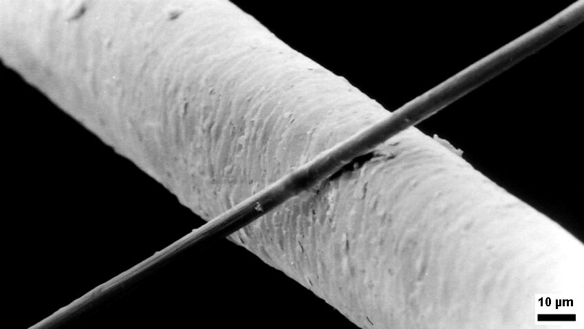
The micrometre ( international spelling as used by the International Bureau of Weights and Measures; SI symbol: μm) or micrometer ( American spelling), also commonly known as a micron, is a unit of length in the International System of Units (SI) equalling (SI standard prefix " micro-" = ); that is, one millionth of a metre (or one thousandth of a millimetre, , or about ). The nearest smaller common SI unit is the nanometre, equivalent to one thousandth of a micrometre, one millionth of a millimetre or one billionth of a metre (). The micrometre is a common unit of measurement for wavelengths of infrared radiation as well as sizes of biological cells and bacteria, and for grading wool by the diameter of the fibres. The width of a single human hair ranges from approximately 20 to . The longest human chromosome, chromosome 1, is approximately in length. Examples Between 1 μm and 10 μm: * 1–10 μm – length of a typical bacterium * 3–8 μm � ... [...More Info...] [...Related Items...] OR: [Wikipedia] [Google] [Baidu] |
Hyaline
A hyaline substance is one with a glassy appearance. The word is derived from el, ὑάλινος, translit=hyálinos, lit=transparent, and el, ὕαλος, translit=hýalos, lit=crystal, glass, label=none. Histopathology Hyaline cartilage is named after its glassy appearance on fresh gross pathology. On light microscopy of H&E stained slides, the extracellular matrix of hyaline cartilage looks homogeneously pink, and the term "hyaline" is used to describe similarly homogeneously pink material besides the cartilage. Hyaline material is usually acellular and proteinaceous. For example, arterial hyaline is seen in aging, high blood pressure, diabetes mellitus Diabetes, also known as diabetes mellitus, is a group of metabolic disorders characterized by a high blood sugar level (hyperglycemia) over a prolonged period of time. Symptoms often include frequent urination, increased thirst and increased ... and in association with some drugs (e.g. calcineurin inhibitors) ... [...More Info...] [...Related Items...] OR: [Wikipedia] [Google] [Baidu] |
Ascospore
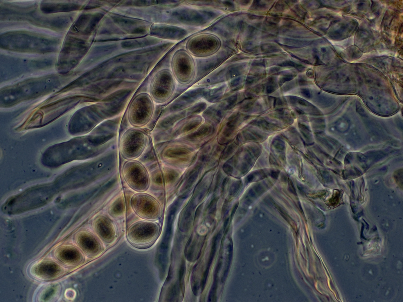
An ascus (; ) is the sexual spore-bearing cell produced in ascomycete fungi. Each ascus usually contains eight ascospores (or octad), produced by meiosis followed, in most species, by a mitotic cell division. However, asci in some genera or species can occur in numbers of one (e.g. '' Monosporascus cannonballus''), two, four, or multiples of four. In a few cases, the ascospores can bud off conidia that may fill the asci (e.g. '' Tympanis'') with hundreds of conidia, or the ascospores may fragment, e.g. some '' Cordyceps'', also filling the asci with smaller cells. Ascospores are nonmotile, usually single celled, but not infrequently may be coenocytic (lacking a septum), and in some cases coenocytic in multiple planes. Mitotic divisions within the developing spores populate each resulting cell in septate ascospores with nuclei. The term ocular chamber, or oculus, refers to the epiplasm (the portion of cytoplasm not used in ascospore formation) that is surrounded by the "bo ... [...More Info...] [...Related Items...] OR: [Wikipedia] [Google] [Baidu] |
Staining
Staining is a technique used to enhance contrast in samples, generally at the microscopic level. Stains and dyes are frequently used in histology (microscopic study of biological tissues), in cytology (microscopic study of cells), and in the medical fields of histopathology, hematology, and cytopathology that focus on the study and diagnoses of diseases at the microscopic level. Stains may be used to define biological tissues (highlighting, for example, muscle fibers or connective tissue), cell populations (classifying different blood cells), or organelles within individual cells. In biochemistry, it involves adding a class-specific ( DNA, proteins, lipids, carbohydrates) dye to a substrate to qualify or quantify the presence of a specific compound. Staining and fluorescent tagging can serve similar purposes. Biological staining is also used to mark cells in flow cytometry, and to flag proteins or nucleic acids in gel electrophoresis. Light microscopes are used for ... [...More Info...] [...Related Items...] OR: [Wikipedia] [Google] [Baidu] |
Ascus
An ascus (; ) is the sexual spore-bearing cell produced in ascomycete fungi. Each ascus usually contains eight ascospores (or octad), produced by meiosis followed, in most species, by a mitotic cell division. However, asci in some genera or species can occur in numbers of one (e.g. '' Monosporascus cannonballus''), two, four, or multiples of four. In a few cases, the ascospores can bud off conidia that may fill the asci (e.g. '' Tympanis'') with hundreds of conidia, or the ascospores may fragment, e.g. some '' Cordyceps'', also filling the asci with smaller cells. Ascospores are nonmotile, usually single celled, but not infrequently may be coenocytic (lacking a septum), and in some cases coenocytic in multiple planes. Mitotic divisions within the developing spores populate each resulting cell in septate ascospores with nuclei. The term ocular chamber, or oculus, refers to the epiplasm (the portion of cytoplasm not used in ascospore formation) that is surrounded by the "bou ... [...More Info...] [...Related Items...] OR: [Wikipedia] [Google] [Baidu] |
Hypha
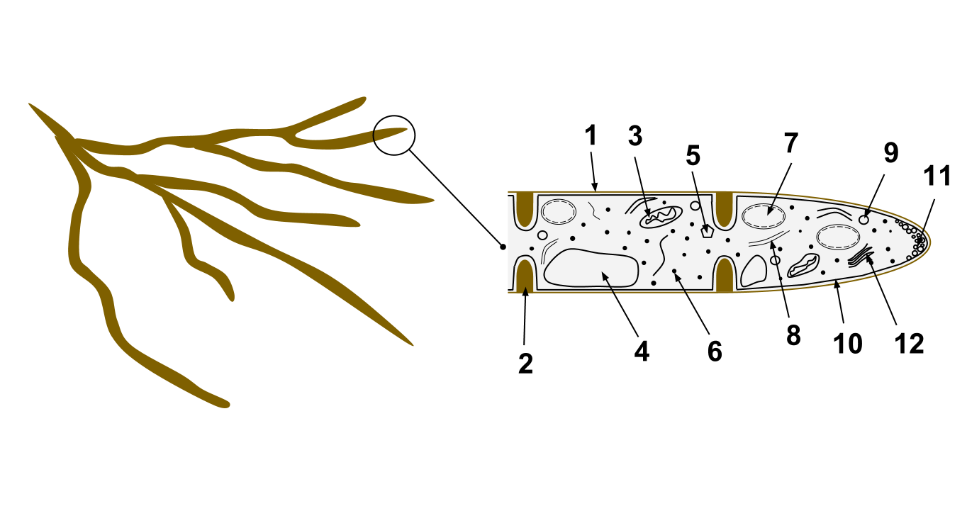
A hypha (; ) is a long, branching, filamentous structure of a fungus, oomycete, or actinobacterium. In most fungi, hyphae are the main mode of vegetative growth, and are collectively called a mycelium. Structure A hypha consists of one or more cells surrounded by a tubular cell wall. In most fungi, hyphae are divided into cells by internal cross-walls called "septa" (singular septum). Septa are usually perforated by pores large enough for ribosomes, mitochondria, and sometimes nuclei to flow between cells. The major structural polymer in fungal cell walls is typically chitin, in contrast to plants and oomycetes that have cellulosic cell walls. Some fungi have aseptate hyphae, meaning their hyphae are not partitioned by septa. Hyphae have an average diameter of 4–6 µm. Growth Hyphae grow at their tips. During tip growth, cell walls are extended by the external assembly and polymerization of cell wall components, and the internal production of new cell membran ... [...More Info...] [...Related Items...] OR: [Wikipedia] [Google] [Baidu] |
Apothecium
An ascocarp, or ascoma (), is the fruiting body ( sporocarp) of an ascomycete phylum fungus. It consists of very tightly interwoven hyphae and millions of embedded asci, each of which typically contains four to eight ascospores. Ascocarps are most commonly bowl-shaped (apothecia) but may take on a spherical or flask-like form that has a pore opening to release spores (perithecia) or no opening (cleistothecia). Classification The ascocarp is classified according to its placement (in ways not fundamental to the basic taxonomy). It is called ''epigeous'' if it grows above ground, as with the morels, while underground ascocarps, such as truffles, are termed ''hypogeous''. The structure enclosing the hymenium is divided into the types described below (apothecium, cleistothecium, etc.) and this character ''is'' important for the taxonomic classification of the fungus. Apothecia can be relatively large and fleshy, whereas the others are microscopic—about the size of flecks ... [...More Info...] [...Related Items...] OR: [Wikipedia] [Google] [Baidu] |

.jpg)


