|
Jeffrey Siewerdsen
Jeffrey Harold Siewerdsen (born 1969) is an American physicist and biomedical engineer who is a Professor of Imaging Physics at The University of Texas MD Anderson Cancer Center as well as Biomedical Engineering, Computer Science, Radiology, and Neurosurgery at Johns Hopkins University.He is among the original inventors of cone-beam CT-guided radiotherapy as well as weight-bearing cone-beam CT for musculoskeletal radiology and orthopedic surgery. His work also includes the early development of flat-panel detectors on mobile C-arms for intraoperative cone-beam CT in image-guided surgery. He developed early models for the signal and noise performance of flat-panel detectors and later extended such analysis to dual-energy imaging and 3D imaging performance in cone-beam CT. He founded the ISTAR Lab (Imaging for Surgery, Therapy, and Radiology) in the Department of Biomedical Engineering, the Carnegie Center for Surgical Innovation at Johns Hopkins Hospital, and the Surgical Data Scienc ... [...More Info...] [...Related Items...] OR: [Wikipedia] [Google] [Baidu] |
Johns Hopkins University
The Johns Hopkins University (often abbreviated as Johns Hopkins, Hopkins, or JHU) is a private university, private research university in Baltimore, Maryland, United States. Founded in 1876 based on the European research institution model, Johns Hopkins is considered to be the first research university in the U.S. The university was named for its first benefactor, the American entrepreneur and Quakers, Quaker philanthropist Johns Hopkins. Hopkins's $7 million bequest (equivalent to $ in ) to establish the university was the largest Philanthropy, philanthropic gift in U.S. history up to that time. Daniel Coit Gilman, who was inaugurated as :Presidents of Johns Hopkins University, Johns Hopkins's first president on February 22, 1876, led the university to revolutionize higher education in the U.S. by integrating teaching and research. In 1900, Johns Hopkins became a founding member of the Association of American Universities. The university has led all Higher education in the U ... [...More Info...] [...Related Items...] OR: [Wikipedia] [Google] [Baidu] |
Homer Neal
Homer Alfred Neal (June 13, 1942 – May 23, 2018) was an American particle physicist and a distinguished professor at the University of Michigan. Neal was president of the American Physical Society in 2016. He was also a board member of Ford Motor Company, a council member of the National Museum of African American History and Culture, and a director of the Richard Lounsbery Foundation. Neal was the interim President of the University of Michigan in 1996. Neal's research group works as part of the ATLAS experiment hosted at CERN in Geneva. Biography Neal grew up an African-American in highly segregated Franklin, Kentucky, and was forced by his neighbors there to break off relations with a white friend with whom he had bonded over a shared interest in ham radio. He received his B.S. in physics from Indiana University Bloomington in 1961, and earned his Ph.D. from the University of Michigan in 1966. From 1976 to 1981, Neal was Dean for Research and Graduate Development at Indiana ... [...More Info...] [...Related Items...] OR: [Wikipedia] [Google] [Baidu] |
Image Registration
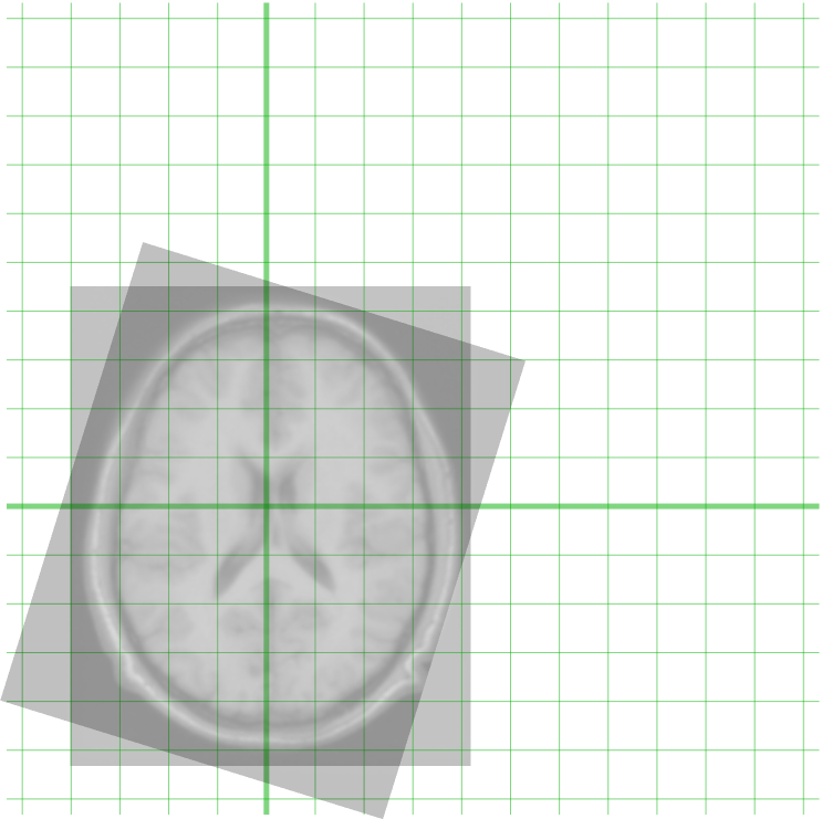
Image registration is the process of transforming different sets of data into one coordinate system. Data may be multiple photographs, data from different sensors, times, depths, or viewpoints. It is used in computer vision, medical imaging, military automatic target recognition, and compiling and analyzing images and data from satellites. Registration is necessary in order to be able to compare or integrate the data obtained from these different measurements. Algorithm classification Intensity-based vs feature-based Image registration or image alignment algorithms can be classified into intensity-based and feature-based.A. Ardeshir Goshtasby2-D and 3-D Image Registration for Medical, Remote Sensing, and Industrial Applications Wiley Press, 2005. One of the images is referred to as the ''target'', ''fixed'' or ''sensed'' image and the others are referred to as the ''moving'' or ''source'' images. Image registration involves spatially transforming the source/moving image(s) ... [...More Info...] [...Related Items...] OR: [Wikipedia] [Google] [Baidu] |
Ontario Cancer Institute
The Ontario Cancer Institute (OCI) is the research division of Princess Margaret Cancer Centre, affiliated to the University Health Network of the University of Toronto Faculty of Medicine. As Canada's first dedicated cancer hospital, it opened officially and began to receive patients in 1958, although its research divisions had begun work a year earlier. Because, at that time, a stigma was associated with the word "cancer", the hospital was soon renamed the Princess Margaret Hospital, although the whole operation was called the Ontario Cancer Institute incorporating the Princess Margaret Hospital, or OCI/PMH. Clinicians usually preferred the hospital name, while the scientists used OCI. The original location of the OCI/PMH was at 500 Sherbourne Street in Toronto. In 1995, the whole operation moved to a new building at 610 University Avenue, and the new Princess Margaret Hospital became part of the University Health Network. The OCI continued as the research arm of the PMH, that ... [...More Info...] [...Related Items...] OR: [Wikipedia] [Google] [Baidu] |
Image-guided Radiation Therapy
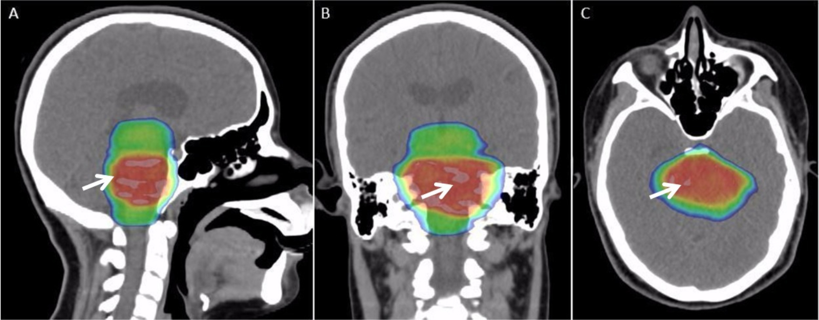
Image-guided radiation therapy (IGRT) is the process of frequent imaging, during a course of radiation treatment, used to direct the treatment, position the patient, and compare to the pre-therapy imaging from the treatment plan. Immediately prior to, or during, a treatment fraction, the patient is localized in the treatment room in the same position as planned from the reference imaging dataset. An example of IGRT would include comparison of a cone beam computed tomography (CBCT) dataset, acquired on the treatment machine, with the computed tomography (CT) dataset from planning. IGRT would also include matching planar kilovoltage (kV) radiographs or megavoltage (MV) images with digital reconstructed radiographs (DRRs) from the planning CT. This process is distinct from the use of imaging to delineate targets and organs in the planning process of radiation therapy. However, there is a connection between the imaging processes as IGRT relies directly on the imaging modalities from pla ... [...More Info...] [...Related Items...] OR: [Wikipedia] [Google] [Baidu] |
Cone Beam Computed Tomography
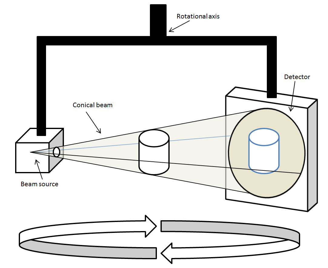
Cone beam computed tomography (or CBCT, also referred to as C-arm CT, cone beam volume CT, flat panel CT or Digital Volume Tomography (DVT)) is a medical imaging technique consisting of X-ray computed tomography where the X-rays are divergent, forming a cone. CBCT has become increasingly important in treatment planning and diagnosis in implant dentistry, ENT, orthopedics, and interventional radiology (IR), among other things. Perhaps because of the increased access to such technology, CBCT scanners are now finding many uses in dentistry, such as in the fields of oral surgery, endodontics and orthodontics. Integrated CBCT is also an important tool for patient positioning and verification in image-guided radiation therapy (IGRT). During dental/orthodontic imaging, the CBCT scanner rotates around the patient's head, obtaining up to nearly 600 distinct images. For interventional radiology, the patient is positioned offset to the table so that the region of interest is centered ... [...More Info...] [...Related Items...] OR: [Wikipedia] [Google] [Baidu] |
David Jaffray
David Anthony Jaffray (born 1966) is a Canadian medical physicist. He is the inaugural chief technology and digital officer at the University of Texas MD Anderson Cancer Center. Jaffray is the inventor, together with John Wong and Jeffrey Siewerdsen, of on-line volumetric kv- imaging guidance system for radiation therapy. Early life and education Jaffray was born in 1966 in Alberta, Canada. He was one of 10 children and grew up on a farm. After completing his Bachelor of Science degree in physics at the University of Alberta, Jaffray was recruited by Jerry Battista to apply to the Schulich School of Medicine and Dentistry The Schulich School of Medicine and Dentistry is the combined medical school and dental school of the University of Western Ontario, a public university in London, Ontario, Canada. The medical education section is one of six in Ontario and one .... References {{DEFAULTSORT:Jaffray, David Canadian physicists Living people 1966 births Scientists from ... [...More Info...] [...Related Items...] OR: [Wikipedia] [Google] [Baidu] |
Detective Quantum Efficiency
The detective quantum efficiency (often abbreviated as DQE) is a measure of the combined effects of the signal (related to image contrast) and noise performance of an imaging system, generally expressed as a function of spatial frequency. This value is used primarily to describe imaging detectors in optical imaging and medical radiography. In medical radiography, the DQE describes how effectively an x-ray imaging system can produce an image with a high signal-to-noise ratio ( SNR) relative to an ideal detector. It is sometimes viewed to be a surrogate measure of the radiation ''dose efficiency'' of a detector since the required radiation exposure to a patient (and therefore the biological risk from that radiation exposure) decreases as the DQE is increased for the same image SNR and exposure conditions. The DQE is also an important consideration for CCDs, especially those used for low-level imaging in light and electron microscopy, because it affects the SNR of the image ... [...More Info...] [...Related Items...] OR: [Wikipedia] [Google] [Baidu] |
Modulation Transfer Function
The optical transfer function (OTF) of an optical system such as a camera, microscope, human eye, or image projector, projector is a scale-dependent description of their imaging contrast. Its magnitude is the image contrast of the Sine and cosine, harmonic intensity pattern, 1 + \cos(2\pi \nu \cdot x), as a function of the spatial frequency, \nu, while its Argument (complex analysis), complex argument indicates a phase shift in the periodic pattern. The optical transfer function is used by optical engineers to describe how the optics project light from the object or scene onto a photographic film, Image sensor, detector array, retina, screen, or simply the next item in the optical transmission chain. Formally, the optical transfer function is defined as the Fourier transform of the point spread function (PSF, that is, the impulse response of the optics, the image of a point source). As a Fourier transform, the OTF is generally complex-valued; however, it is real-valued in the comm ... [...More Info...] [...Related Items...] OR: [Wikipedia] [Google] [Baidu] |
Radiation Therapy
Radiation therapy or radiotherapy (RT, RTx, or XRT) is a therapy, treatment using ionizing radiation, generally provided as part of treatment of cancer, cancer therapy to either kill or control the growth of malignancy, malignant cell (biology), cells. It is normally delivered by a linear particle accelerator. Radiation therapy may be cure, curative in a number of types of cancer if they are localized to one area of the body, and have not metastasis, spread to other parts. It may also be used as part of adjuvant therapy, to prevent tumor recurrence after surgery to remove a primary malignant tumor (for example, early stages of breast cancer). Radiation therapy is synergistic with chemotherapy, and has been used before, during, and after chemotherapy in susceptible cancers. The subspecialty of oncology concerned with radiotherapy is called radiation oncology. A physician who practices in this subspecialty is a radiation oncologist. Radiation therapy is commonly applied to the canc ... [...More Info...] [...Related Items...] OR: [Wikipedia] [Google] [Baidu] |
Mammography
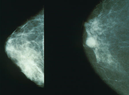
Mammography (also called mastography; DICOM modality: MG) is the process of using low-energy X-rays (usually around 30 kVp) to examine the human breast for diagnosis and screening. The goal of mammography is the early detection of breast cancer, typically through detection of characteristic masses, microcalcifications, asymmetries, and distortions. As with all X-rays, mammograms use doses of ionizing radiation to create images. These images are then analyzed for abnormal findings. It is usual to employ lower-energy X-rays, typically Mo (K-shell X-ray energies of 17.5 and 19.6 keV) and Rh (20.2 and 22.7 keV) than those used for radiography of bones. Mammography may be 2D or 3D ( tomosynthesis), depending on the available equipment or purpose of the examination. Ultrasound, ductography, positron emission mammography (PEM), and magnetic resonance imaging (MRI) are adjuncts to mammography. Ultrasound is typically used for further evaluation of masses found on mammography or palp ... [...More Info...] [...Related Items...] OR: [Wikipedia] [Google] [Baidu] |
Fluoroscopy
Fluoroscopy (), informally referred to as "fluoro", is an imaging technique that uses X-rays to obtain real-time moving images of the interior of an object. In its primary application of medical imaging, a fluoroscope () allows a surgeon to see the internal anatomy, structure and physiology, function of a patient, so that the pumping action of the heart or the motion of swallowing, for example, can be watched. This is useful for both medical diagnosis, diagnosis and therapy and occurs in general radiology, interventional radiology, and image-guided surgery. In its simplest form, a fluoroscope consists of an X-ray generator, X-ray source and a fluorescence, fluorescent screen, between which a patient is placed. However, since the 1950s most fluoroscopes have included X-ray image intensifiers and cameras as well, to improve the image's visibility and make it available on a remote display screen. For many decades, fluoroscopy tended to produce live pictures that were not recorded, bu ... [...More Info...] [...Related Items...] OR: [Wikipedia] [Google] [Baidu] |


