|
Iris Sphincter Muscle
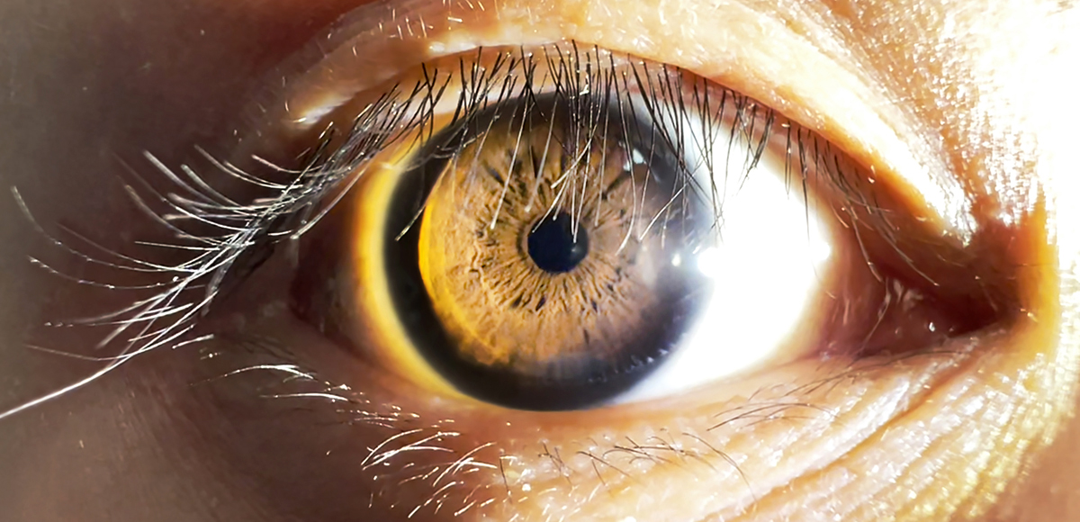
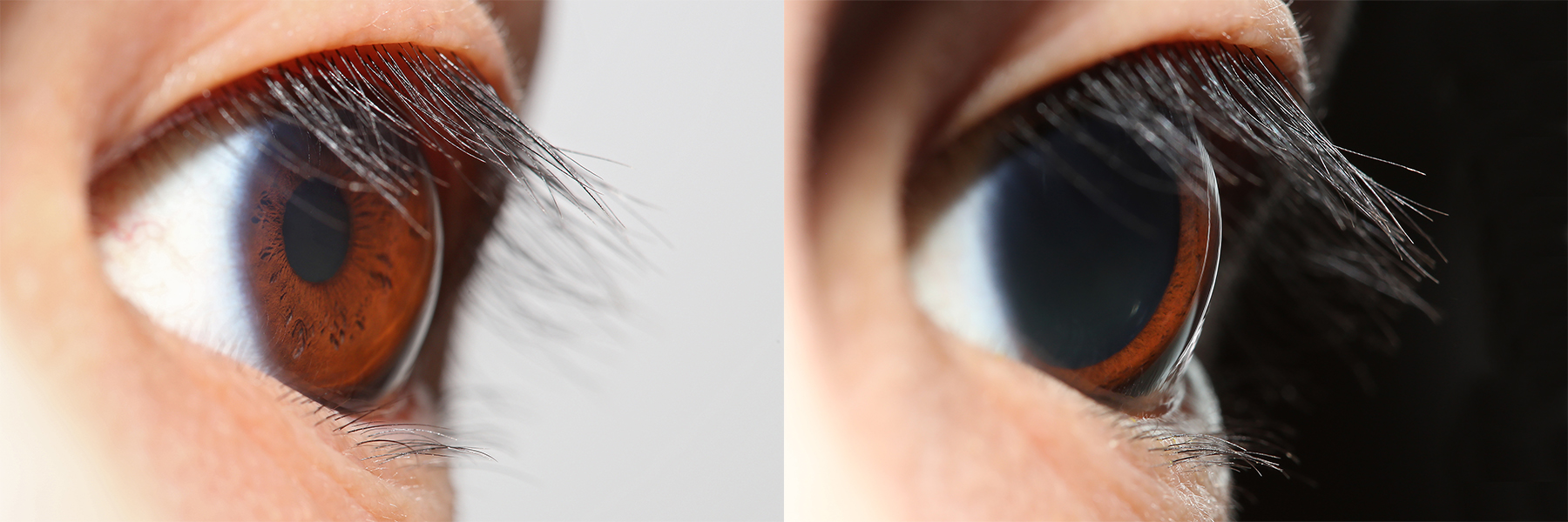
The iris sphincter muscle (pupillary sphincter, pupillary constrictor, circular muscle of iris, circular fibers) is a muscle in the part of the eye called the iris. It encircles the pupil of the iris, appropriate to its function as a constrictor of the pupil. The ciliary muscle, pupillary sphincter muscle and pupillary dilator muscle sometimes are called intrinsic ocular muscles or intraocular muscles. Comparative anatomy This structure is found in vertebrates and in some cephalopods. General structure All the myocytes are of the smooth muscle type. Its dimensions are about 0.75 mm wide by 0.15 mm thick. Mode of action In humans, it functions to constrict the pupil in bright light (pupillary light reflex) or during accommodation. In , the muscle cells themselves are photosensitive causing iris action without brain input. Innervation It is controlled by parasympathetic postganglionic fibers releasing acetylcholine acting primarily on the muscarinic acetylc ... [...More Info...] [...Related Items...] OR: [Wikipedia] [Google] [Baidu] |
Iris (anatomy)
The iris (: irides or irises) is a thin, annular structure in the eye in most mammals and birds that is responsible for controlling the diameter and size of the pupil, and thus the amount of light reaching the retina. In optical terms, the pupil is the eye's aperture, while the iris is the diaphragm (optics), diaphragm. Eye color is defined by the iris. Etymology The word "iris" is derived from the Greek word for "rainbow", also Iris (mythology), its goddess plus messenger of the gods in the ''Iliad'', because of the many eye color, colours of this eye part. Structure The iris consists of two layers: the front pigmented Wikt:fibrovascular, fibrovascular layer known as a stroma of iris, stroma and, behind the stroma, pigmented epithelial cells. The stroma is connected to a sphincter muscle (sphincter pupillae), which contracts the pupil in a circular motion, and a set of dilator muscles (dilator pupillae), which pull the iris radially to enlarge the pupil, pulling it in folds. ... [...More Info...] [...Related Items...] OR: [Wikipedia] [Google] [Baidu] |
Human Eye In Bright Light
Humans (''Homo sapiens'') or modern humans are the most common and widespread species of primate, and the last surviving species of the genus ''Homo''. They are Hominidae, great apes characterized by their Prehistory of nakedness and clothing#Evolution of hairlessness, hairlessness, bipedality, bipedalism, and high Human intelligence, intelligence. Humans have large Human brain, brains, enabling more advanced cognitive skills that facilitate successful adaptation to varied environments, development of sophisticated tools, and formation of complex social structures and civilizations. Humans are Sociality, highly social, with individual humans tending to belong to a Level of analysis, multi-layered network of distinct social groups — from families and peer groups to corporations and State (polity), political states. As such, social interactions between humans have established a wide variety of Value theory, values, norm (sociology), social norms, languages, and traditions (co ... [...More Info...] [...Related Items...] OR: [Wikipedia] [Google] [Baidu] |
Miosis
Miosis, or myosis (), is excessive constriction of the pupil. citing: Mosby's Medical Dictionary, 8th ed. The opposite condition, mydriasis, is the dilation of the pupil. Anisocoria is the condition of one pupil being more dilated than the other. Causes Age * Senile miosis (a reduction in the size of a person's pupil in old age)Diseases *[...More Info...] [...Related Items...] OR: [Wikipedia] [Google] [Baidu] |
Choroid
The choroid, also known as the choroidea or choroid coat, is a part of the uvea, the vascular layer of the eye. It contains connective tissues, and lies between the retina and the sclera. The human choroid is thickest at the far extreme rear of the eye (at 0.2 mm), while in the outlying areas it narrows to 0.1 mm. The choroid provides oxygen and nourishment to the outer layers of the retina. Along with the ciliary body and iris, the choroid forms the uveal tract. The structure of the choroid is generally divided into four layers (classified in order of furthest away from the retina to closest): *Haller's layer – outermost layer of the choroid consisting of larger diameter blood vessels; * Sattler's layer – layer of medium diameter blood vessels; * Choriocapillaris – layer of capillaries; and * Bruch's membrane (synonyms: Lamina basalis, Complexus basalis, Lamina vitra) – innermost layer of the choroid. Blood supply There are two circulations of the eye: ... [...More Info...] [...Related Items...] OR: [Wikipedia] [Google] [Baidu] |
Sclera
The sclera, also known as the white of the eye or, in older literature, as the tunica albuginea oculi, is the opaque, fibrous, protective outer layer of the eye containing mainly collagen and some crucial elastic fiber. In the development of the embryo, the sclera is derived from the neural crest. In children, it is thinner and shows some of the underlying pigment, appearing slightly blue. In the elderly, fatty deposits on the sclera can make it appear slightly yellow. People with dark skin can have naturally darkened sclerae, the result of melanin pigmentation. In humans, and some other vertebrates, the whole sclera is white or pale, contrasting with the coloured iris (anatomy), iris. The cooperative eye hypothesis suggests that the pale sclera evolved as a method of nonverbal communication that makes it easier for one individual to identify where another individual is looking. Other mammals with white or pale sclera include chimpanzees, many orangutans, some gorillas, and bon ... [...More Info...] [...Related Items...] OR: [Wikipedia] [Google] [Baidu] |
Short Ciliary Nerves
The short ciliary nerves are nerves of the orbit around the eye. They are branches of the ciliary ganglion. They supply parasympathetic and sympathetic nerve fibers to the ciliary muscle, iris, and cornea. Damage to the short ciliary nerve may result in loss of the pupillary light reflex, or mydriasis. Structure The short ciliary nerves are branches of the ciliary ganglion. They arise from the forepart of the ganglion in two bundles connected with its superior and inferior angles. The lower bundle is the larger than the upper bundle. These split into between 6 and 10 filaments. They run forward with the ciliary arteries in a wavy course. One bundle is set above the optic nerve, while the other bundle is set below it. They are accompanied by the long ciliary nerves from the nasociliary. They pierce the sclera at the back part of the bulb of the eye, pass forward in delicate grooves on the inner surface of the sclera, and are distributed to the ciliary muscle, iris, and corne ... [...More Info...] [...Related Items...] OR: [Wikipedia] [Google] [Baidu] |
Springer Berlin Heidelberg
Springer Science+Business Media, commonly known as Springer, is a German multinational publishing company of books, e-books and peer-reviewed journals in science, humanities, technical and medical (STM) publishing. Originally founded in 1842 in Berlin, it expanded internationally in the 1960s, and through mergers in the 1990s and a sale to venture capitalists it fused with Wolters Kluwer and eventually became part of Springer Nature in 2015. Springer has major offices in Berlin, Heidelberg, Dordrecht, and New York City. History Julius Springer founded Springer-Verlag in Berlin in 1842 and his son Ferdinand Springer grew it from a small firm of 4 employees into Germany's then second-largest academic publisher with 65 staff in 1872.Chronology ". Springer Science+Business Media. In 1964, Springer expanded its business internationally, ... [...More Info...] [...Related Items...] OR: [Wikipedia] [Google] [Baidu] |
Ciliary Ganglion
The ciliary ganglion is a parasympathetic ganglion located just behind the eye in the posterior orbit. It is 1–2 mm in diameter and in humans contains approximately 2,500 neurons. The ganglion contains postganglionic parasympathetic neurons. These neurons supply the pupillary sphincter muscle, which constricts the pupil, and the ciliary muscle which contracts to make the lens more convex. Both of these muscles are involuntary since they are controlled by the parasympathetic division of the autonomic nervous system. The ciliary ganglion is one of four parasympathetic ganglia of the head. The others are the submandibular ganglion, pterygopalatine ganglion, and otic ganglion. Structure The ciliary ganglion contains postganglionic parasympathetic neurons that supply the ciliary muscle and the pupillary sphincter muscle. Because of the much larger size of the ciliary muscle, 95% of the neurons in the ciliary ganglion innervate it compared to the pupillary sphincter. Roots T ... [...More Info...] [...Related Items...] OR: [Wikipedia] [Google] [Baidu] |
Oculomotor Nerve
The oculomotor nerve, also known as the third cranial nerve, cranial nerve III, or simply CN III, is a cranial nerve that enters the orbit through the superior orbital fissure and innervates extraocular muscles that enable most movements of the eye and that raise the eyelid. The nerve also contains fibers that innervate the intrinsic eye muscles that enable pupillary constriction and accommodation (ability to focus on near objects as in reading). The oculomotor nerve is derived from the basal plate of the embryonic midbrain. Cranial nerves IV and VI also participate in control of eye movement. Structure The oculomotor nerve originates from the third nerve nucleus at the level of the superior colliculus in the midbrain. The third nerve nucleus is located ventral to the cerebral aqueduct, on the pre-aqueductal grey matter. The fibers from the two third nerve nuclei located laterally on either side of the cerebral aqueduct then pass through the red nucleus. From the ... [...More Info...] [...Related Items...] OR: [Wikipedia] [Google] [Baidu] |
Edinger–Westphal Nucleus
The Edinger–Westphal nucleus also called the accessory or visceral oculomotor nerve, is one of the two nuclei of the oculomotor nerve (CN III) located in the midbrain. It receives afferents from both pretectal nuclei (which have in turn received afferents from the optic tract). It contains parasympathetic pre-ganglionic neuron cell bodies that synapse in the ciliary ganglion. It contributes the autonomic, parasympathetic component to the oculomotor nerve (CN III), ultimately providing innervation to the iris sphincter muscle and ciliary muscle to mediate the pupillary light reflex and accommodation, respectively. The Edinger–Westphal nucleus has two parts. The first is of preganglionic fibers (EWpg) that terminate in the ciliary ganglion. The second is of centrally projecting cells (EWcp) that project to a number of brainstem structures. Structure The Edinger–Westphal nucleus refers to the adjacent population of non-preganglionic neurons that do not project to the cilia ... [...More Info...] [...Related Items...] OR: [Wikipedia] [Google] [Baidu] |
Muscarinic Acetylcholine Receptor
Muscarinic acetylcholine receptors (mAChRs) are acetylcholine receptors that form G protein-coupled receptor, G protein-coupled receptor complexes in the cell membranes of certain neurons and other Cell (biology), cells. They play several roles, including acting as the main end-receptor stimulated by acetylcholine released from postganglionic fibers. They are mainly found in the parasympathetic nervous system, but also have a role in the sympathetic nervous system in the control of Sweat gland, sweat glands. Muscarinic receptors are so named because they are more sensitive to muscarine than to nicotine. Their counterparts are nicotinic acetylcholine receptors (nAChRs), receptor ion channels that are also important in the autonomic nervous system. Many drugs and other substances (for example pilocarpine and Hyoscine hydrobromide, scopolamine) manipulate these two distinct receptors by acting as selective agonists or receptor antagonist, antagonists. Function acetylcholine, Ac ... [...More Info...] [...Related Items...] OR: [Wikipedia] [Google] [Baidu] |
Parasympathetic Nervous System
The parasympathetic nervous system (PSNS) is one of the three divisions of the autonomic nervous system, the others being the sympathetic nervous system and the enteric nervous system. The autonomic nervous system is responsible for regulating the body's unconscious actions. The parasympathetic system is responsible for stimulation of "rest-and-digest" or "feed-and-breed" activities that occur when the body is at rest, especially after eating, including sexual arousal, salivation, lacrimation (tears), urination, digestion, and defecation. Its action is described as being complementary to that of the sympathetic nervous system, which is responsible for stimulating activities associated with the fight-or-flight response. Nerve fibres of the parasympathetic nervous system arise from the central nervous system. Specific nerves include several cranial nerves, specifically the oculomotor nerve, facial nerve, glossopharyngeal nerve, and vagus nerve. Three spinal nerves ... [...More Info...] [...Related Items...] OR: [Wikipedia] [Google] [Baidu] |


