|
Intercellular Cleft
An intercellular cleft is a channel between two cells through which molecules may travel and gap junctions and tight junctions may be present. Most notably, intercellular clefts are often found between epithelial cells and the endothelium of blood vessels and lymphatic vessels, also helping to form the blood-nerve barrier surrounding nerves. Intercellular clefts are important for allowing the transportation of fluids and small solute matter through the endothelium. Dimensions of intercellular cleft The dimensions of intercellular clefts vary throughout the body, however cleft lengths have been determined for a series of capillaries. The average cleft length for capillaries is about 20m/cm2. The depths of the intercellular clefts, measured from the luminal to the abluminal openings, vary among different types of capillaries, but the average is about 0.7 μm. The width of the intercellular clefts is about 20 nm outside the junctional region (i.e. in the larger part of the cl ... [...More Info...] [...Related Items...] OR: [Wikipedia] [Google] [Baidu] |
Gap Junction
Gap junctions are membrane channels between adjacent cells that allow the direct exchange of cytoplasmic substances, such small molecules, substrates, and metabolites. Gap junctions were first described as ''close appositions'' alongside tight junctions, however, electron microscopy studies in 1967 led to gap junctions being named as such to be distinguished from tight junctions. They bridge a 2-4 nm gap between cell membranes. Gap junctions use protein complexes known as connexons, composed of connexin proteins to connect one cell to another. Gap junction proteins include the more than 26 types of connexin, as well as at least 12 non-connexin components that make up the gap junction complex or ''nexus,'' including the tight junction protein ZO-1—a protein that holds membrane content together and adds structural clarity to a cell, sodium channels, and aquaporin. More gap junction proteins have become known due to the development of next-generation sequencing. Connexins ... [...More Info...] [...Related Items...] OR: [Wikipedia] [Google] [Baidu] |
Neutrophil Granulocyte
Neutrophils are a type of phagocytic white blood cell and part of innate immunity. More specifically, they form the most abundant type of granulocytes and make up 40% to 70% of all white blood cells in humans. Their functions vary in different animals. They are also known as neutrocytes, heterophils or polymorphonuclear leukocytes. They are formed from stem cells in the bone marrow and differentiated into subpopulations of neutrophil-killers and neutrophil-cagers. They are short-lived (between 5 and 135 hours, see ) and highly mobile, as they can enter parts of tissue where other cells/molecules cannot. Neutrophils may be subdivided into segmented neutrophils and banded neutrophils (or bands). They form part of the polymorphonuclear cells family (PMNs) together with basophils and eosinophils. The name ''neutrophil'' derives from staining characteristics on hematoxylin and eosin ( H&E) histological or cytological preparations. Whereas basophilic white blood cells st ... [...More Info...] [...Related Items...] OR: [Wikipedia] [Google] [Baidu] |
Cardiac Muscle
Cardiac muscle (also called heart muscle or myocardium) is one of three types of vertebrate muscle tissues, the others being skeletal muscle and smooth muscle. It is an involuntary, striated muscle that constitutes the main tissue of the wall of the heart. The cardiac muscle (myocardium) forms a thick middle layer between the outer layer of the heart wall (the pericardium) and the inner layer (the endocardium), with blood supplied via the coronary circulation. It is composed of individual cardiac muscle cells joined by intercalated discs, and encased by collagen fibers and other substances that form the extracellular matrix. Cardiac muscle contracts in a similar manner to skeletal muscle, although with some important differences. Electrical stimulation in the form of a cardiac action potential triggers the release of calcium from the cell's internal calcium store, the sarcoplasmic reticulum. The rise in calcium causes the cell's myofilaments to slide past each other i ... [...More Info...] [...Related Items...] OR: [Wikipedia] [Google] [Baidu] |
Endocardial
The endocardium (: endocardia) is the innermost layer of tissue that lines the chambers of the heart. Its cells are embryologically and biologically similar to the endothelial cells that line blood vessels. The endocardium also provides protection to the valves and heart chambers. The endocardium underlies the much more voluminous myocardium, the muscular tissue responsible for the contraction of the heart. The outer layer of the heart is termed epicardium and the heart is surrounded by a small amount of fluid enclosed by a fibrous sac called the pericardium. Function The endocardium, which is primarily made up of endothelial cells, controls myocardial function. This modulating role is separate from the homeometric and heterometric regulatory mechanisms that control myocardial contractility. Moreover, the endothelium of the myocardial (heart muscle) capillaries, which is also closely appositioned to the cardiomyocytes (heart muscle cells), is involved in this modulatory ro ... [...More Info...] [...Related Items...] OR: [Wikipedia] [Google] [Baidu] |
Glomerulus (kidney)
The glomerulus (: glomeruli) is a network of small blood vessels (capillaries) known as a ''tuft'', located at the beginning of a nephron in the kidney. Each of the two kidneys contains about one million nephrons. The tuft is structurally supported by the mesangium (the space between the blood vessels), composed of Intraglomerular mesangial cell, intraglomerular mesangial cells. The blood is filtered across the capillary walls of this tuft through the glomerular filtration barrier, which yields its Ultrafiltration (renal), filtrate of water and soluble substances to a cup-like sac known as Bowman's capsule. The filtrate then enters the Nephron#Renal tubule, renal tubule of the nephron. The glomerulus receives its blood supply from an afferent arteriole of the renal arterial circulation. Unlike most capillary beds, the glomerular capillaries exit into efferent arterioles rather than venules. The resistance of the efferent arterioles causes sufficient hydrostatic pressure within th ... [...More Info...] [...Related Items...] OR: [Wikipedia] [Google] [Baidu] |
Bradykinin
Bradykinin (BK) (from Greek ''brady-'' 'slow' + ''-kinin'', ''kīn(eîn)'' 'to move') is a peptide that promotes inflammation. It causes arterioles to dilate (enlarge) via the release of prostacyclin, nitric oxide, and endothelium-derived hyperpolarizing factor and makes veins constrict, via prostaglandin F2, thereby leading to leakage into capillary beds, due to the increased pressure in the capillaries. Bradykinin consists of nine amino acids, and is a physiologically and pharmacologically active peptide of the kinin group of proteins. A class of drugs called angiotensin-converting-enzyme inhibitors (ACE inhibitors) increase bradykinin levels by inhibiting its degradation, thereby increasing its blood pressure lowering effect. ACE inhibitors are FDA approved for the treatment of hypertension and heart failure. Structure Bradykinin, sometimes referred to as BK, is a 9–amino acid Translation (genetics), peptide chain. The amino acid sequence of bradykinin is: arginine, Arg-pr ... [...More Info...] [...Related Items...] OR: [Wikipedia] [Google] [Baidu] |
Histamine
Histamine is an organic nitrogenous compound involved in local immune responses communication, as well as regulating physiological functions in the gut and acting as a neurotransmitter for the brain, spinal cord, and uterus. Discovered in 1910, histamine has been considered a local hormone ( autocoid) because it is produced without involvement of the classic endocrine glands; however, in recent years, histamine has been recognized as a central neurotransmitter. Histamine is involved in the inflammatory response and has a central role as a mediator of itching. As part of an immune response to foreign pathogens, histamine is produced by basophils and by mast cells found in nearby connective tissues. Histamine increases the permeability of the capillaries to white blood cells and some proteins, to allow them to engage pathogens in the infected tissues. It consists of an imidazole ring attached to an ethylamine chain; under physiological conditions, the amino grou ... [...More Info...] [...Related Items...] OR: [Wikipedia] [Google] [Baidu] |
Colloid Osmotic Pressure
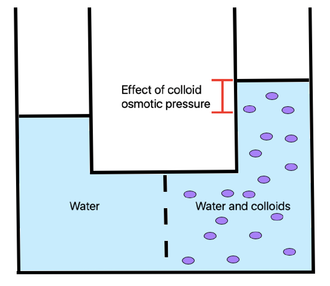
Oncotic pressure, or colloid osmotic-pressure, is a type of osmotic pressure induced by the plasma proteins, notably albumin, in a blood vessel's plasma (or any other body fluid such as blood and lymph) that causes a pull on fluid back into the capillary. It has an effect opposing both the hydrostatic blood pressure, which pushes water and small molecules out of the blood into the interstitial spaces at the arterial end of capillaries, and the interstitial colloidal osmotic pressure. These interacting factors determine the partitioning of extracellular water between the blood plasma and the extravascular space. Oncotic pressure strongly affects the physiological function of the circulatory system. It is suspected to have a major effect on the pressure across the glomerular filter. However, this concept has been strongly criticised and attention has shifted to the impact of the intravascular glycocalyx layer as the major player. Etymology The word 'oncotic' by definiti ... [...More Info...] [...Related Items...] OR: [Wikipedia] [Google] [Baidu] |
Hydrostatic Pressure
Hydrostatics is the branch of fluid mechanics that studies fluids at hydrostatic equilibrium and "the pressure in a fluid or exerted by a fluid on an immersed body". The word "hydrostatics" is sometimes used to refer specifically to water and other liquids, but more often it includes both gases and liquids, whether Compressible flow, compressible or Incompressible flow, incompressible. It encompasses the study of the conditions under which fluids are at rest in Mechanical equilibrium, stable equilibrium. It is opposed to ''fluid dynamics'', the study of fluids in motion. Hydrostatics is fundamental to ''hydraulics'', the engineering of equipment for storing, transporting and using fluids. It is also relevant to geophysics and astrophysics (for example, in understanding plate tectonics and the anomalies of the Gravity of Earth, Earth's gravitational field), to meteorology, to medicine (in the context of blood pressure), and many other fields. Hydrostatics offers physical expl ... [...More Info...] [...Related Items...] OR: [Wikipedia] [Google] [Baidu] |
Capillary
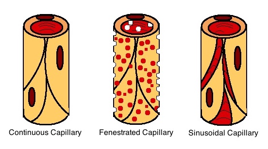
A capillary is a small blood vessel, from 5 to 10 micrometres in diameter, and is part of the microcirculation system. Capillaries are microvessels and the smallest blood vessels in the body. They are composed of only the tunica intima (the innermost layer of an artery or vein), consisting of a thin wall of simple squamous endothelial cells. They are the site of the exchange of many substances from the surrounding interstitial fluid, and they convey blood from the smallest branches of the arteries (arterioles) to those of the veins (venules). Other substances which cross capillaries include water, oxygen, carbon dioxide, urea, glucose, uric acid, lactic acid and creatinine. Lymph capillaries connect with larger lymph vessels to drain lymphatic fluid collected in microcirculation. Etymology ''Capillary'' comes from the Latin word , meaning "of or resembling hair", with use in English beginning in the mid-17th century. The meaning stems from the tiny, hairlike diameter of a capi ... [...More Info...] [...Related Items...] OR: [Wikipedia] [Google] [Baidu] |
Platelet
Platelets or thrombocytes () are a part of blood whose function (along with the coagulation#Coagulation factors, coagulation factors) is to react to bleeding from blood vessel injury by clumping to form a thrombus, blood clot. Platelets have no cell nucleus; they are fragments of cytoplasm from megakaryocytes which reside in bone marrow or Lung, lung tissue, and then enter the circulation. Platelets are found only in mammals, whereas in other vertebrates (e.g. birds, amphibians), thrombocytes circulate as intact agranulocyte, mononuclear cells. One major function of platelets is to contribute to hemostasis: the process of stopping bleeding at the site where the lining of vessels (endothelium) has been interrupted. Platelets gather at the site and, unless the interruption is physically too large, they plug the hole. First, platelets attach to substances outside the interrupted endothelium: ''adhesion (medicine), adhesion''. Second, they change shape, turn on receptors and secret ... [...More Info...] [...Related Items...] OR: [Wikipedia] [Google] [Baidu] |
Red Blood Cell
Red blood cells (RBCs), referred to as erythrocytes (, with -''cyte'' translated as 'cell' in modern usage) in academia and medical publishing, also known as red cells, erythroid cells, and rarely haematids, are the most common type of blood cell and the vertebrate's principal means of delivering oxygen () to the body tissue (biology), tissues—via blood flow through the circulatory system. Erythrocytes take up oxygen in the lungs, or in fish the gills, and release it into tissues while squeezing through the body's capillary, capillaries. The cytoplasm of a red blood cell is rich in hemoglobin (Hb), an iron-containing biomolecule that can bind oxygen and is responsible for the red color of the cells and the blood. Each human red blood cell contains approximately 270 million hemoglobin molecules. The cell membrane is composed of proteins and lipids, and this structure provides properties essential for physiological Cell (biology), cell function such as erythrocyte deformabil ... [...More Info...] [...Related Items...] OR: [Wikipedia] [Google] [Baidu] |


