|
Heinrich Müller (physiologist)
Heinrich Müller (17 December 1820 – 10 May 1864) was a German anatomist and professor at the University of Würzburg. He is best known for his work in comparative anatomy and his studies involving the eye. He was a native of Castell, Lower Franconia. He was a student at several universities, being influenced by Ignaz Dollinger (1770–1841) in Munich, Friedrich Arnold (1803–1890) in Freiburg, Jakob Henle (1809–1895) in Heidelberg and Carl von Rokitansky (1804–1878) in Vienna. In 1847 he received his habilitation at Würzburg, where from 1858 he served as a full professor of topographical and comparative anatomy. As an instructor, he also taught classes in systematic anatomy, histology and microscopy. In 1851 Müller noticed the red color in rod cells now known as rhodopsin or visual purple, which is a pigment that is present in the rods of the retina. However, Franz Christian Boll (1849–1879) is credited as the discoverer of rhodopsin because he was able to desc ... [...More Info...] [...Related Items...] OR: [Wikipedia] [Google] [Baidu] |
Anatomist
Anatomy () is the branch of morphology concerned with the study of the internal structure of organisms and their parts. Anatomy is a branch of natural science that deals with the structural organization of living things. It is an old science, having its beginnings in prehistoric times. Anatomy is inherently tied to developmental biology, embryology, comparative anatomy, evolutionary biology, and phylogeny, as these are the processes by which anatomy is generated, both over immediate and long-term timescales. Anatomy and physiology, which study the structure and function of organisms and their parts respectively, make a natural pair of related disciplines, and are often studied together. Human anatomy is one of the essential basic sciences that are applied in medicine, and is often studied alongside physiology. Anatomy is a complex and dynamic field that is constantly evolving as discoveries are made. In recent years, there has been a significant increase in the use of ... [...More Info...] [...Related Items...] OR: [Wikipedia] [Google] [Baidu] |
Visual Purple
Rhodopsin, also known as visual purple, is a protein encoded by the ''RHO'' gene and a G-protein-coupled receptor (GPCR). It is a light-sensitive receptor protein that triggers visual phototransduction in rod cells. Rhodopsin mediates dim light vision and thus is extremely sensitive to light. When rhodopsin is exposed to light, it immediately photobleaches. In humans, it is fully regenerated in about 30 minutes, after which the rods are more sensitive. Defects in the rhodopsin gene cause eye diseases such as retinitis pigmentosa and congenital stationary night blindness. History Rhodopsin was discovered by Franz Christian Boll in 1876. The name rhodopsin derives from Ancient Greek () for "rose", due to its pinkish color, and () for "sight". It was coined in 1878 by the German physiologist Wilhelm Friedrich Kühne (1837–1900). When George Wald discovered that rhodopsin is a holoprotein, consisting of retinal and an apoprotein, he called it opsin, which today would ... [...More Info...] [...Related Items...] OR: [Wikipedia] [Google] [Baidu] |
Smooth Muscle
Smooth muscle is one of the three major types of vertebrate muscle tissue, the others being skeletal and cardiac muscle. It can also be found in invertebrates and is controlled by the autonomic nervous system. It is non- striated, so-called because it has no sarcomeres and therefore no striations (''bands'' or ''stripes''). It can be divided into two subgroups, ''single-unit'' and ''multi-unit'' smooth muscle. Within single-unit muscle, the whole bundle or sheet of smooth muscle cells contracts as a syncytium. Smooth muscle is found in the walls of hollow organs, including the stomach, intestines, bladder and uterus. In the walls of blood vessels, and lymph vessels, (excluding blood and lymph capillaries) it is known as vascular smooth muscle. There is smooth muscle in the tracts of the respiratory, urinary, and reproductive systems. In the eyes, the ciliary muscles, iris dilator muscle, and iris sphincter muscle are types of smooth muscles. The iris dilator and s ... [...More Info...] [...Related Items...] OR: [Wikipedia] [Google] [Baidu] |
Superior Tarsal Muscle
The superior tarsal muscle is a smooth muscle adjoining the levator palpebrae superioris muscle muscle that helps to raise the upper eyelid. Structure The superior tarsal muscle originates on the underside of levator palpebrae superioris muscle and inserts on the superior tarsal plate of the eyelid. Nerve supply The superior tarsal muscle receives its innervation from the sympathetic nervous system. Postganglionic sympathetic fibers originate in the superior cervical ganglion, and travel via the internal carotid plexus, where small branches communicate with the oculomotor nerve as it passes through the cavernous sinus. The sympathetic fibres continue to the superior division of the oculomotor nerve The oculomotor nerve, also known as the third cranial nerve, cranial nerve III, or simply CN III, is a cranial nerve that enters the orbit through the superior orbital fissure and innervates extraocular muscles that enable most movements o ..., where they enter th ... [...More Info...] [...Related Items...] OR: [Wikipedia] [Google] [Baidu] |
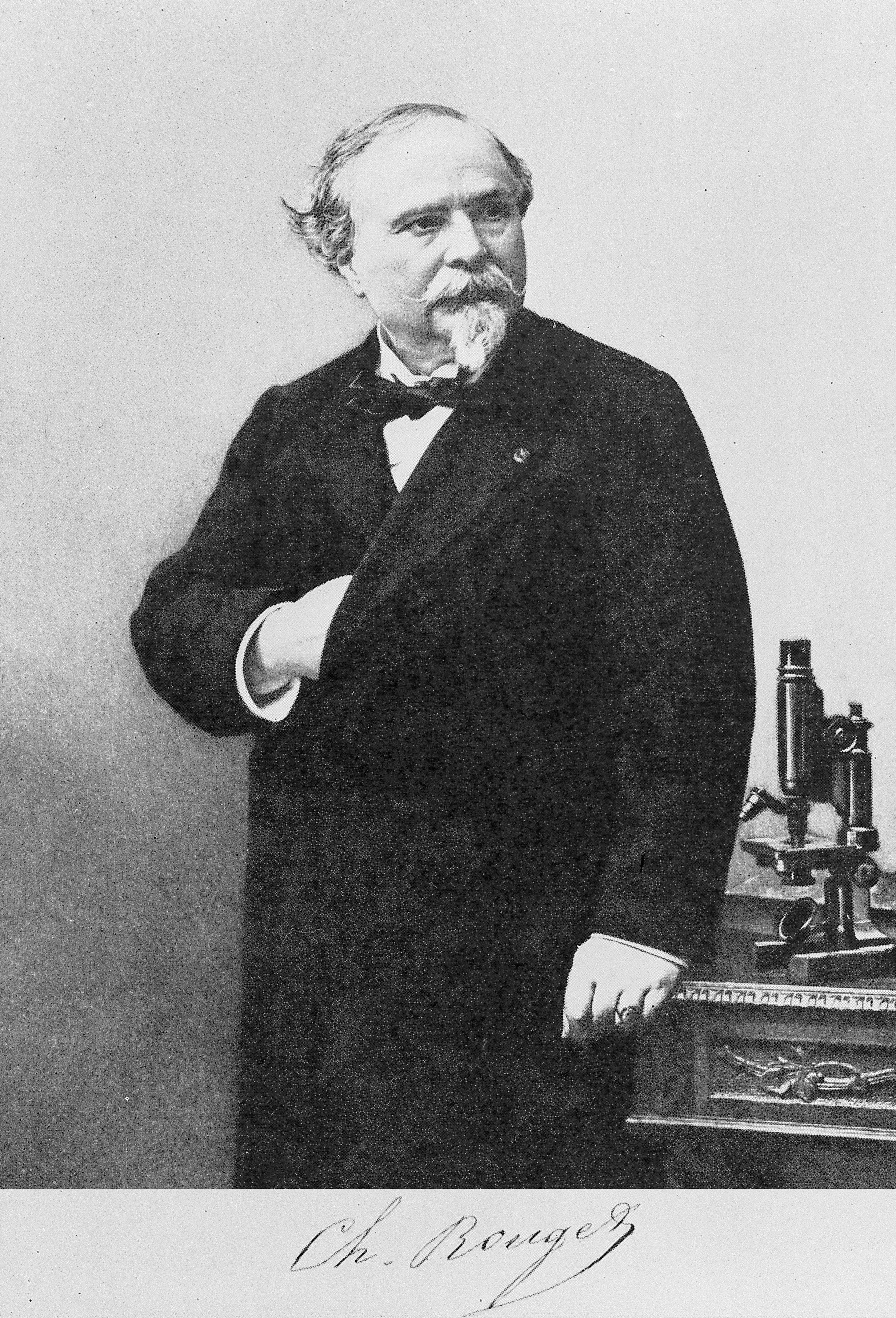
Charles Marie Benjamin Rouget
Charles Marie Benjamin Rouget (19 August 1824 – 1904, Paris) was a French physiologist born in Gisors, Gisors, Eure. He studied at the Collège Sainte-Barbe with medical training at hospitals in Paris. He was later a professor of physiology at the University of Montpellier (1860). From 1879 to 1893, he was a professor of physiology at the National Museum of Natural History (France), Muséum d’Histoire Naturelle in Paris. Rouget is largely remembered for his correlation of physiology to microscopic anatomy, anatomical structure. He was the first to discover the branching contractile cells on the external wall of the capillary, capillaries in amphibians, structures that are now known as "Pericyte, Rouget cells". edited by Benjamin W. Zweifach, Lester Grant, Robert T. McCluskey [...More Info...] [...Related Items...] OR: [Wikipedia] [Google] [Baidu] |
Physiologist
Physiology (; ) is the scientific study of functions and mechanisms in a living system. As a subdiscipline of biology, physiology focuses on how organisms, organ systems, individual organs, cells, and biomolecules carry out chemical and physical functions in a living system. According to the classes of organisms, the field can be divided into medical physiology, animal physiology, plant physiology, cell physiology, and comparative physiology. Central to physiological functioning are biophysical and biochemical processes, homeostatic control mechanisms, and communication between cells. ''Physiological state'' is the condition of normal function. In contrast, '' pathological state'' refers to abnormal conditions, including human diseases. The Nobel Prize in Physiology or Medicine is awarded by the Royal Swedish Academy of Sciences for exceptional scientific achievements in physiology related to the field of medicine. Foundations Because physiology focuses on the ... [...More Info...] [...Related Items...] OR: [Wikipedia] [Google] [Baidu] |
Ciliary Muscle
The ciliary muscle is an intrinsic muscle of the eye formed as a ring of smooth muscleSchachar, Ronald A. (2012). "Anatomy and Physiology." (Chapter 4) . in the eye's middle layer, the uvea ( vascular layer). It controls accommodation for viewing objects at varying distances and regulates the flow of aqueous humor into Schlemm's canal. It also changes the shape of the lens within the eye but not the size of the pupil which is carried out by the sphincter pupillae muscle and dilator pupillae. The ciliary muscle, pupillary sphincter muscle and pupillary dilator muscle sometimes are called intrinsic ocular muscles or intraocular muscles. Structure Development The ciliary muscle develops from mesenchyme within the choroid and is considered a cranial neural crest derivative. Nerve supply The ciliary muscle receives parasympathetic fibers from the short ciliary nerves that arise from the ciliary ganglion. The parasympathetic postganglionic fibers are part of cranial n ... [...More Info...] [...Related Items...] OR: [Wikipedia] [Google] [Baidu] |
Albert Von Kölliker
Albert von Kölliker (born Rudolf Albert Kölliker'';'' 6 July 1817 – 2 November 1905) was a Swiss anatomist, physiologist, and histologist. Biography Albert Kölliker was born in Zürich, Switzerland. His early education was carried on in Zürich, and he entered the university there in 1836. After two years, however, he moved to the University of Bonn, and later to that of Berlin, becoming a pupil of noted physiologists Johannes Peter Müller and of Friedrich Gustav Jakob Henle. He graduated in philosophy at Zürich in 1841, and in medicine at Heidelberg in 1842 The first academic post which he held was that of prosector of anatomy under Henle, but his tenure of this office was briefin 1844 he returned to University of Zurich to occupy a chair as professor extraordinary of physiology and comparative anatomy. His stay here was also brief; in 1847 the University of Würzburg, attracted by his rising fame, offered him the post of professor of physiology and of microscopical an ... [...More Info...] [...Related Items...] OR: [Wikipedia] [Google] [Baidu] |
Motion Parallax
Parallax is a displacement or difference in the apparent position of an object viewed along two different lines of sight and is measured by the angle or half-angle of inclination between those two lines. Due to foreshortening, nearby objects show a larger parallax than farther objects, so parallax can be used to determine distances. To measure large distances, such as the distance of a planet or a star from Earth, astronomers use the principle of parallax. Here, the term ''parallax'' is the semi-angle of inclination between two sight-lines to the star, as observed when Earth is on opposite sides of the Sun in its orbit. These distances form the lowest rung of what is called "the cosmic distance ladder", the first in a succession of methods by which astronomers determine the distances to celestial objects, serving as a basis for other distance measurements in astronomy forming the higher rungs of the ladder. Because parallax is weak if the triangle formed with an object under ... [...More Info...] [...Related Items...] OR: [Wikipedia] [Google] [Baidu] |
Sclera
The sclera, also known as the white of the eye or, in older literature, as the tunica albuginea oculi, is the opaque, fibrous, protective outer layer of the eye containing mainly collagen and some crucial elastic fiber. In the development of the embryo, the sclera is derived from the neural crest. In children, it is thinner and shows some of the underlying pigment, appearing slightly blue. In the elderly, fatty deposits on the sclera can make it appear slightly yellow. People with dark skin can have naturally darkened sclerae, the result of melanin pigmentation. In humans, and some other vertebrates, the whole sclera is white or pale, contrasting with the coloured iris (anatomy), iris. The cooperative eye hypothesis suggests that the pale sclera evolved as a method of nonverbal communication that makes it easier for one individual to identify where another individual is looking. Other mammals with white or pale sclera include chimpanzees, many orangutans, some gorillas, and bon ... [...More Info...] [...Related Items...] OR: [Wikipedia] [Google] [Baidu] |
Carl Bergmann (anatomist)
Carl Georg Lucas Christian Bergmann (18 May 1814 – 30 April 1865), also known as Karl Georg Lucas Christian Bergmann, was a German Anatomy, anatomist, physiologist, and biologist. He developed Bergmann's rule (that populations and species of animals of larger size are found in colder environments). He Histology, microscopically examined the cells of the retina to determine which of them convert light into Nervous system, neural signals that lead ultimately to visual perception: the Cone cell, cones and the Rod cell, rods. Bergmann also coined the terms fovea centralis (for the very center of the retina), homoiothermic (referring to warm-blooded animals), and poikilothermic (referring to non-homoiothermic animals). Biography Bergmann was born in Göttingen, then in the Electorate of Hanover. His father was Friedrich Christian Bergmann (1785–1845), a lawyer and professor. His mother was Henriette Christine, née Mejer. After graduating from high school in Holzminden in 1832, h ... [...More Info...] [...Related Items...] OR: [Wikipedia] [Google] [Baidu] |
Neuroglia
Glia, also called glial cells (gliocytes) or neuroglia, are non- neuronal cells in the central nervous system (the brain and the spinal cord) and in the peripheral nervous system that do not produce electrical impulses. The neuroglia make up more than one half the volume of neural tissue in the human body. They maintain homeostasis, form myelin, and provide support and protection for neurons. In the central nervous system, glial cells include oligodendrocytes (that produce myelin), astrocytes, ependymal cells and microglia, and in the peripheral nervous system they include Schwann cells (that produce myelin), and satellite cells. Function They have four main functions: * to surround neurons and hold them in place * to supply nutrients and oxygen to neurons * to insulate one neuron from another * to destroy pathogens and remove dead neurons. They also play a role in neurotransmission and synaptic connections, and in physiological processes such as breathing. While ... [...More Info...] [...Related Items...] OR: [Wikipedia] [Google] [Baidu] |


