|
G-banding
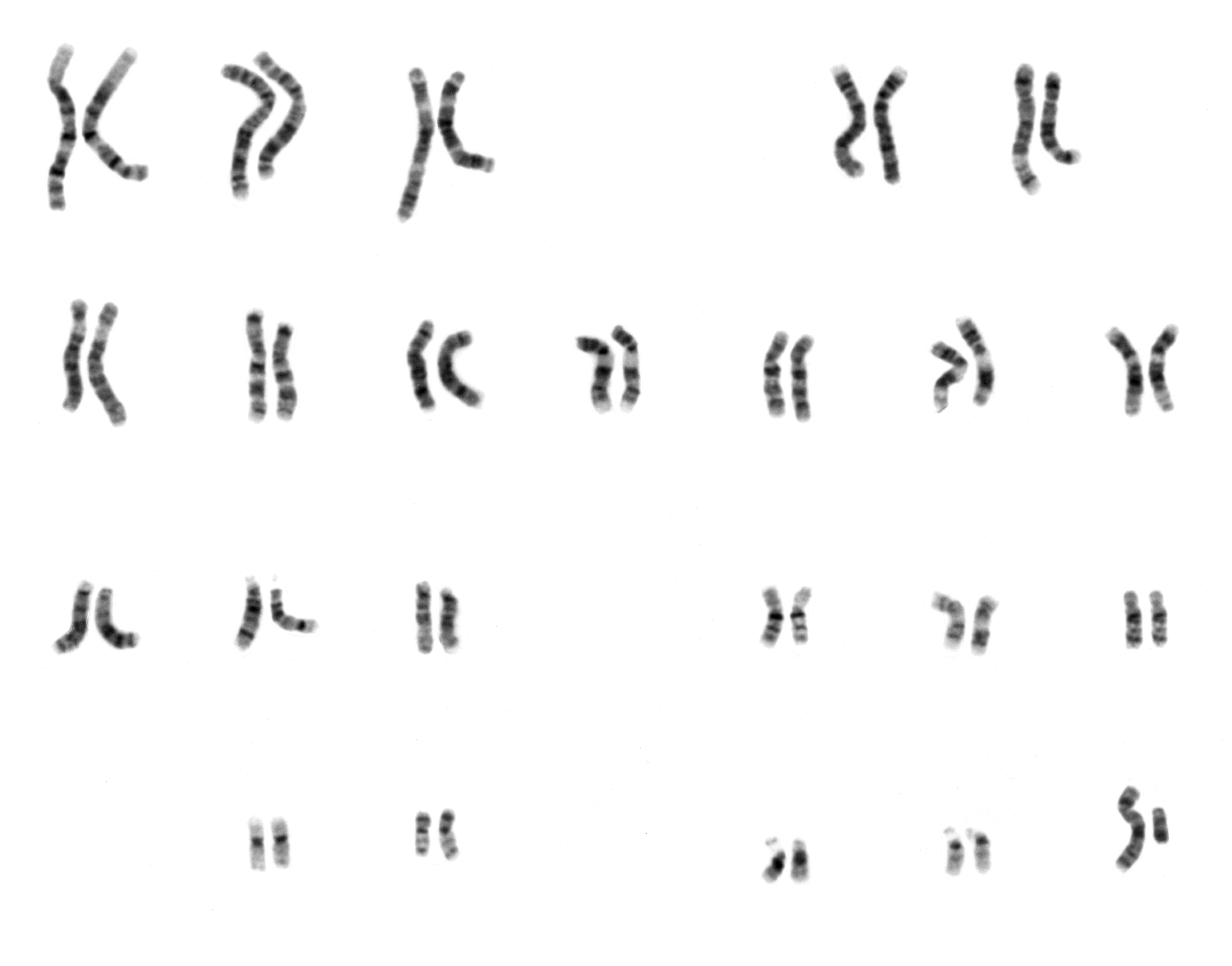
G-banding, G banding or Giemsa banding is a technique used in cytogenetics to produce a visible karyotype by staining condensed chromosomes. It is the most common chromosome banding method. It is useful for identifying genetic diseases (mainly chromosomal abnormalities) through the photographic representation of the entire chromosome complement.Speicher, Michael R. and Nigel P. Carter. "The New Cytogenetics: Blurring the Boundaries with Molecular Biology." ''Nature'' Reviews Genetics, Vol 6. Oct 2005. Method The metaphase chromosomes are treated with trypsin (to partially digest the chromosome) and stained with Giemsa stain. Heterochromatic regions, which tend to be rich with adenine and thymine (AT-rich) DNA and relatively gene-poor, stain more darkly in G-banding. In contrast, less condensed chromatin (Euchromatin)—which tends to be rich with guanine and cytosine ( GC-rich) and more transcriptionally active—incorporates less Giemsa stain, and these regions appear as light ... [...More Info...] [...Related Items...] OR: [Wikipedia] [Google] [Baidu] |
Prophase
Prophase () is the first stage of cell division in both mitosis and meiosis. Beginning after interphase, DNA has already been replicated when the cell enters prophase. The main occurrences in prophase are the condensation of the chromatin reticulum and the disappearance of the nucleolus. Staining and microscopy Microscopy can be used to visualize condensed chromosomes as they move through meiosis and mitosis. Various DNA stains are used to treat cells such that condensing chromosomes can be visualized as the move through prophase. The giemsa G-banding technique is commonly used to identify mammalian chromosomes, but utilizing the technology on plant cells was originally difficult due to the high degree of chromosome compaction in plant cells. G-banding was fully realized for plant chromosomes in 1990. During both meiotic and mitotic prophase, giemsa staining can be applied to cells to elicit G-banding in chromosomes. Silver staining, a more modern technology, in conj ... [...More Info...] [...Related Items...] OR: [Wikipedia] [Google] [Baidu] |
Karyotype
A karyotype is the general appearance of the complete set of chromosomes in the cells of a species or in an individual organism, mainly including their sizes, numbers, and shapes. Karyotyping is the process by which a karyotype is discerned by determining the chromosome complement of an individual, including the number of chromosomes and any abnormalities. A karyogram or idiogram is a graphical depiction of a karyotype, wherein chromosomes are generally organized in pairs, ordered by size and position of centromere for chromosomes of the same size. Karyotyping generally combines light microscopy and photography in the metaphase of the cell cycle, and results in a photomicrographic (or simply micrographic) karyogram. In contrast, a schematic karyogram is a designed graphic representation of a karyotype. In schematic karyograms, just one of the sister chromatids of each chromosome is generally shown for brevity, and in reality they are generally so close together that they look as ... [...More Info...] [...Related Items...] OR: [Wikipedia] [Google] [Baidu] |
Cytogenetics
Cytogenetics is essentially a branch of genetics, but is also a part of cell biology/cytology (a subdivision of human anatomy), that is concerned with how the chromosomes relate to cell behaviour, particularly to their behaviour during mitosis and meiosis. Techniques used include Karyotype, karyotyping, analysis of G banding, G-banded chromosomes, other cytogenetic banding techniques, as well as molecular cytogenetics such as Fluorescence in situ hybridization, fluorescence ''in situ'' hybridization (FISH) and comparative genomic hybridization (CGH). History Beginnings Chromosomes were first observed in plant cells by Carl Nägeli in 1842. Their behavior in animal (salamander) cells was described by Walther Flemming, the discoverer of mitosis, in 1882. The name was coined by another German anatomist, Heinrich Wilhelm Gottfried von Waldeyer-Hartz, von Waldeyer in 1888. The next stage took place after the development of genetics in the early 20th century, when it was appreciated ... [...More Info...] [...Related Items...] OR: [Wikipedia] [Google] [Baidu] |
Giemsa Stain
Giemsa stain (), named after German chemist and bacteriologist Gustav Giemsa, is a nucleic acid stain used in cytogenetics and for the histopathological diagnosis of malaria and other parasites. Uses It is specific for the phosphate groups of DNA and attaches itself to regions of DNA where there are high amounts of adenine-thymine bonding. Giemsa stain is used in Giemsa banding, commonly called G-banding, to stain chromosomes and often used to create a karyogram (chromosome map). It can identify chromosomal aberrations such as translocations and rearrangements. It stains the trophozoite ''Trichomonas vaginalis'', which presents with greenish discharge and motile cells on wet prep. Giemsa stain is also a differential stain, such as when it is combined with Wright stain to form Wright–Giemsa stain. It can be used to study the adherence of pathogenic bacteria to human cells. It differentially stains human and bacterial cells purple and pink respectively. It can be used fo ... [...More Info...] [...Related Items...] OR: [Wikipedia] [Google] [Baidu] |
R‑banding
R-banding is a cytogenetics technique that produces the reverse of the G-band stain on chromosomes. R-banding is obtained by incubating the slides in hot phosphate buffer, then a subsequent treatment of giemsa dye. Resulting chromosome patterns shows darkly stained R bands, the complement to G-bands. Darkly colored R bands are guanine-cytosine rich, and adenine-thymine rich regions are more readily denatured by heat. The technique is useful for analyzing genetic deletions or chromosomal translocations that involve the telomeres of chromosomes A chromosome is a package of DNA containing part or all of the genetic material of an organism. In most chromosomes, the very long thin DNA fibers are coated with nucleosome-forming packaging proteins; in eukaryotic cells, the most importa .... Cytogenetics References {{genetics-stub ... [...More Info...] [...Related Items...] OR: [Wikipedia] [Google] [Baidu] |
Chromosomes
A chromosome is a package of DNA containing part or all of the genetic material of an organism. In most chromosomes, the very long thin DNA fibers are coated with nucleosome-forming packaging proteins; in eukaryotic cells, the most important of these proteins are the histones. Aided by chaperone proteins, the histones bind to and condense the DNA molecule to maintain its integrity. These eukaryotic chromosomes display a complex three-dimensional structure that has a significant role in transcriptional regulation. Normally, chromosomes are visible under a light microscope only during the metaphase of cell division, where all chromosomes are aligned in the center of the cell in their condensed form. Before this stage occurs, each chromosome is duplicated ( S phase), and the two copies are joined by a centromere—resulting in either an X-shaped structure if the centromere is located equatorially, or a two-armed structure if the centromere is located distally; the joined ... [...More Info...] [...Related Items...] OR: [Wikipedia] [Google] [Baidu] |
Chromosome
A chromosome is a package of DNA containing part or all of the genetic material of an organism. In most chromosomes, the very long thin DNA fibers are coated with nucleosome-forming packaging proteins; in eukaryotic cells, the most important of these proteins are the histones. Aided by chaperone proteins, the histones bind to and condense the DNA molecule to maintain its integrity. These eukaryotic chromosomes display a complex three-dimensional structure that has a significant role in transcriptional regulation. Normally, chromosomes are visible under a light microscope only during the metaphase of cell division, where all chromosomes are aligned in the center of the cell in their condensed form. Before this stage occurs, each chromosome is duplicated ( S phase), and the two copies are joined by a centromere—resulting in either an X-shaped structure if the centromere is located equatorially, or a two-armed structure if the centromere is located distally; the jo ... [...More Info...] [...Related Items...] OR: [Wikipedia] [Google] [Baidu] |
Eukaryotic Chromosome Fine Structure
In genetics, eukaryotic chromosome fine structure refers to the structure of sequences for the chromosomes of eukaryotic organisms. Some fine sequences are included in more than one class, so the classification listed is not intended to be completely separate. Chromosomal characteristics Some sequences are required for a properly functioning chromosome: * Centromere: Used during cell division as the attachment point for the spindle fibers. * Telomere: Used to maintain chromosomal integrity by capping off the ends of the linear chromosomes. This region is a microsatellite, but its function is more specific than a simple tandem repeat. Throughout the eukaryotic kingdom, the overall structure of chromosome ends is conserved and is characterized by the telomeric tract - a series of short G-rich repeats. This is succeeded by an extensive subtelomeric region consisting of various types and lengths of repeats - the telomere associated sequences (TAS).Pryde FE, Gorham HC, Louis EJ (199 ... [...More Info...] [...Related Items...] OR: [Wikipedia] [Google] [Baidu] |
Telomeric
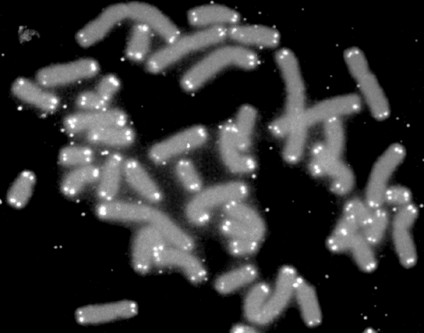
A telomere (; ) is a region of repetitive nucleotide sequences associated with specialized proteins at the ends of linear chromosomes (see Sequences). Telomeres are a widespread genetic feature most commonly found in eukaryotes. In most, if not all species possessing them, they protect the terminal regions of chromosomal DNA from progressive degradation and ensure the integrity of linear chromosomes by preventing DNA repair systems from mistaking the very ends of the DNA strand for a double-strand break. Discovery The existence of a special structure at the ends of chromosomes was independently proposed in 1938 by Hermann Joseph Muller, studying the fruit fly ''Drosophila melanogaster'', and in 1939 by Barbara McClintock, working with maize. Muller observed that the ends of irradiated fruit fly chromosomes did not present alterations such as deletions or inversions. He hypothesized the presence of a protective cap, which he coined "telomeres", from the Greek ''telos'' (end) a ... [...More Info...] [...Related Items...] OR: [Wikipedia] [Google] [Baidu] |
Telomere
A telomere (; ) is a region of repetitive nucleotide sequences associated with specialized proteins at the ends of linear chromosomes (see #Sequences, Sequences). Telomeres are a widespread genetic feature most commonly found in eukaryotes. In most, if not all species possessing them, they protect the terminal regions of DNA, chromosomal DNA from progressive degradation and ensure the integrity of linear chromosomes by preventing DNA repair systems from mistaking the very ends of the DNA strand for a double-strand break. Discovery The existence of a special structure at the ends of chromosomes was independently proposed in 1938 by Hermann Joseph Muller, studying the fruit fly ''Drosophila melanogaster'', and in 1939 by Barbara McClintock, working with maize. Muller observed that the ends of irradiated fruit fly chromosomes did not present alterations such as deletions or inversions. He hypothesized the presence of a protective cap, which he coined "telomeres", from the Greek ' ... [...More Info...] [...Related Items...] OR: [Wikipedia] [Google] [Baidu] |
Gene Mapping
Gene mapping or genome mapping describes the methods used to identify the location of a gene on a chromosome and the distances between genes. Gene mapping can also describe the distances between different sites within a gene. The essence of all genome mapping is to place a collection of molecular markers onto their respective positions on the genome. Molecular markers come in all forms. Genes can be viewed as one special type of genetic markers in the construction of genome maps, and mapped the same way as any other markers. In some areas of study, gene mapping contributes to the creation of new recombinants within an organism. Gene maps help describe the spatial arrangement of genes on a chromosome. Genes are designated to a specific location on a chromosome known as the locus and can be used as molecular markers to find the distance between other genes on a chromosome. Maps provide researchers with the opportunity to predict the inheritance patterns of specific traits, which ... [...More Info...] [...Related Items...] OR: [Wikipedia] [Google] [Baidu] |
Constitutive Heterochromatin
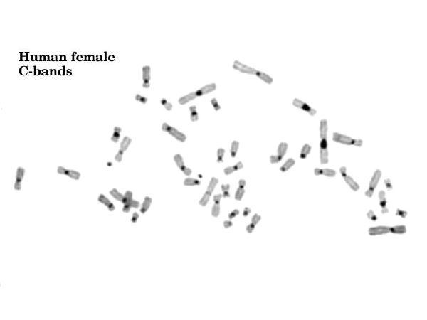
Constitutive heterochromatin domains are regions of DNA found throughout the chromosomes of eukaryotes. The majority of constitutive heterochromatin is found at the pericentromeric regions of chromosomes, but is also found at the telomeres and throughout the chromosomes. In humans there is significantly more constitutive heterochromatin found on chromosomes 1, 9, 16, 19 and Y. Constitutive heterochromatin is composed mainly of high copy number tandem repeats known as satellite repeats, minisatellite and microsatellite repeats, and Transposable element, transposon repeats. In humans these regions account for about 200Mb or 6.5% of the total human genome, but their repeat composition makes them difficult to sequence, so only small regions have been sequenced. Visualization of constitutive heterochromatin is possible by using the Cytogenetics, C-banding technique. The regions that stain darker are regions of constitutive heterochromatin. The constitutive heterochromatin stains darker ... [...More Info...] [...Related Items...] OR: [Wikipedia] [Google] [Baidu] |


