|
Forearm Pronators
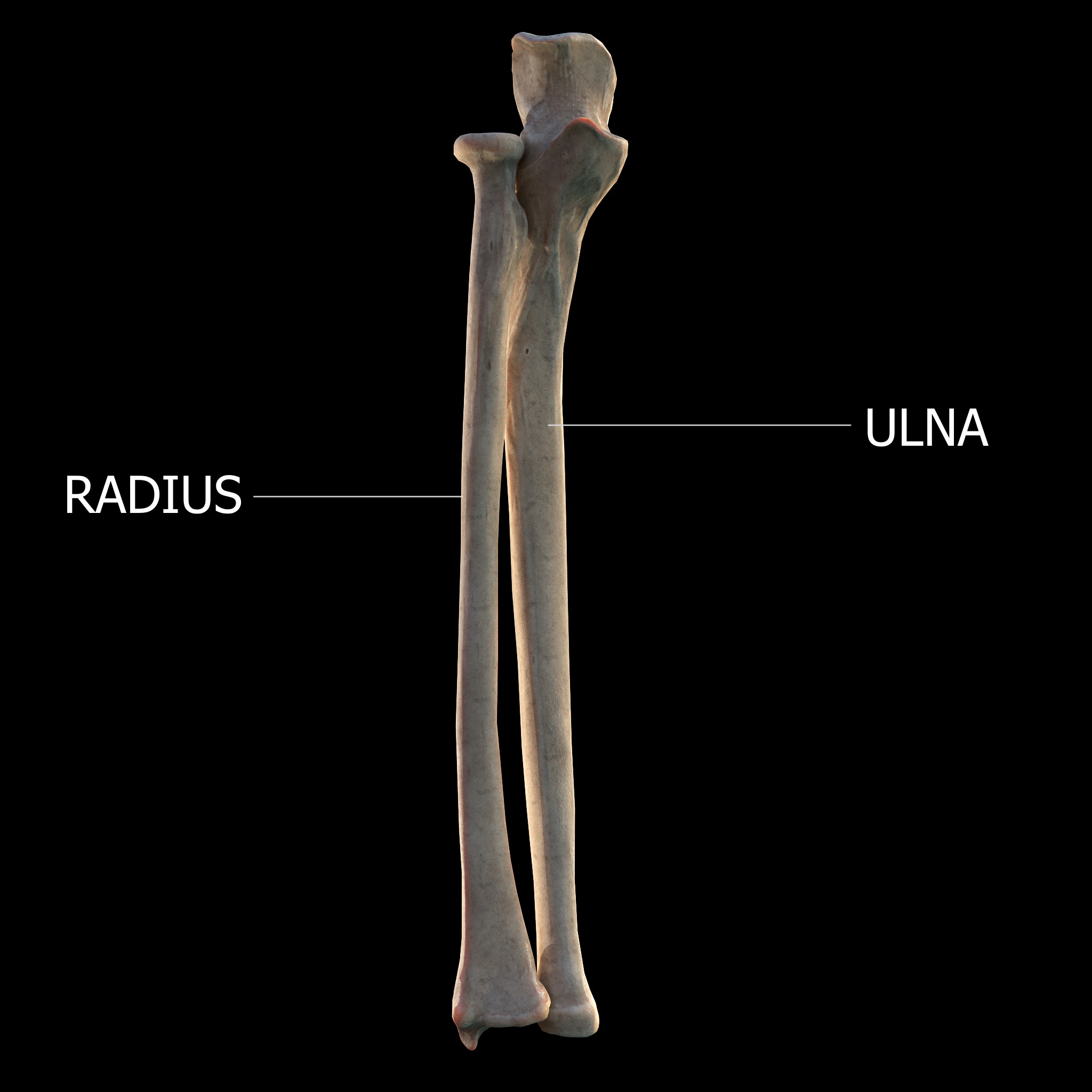
The forearm is the region of the upper limb between the elbow and the wrist. The term forearm is used in anatomy to distinguish it from the arm, a word which is used to describe the entire appendage of the upper limb, but which in anatomy, technically, means only the region of the upper arm, whereas the lower "arm" is called the forearm. It is homologous to the region of the leg that lies between the knee and the ankle joints, the crus. The forearm contains two long bones, the radius and the ulna, forming the two radioulnar joints. The interosseous membrane connects these bones. Ultimately, the forearm is covered by skin, the anterior surface usually being less hairy than the posterior surface. The forearm contains many muscles, including the flexors and extensors of the wrist, flexors and extensors of the digits, a flexor of the elbow (brachioradialis), and pronators and supinators that turn the hand to face down or upwards, respectively. In cross-section, the forearm can be ... [...More Info...] [...Related Items...] OR: [Wikipedia] [Google] [Baidu] |
Upper Limb
The upper Limb (anatomy), limbs or upper extremities are the forelimbs of an upright posture, upright-postured tetrapod vertebrate, extending from the scapulae and clavicles down to and including the digit (anatomy), digits, including all the musculatures and ligaments involved with the shoulder, elbow, wrist and knuckle joints. In humans, each upper limb is divided into the shoulder, arm, elbow, forearm, wrist and hand, and is primarily used for climbing, manual handling of loads, lifting and dexterity, manipulating objects. In anatomy, just as arm refers to the upper arm, leg refers to the lower leg. Definition In formal usage, the term "arm" only refers to the structures from the shoulder to the elbow, explicitly excluding the forearm, and thus "upper limb" and "arm" are not synonymous. However, in casual usage, the terms are often used interchangeably. The term "upper arm" is redundant in anatomy, but in informal usage is used to distinguish between the two terms. Structure I ... [...More Info...] [...Related Items...] OR: [Wikipedia] [Google] [Baidu] |
Median Nerve
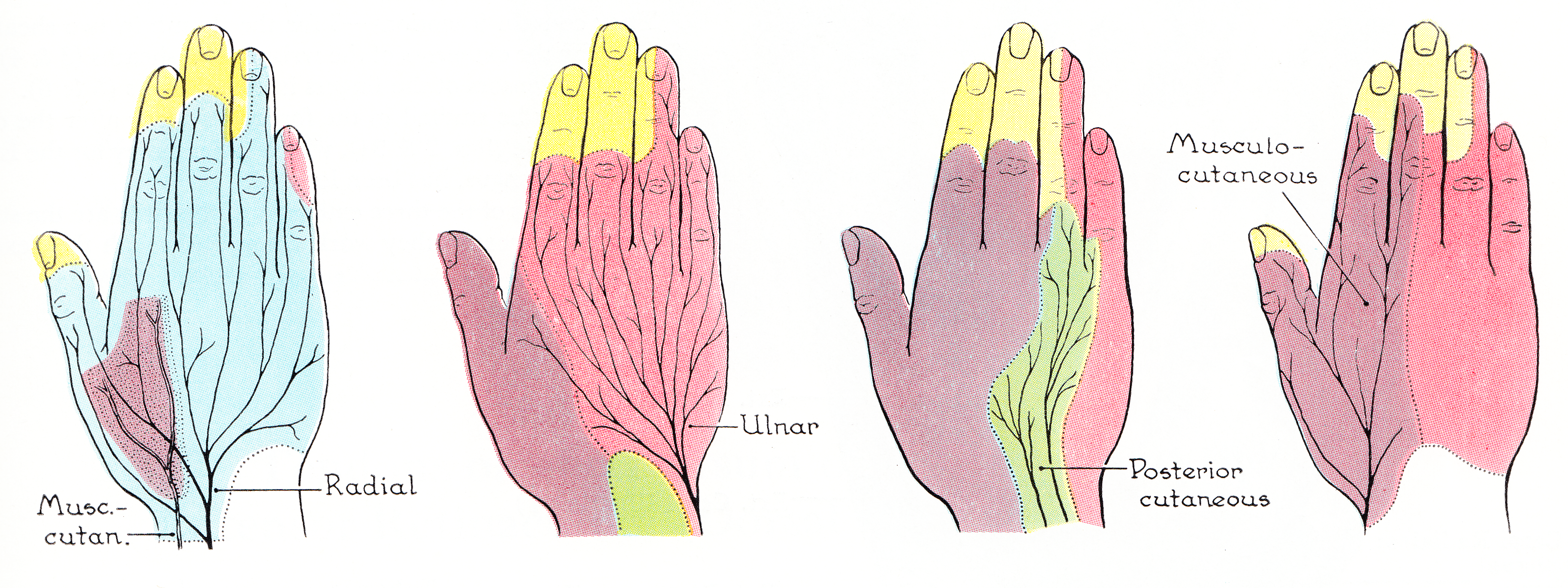
The median nerve is a nerve in humans and other animals in the upper limb. It is one of the five main nerves originating from the brachial plexus. The median nerve originates from the lateral and medial cords of the brachial plexus, and has contributions from ventral roots of C6-C7 (lateral cord) and C8 and T1 (medial cord). The median nerve is the only nerve that passes through the carpal tunnel. Carpal tunnel syndrome is the disability that results from the median nerve being pressed in the carpal tunnel. Structure The median nerve arises from the branches from lateral and medial cords of the brachial plexus, courses through the anterior part of arm, forearm, and hand, and terminates by supplying the muscles of the hand. Arm After receiving inputs from both the lateral and medial cords of the brachial plexus, the median nerve enters the arm from the axilla at the inferior margin of the teres major muscle. It then passes vertically down and courses lateral to the brac ... [...More Info...] [...Related Items...] OR: [Wikipedia] [Google] [Baidu] |
Proximal Radioulnar Articulation
The proximal radioulnar articulation, also known as the proximal radioulnar joint (PRUJ), is a synovial pivot joint between the circumference of the head of the radius and the ring formed by the radial notch of the ulna and the annular ligament. Structure The proximal radioulnar joint is a synovial pivot joint. It occurs between the circumference of the head of the radius and the ring formed by the radial notch of the ulna and the annular ligament. The interosseous membrane of the forearm and the annular ligament stabilise the joint. A number of nerves run close to the proximal radioulnar joint, including: *median nerve The median nerve is a nerve in humans and other animals in the upper limb. It is one of the five main nerves originating from the brachial plexus. The median nerve originates from the lateral and medial cords of the brachial plexus, and has cont ... * musculocutaneous nerve * radial nerve See also * Distal radioulnar articulation * Supination ... [...More Info...] [...Related Items...] OR: [Wikipedia] [Google] [Baidu] |
Articulation (anatomy)
A joint or articulation (or articular surface) is the connection made between bones, ossicles, or other hard structures in the body which link an animal's skeletal system into a functional whole.Saladin, Ken. Anatomy & Physiology. 7th ed. McGraw-Hill Connect. Webp.274/ref> They are constructed to allow for different degrees and types of movement. Some joints, such as the knee, elbow, and shoulder, are self-lubricating, almost frictionless, and are able to withstand compression and maintain heavy loads while still executing smooth and precise movements. Other joints such as sutures between the bones of the skull permit very little movement (only during birth) in order to protect the brain and the sense organs. The connection between a tooth and the jawbone is also called a joint, and is described as a fibrous joint known as a gomphosis. Joints are classified both structurally and functionally. Joints play a vital role in the human body, contributing to movement, stability ... [...More Info...] [...Related Items...] OR: [Wikipedia] [Google] [Baidu] |
Elbow
The elbow is the region between the upper arm and the forearm that surrounds the elbow joint. The elbow includes prominent landmarks such as the olecranon, the cubital fossa (also called the chelidon, or the elbow pit), and the lateral and the medial epicondyles of the humerus. The elbow joint is a hinge joint between the arm and the forearm; more specifically between the humerus in the upper arm and the radius and ulna in the forearm which allows the forearm and hand to be moved towards and away from the body. The term ''elbow'' is specifically used for humans and other primates, and in other vertebrates it is not used. In those cases, forelimb plus joint is used. The name for the elbow in Latin is ''cubitus'', and so the word cubital is used in some elbow-related terms, as in ''cubital nodes'' for example. Structure Joint The elbow joint has three different portions surrounded by a common joint capsule. These are joints between the three bones of the elbow, the ... [...More Info...] [...Related Items...] OR: [Wikipedia] [Google] [Baidu] |
Capitulum Of The Humerus
In human anatomy of the arm, the capitulum of the humerus is a smooth, rounded eminence on the lateral portion of the distal articular surface of the humerus. It articulates with the cup-shaped depression on the head of the radius, and is limited to the front and lower part of the bone. In non-human tetrapods, the name capitellum is generally used, with "capitulum" limited to the anteroventral articular facet of the rib (in archosauromorphs). Lepidosauromorpha Lepidosaurs show a distinct capitellum and trochlea on the centre of the ventral (anterior in upright taxa) surface of the humerus at the distal end. Archosauromorpha In non-avian archosaurs, including crocodiles, the capitellum and the trochlea are no longer bordered by distinct etc.- and entepicondyles respectively, and the distal humerus consists two gently expanded condyles, one lateral and one medial, separated by a shallow groove and a supinator process. Romer (1976) homologizes the capitellum in Archosauromo ... [...More Info...] [...Related Items...] OR: [Wikipedia] [Google] [Baidu] |
Radius And Ulna - Forearm Bones
In classical geometry, a radius (: radii or radiuses) of a circle or sphere is any of the line segments from its center to its perimeter, and in more modern usage, it is also their length. The radius of a regular polygon is the line segment or distance from its center to any of its vertices. The name comes from the Latin ''radius'', meaning ray but also the spoke of a chariot wheel.Definition of Radius at dictionary.reference.com. Accessed on 2009-08-08. The typical abbreviation and mathematical symbol for radius is ''R'' or ''r''. By extension, the diameter ''D'' is defined as twice the radius:Definition of radius at mathwords.com. Accessed on 2009-08-08. : [...More Info...] [...Related Items...] OR: [Wikipedia] [Google] [Baidu] |
Cubital Fossa
The cubital fossa, antecubital fossa, chelidon, inside of elbow, or, humorously, wagina, is the area on the anterior side of the upper part between the arm and forearm of a human or other hominid animals. It lies anteriorly to the elbow (antecubital) (Latin ) when in standard anatomical position. The cubital fossa is a triangular area having three borders. Boundaries * superior (proximal) boundary – an imaginary horizontal line connecting the medial epicondyle of the humerus to the lateral epicondyle of the humerus * medial (ulnar) boundary – lateral border of pronator teres muscle originating from the medial epicondyle of the humerus. * lateral (radial) boundary – medial border of brachioradialis muscle originating from the lateral supraepicondylar ridge of the humerus. * apex – it is directed inferiorly, and is formed by the meeting point of the lateral and medial boundaries * superficial boundary (roof) – skin, superficial fascia containing the median cubital vein, ... [...More Info...] [...Related Items...] OR: [Wikipedia] [Google] [Baidu] |
Venipuncture
In medicine, venipuncture or venepuncture is the process of obtaining intravenous access for the purpose of venous Sampling (medicine)#blood, blood sampling (also called ''phlebotomy'') or intravenous therapy. In healthcare, this procedure is performed by Medical Laboratory Scientist, medical laboratory scientists, physician, medical practitioners, some EMTs, paramedics, phlebotomists, Kidney dialysis, dialysis technicians, and other nursing staff. In veterinary medicine, the procedure is performed by veterinarians and Paraveterinary worker, veterinary technicians. It is essential to follow a standard procedure for the collection of blood specimens to get accurate laboratory results. Any error in collecting the blood or filling the test tubes may lead to erroneous laboratory results. Venipuncture is one of the most routinely performed invasive procedures and is carried out for any of five reasons: # to obtain blood for diagnostic purposes; # to monitor levels of blood component ... [...More Info...] [...Related Items...] OR: [Wikipedia] [Google] [Baidu] |
Basilic Vein
The basilic vein is a large superficial vein of the upper limb that helps drain parts of the hand and forearm. It originates on the medial ( ulnar) side of the dorsal venous network of the hand and travels up the base of the forearm, where its course is generally visible through the skin as it travels in the subcutaneous fat and fascia lying superficial to the muscles. The basilic vein terminates by uniting with the brachial veins to form the axillary vein. Anatomy Course As it ascends the medial side of the biceps in the arm proper (between the elbow and shoulder), the basilic vein normally perforates the brachial fascia ( deep fascia) in the middle of the medial bicipital groove, and run upwards medial to the brachial artery to the lower border of teres major, continuing as the axillary vein. Tributaries and anastomoses Near the region anterior to the cubital fossa (in the bend of the elbow joint), the basilic vein usually communicates with the cephalic vein (th ... [...More Info...] [...Related Items...] OR: [Wikipedia] [Google] [Baidu] |
Median Antebrachial Vein
The median antebrachial vein, also known as median vein of forearm, is a superficial vein of the (anterior) forearm. It arises from - and drains - the superficial palmar venous arch, ascending superficially along the anterior forearm before ending by opening into the median cubital vein near the junction with the basilic vein within the cubital fossa; alternately, it may fork distal to the elbow and proceed to drain into both aforementioned veins. A bifurcation of the median antebrachial vein produces the (medial) intermediate basilic vein and the (lateral) intermediate cephalic vein; the two veins produced by such a split may replace the median cubital vein In human anatomy, the median cubital vein (or median basilic vein) is a superficial vein of the arm on the anterior aspect of the elbow. It classically shunts blood from the cephalic to the basilic vein at the roof of the cubital fossa. It is ty .... References Veins of the upper limb {{circulatory-stub ... [...More Info...] [...Related Items...] OR: [Wikipedia] [Google] [Baidu] |
Cephalic Vein
In human anatomy, the cephalic vein (also called the antecubital vein) is a superficial vein in the arm. It is the longest vein of the upper limb. It starts at the anatomical snuffbox from the radial end of the dorsal venous network of hand, and ascends along the radial (lateral) side of the arm before emptying into the axillary vein. At the elbow, it communicates with the basilic vein via the median cubital vein. Anatomy The cephalic vein is situated within the superficial fascia along the anterolateral surface of the biceps. Origin The cephalic vein forms at the roof of the anatomical snuffbox at the radial end of the dorsal venous network of hand. Course and relations From its origin, it ascends up the lateral aspect of the radius. Near the shoulder, the cephalic vein passes between the deltoid and pectoralis major muscles ( deltopectoral groove) through the clavipectoral triangle, where it empties into the axillary vein. Anastomoses It communicates wit ... [...More Info...] [...Related Items...] OR: [Wikipedia] [Google] [Baidu] |


