|
Epithelial Membrane
A tissue membrane is a thin layer or sheet of cells that covers the outside of the body (for example, skin), the organs (for example, pericardium), internal passageways that lead to the exterior of the body (for example, mucosa of stomach), and the lining of the moveable joint cavities. There are two basic types of tissue membranes: connective tissue and epithelial membranes. Connective tissue membrane The connective tissue membrane is formed solely from connective tissue. These membranes encapsulate organs, such as the kidneys, and line our movable joints. A synovial membrane is a type of connective tissue membrane that lines the cavity of a freely movable joint. For example, synovial membranes surround the joints of the shoulder, elbow, and knee. Fibroblasts in the inner layer of the synovial membrane release hyaluronan into the joint cavity. The hyaluronan effectively traps available water to form the synovial fluid, a natural lubricant that enables the bones of a joint to move ... [...More Info...] [...Related Items...] OR: [Wikipedia] [Google] [Baidu] |
Pericardium
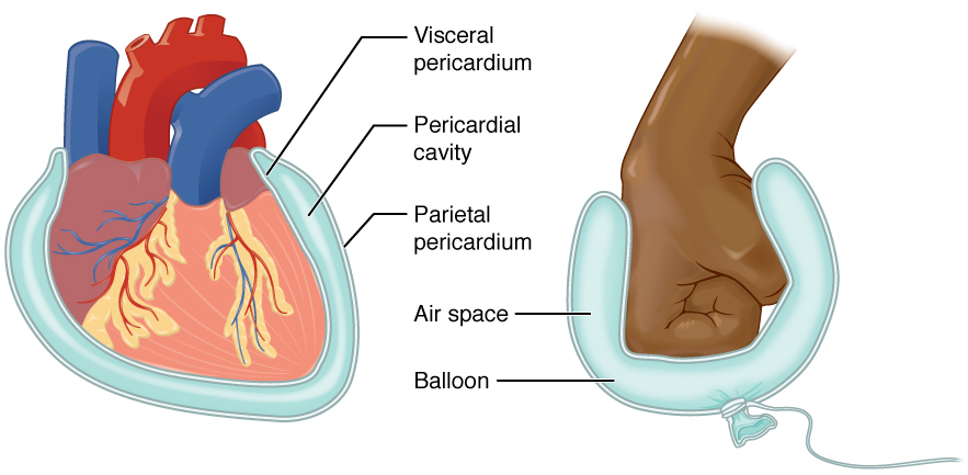
The pericardium (: pericardia), also called pericardial sac, is a double-walled sac containing the heart and the roots of the great vessels. It has two layers, an outer layer made of strong inelastic connective tissue (fibrous pericardium), and an inner layer made of serous membrane (serous pericardium). It encloses the pericardial cavity, which contains pericardial fluid, and defines the middle mediastinum. It separates the heart from interference of other structures, protects it against infection and blunt trauma, and lubricates the heart's movements. The English name originates from the Ancient Greek prefix ''peri-'' (περί) 'around' and the suffix ''-cardion'' (κάρδιον) 'heart'. Anatomy The pericardium is a tough fibroelastic sac which covers the heart from all sides except at the cardiac root (where the great vessels join the heart) and the bottom (where only the serous pericardium exists to cover the upper surface of the central tendon of diaphragm). ... [...More Info...] [...Related Items...] OR: [Wikipedia] [Google] [Baidu] |
Mucosa
A mucous membrane or mucosa is a membrane that lines various cavities in the body of an organism and covers the surface of internal organs. It consists of one or more layers of epithelial cells overlying a layer of loose connective tissue. It is mostly of endodermal origin and is continuous with the skin at body openings such as the eyes, eyelids, ears, inside the nose, inside the mouth, lips, the genital areas, the urethral opening and the anus. Some mucous membranes secrete mucus, a thick protective fluid. The function of the membrane is to stop pathogens and dirt from entering the body and to prevent bodily tissues from becoming dehydrated. Structure The mucosa is composed of one or more layers of epithelial cells that secrete mucus, and an underlying lamina propria of loose connective tissue. The type of cells and type of mucus secreted vary from organ to organ and each can differ along a given tract. Mucous membranes line the digestive, respiratory and rep ... [...More Info...] [...Related Items...] OR: [Wikipedia] [Google] [Baidu] |
Synovial Membrane
Synovial () may refer to: * Synovial fluid * Synovial joint A synovial joint, also known as diarthrosis, joins bones or cartilage with a fibrous joint capsule that is continuous with the periosteum of the joined bones, constitutes the outer boundary of a synovial cavity, and surrounds the bones' articulati ... * Synovial membrane * Synovial bursa {{disambiguation ... [...More Info...] [...Related Items...] OR: [Wikipedia] [Google] [Baidu] |
Hyaluronan
Hyaluronic acid (; abbreviated HA; conjugate acid, conjugate base hyaluronate), also called hyaluronan, is an anion#Anions and cations, anionic, Sulfation, nonsulfated glycosaminoglycan distributed widely throughout connective tissue, connective, epithelial tissue, epithelial, and neural tissues. It is unique among glycosaminoglycans as it is non-sulfated, forms in the plasma membrane instead of the Golgi apparatus, and can be very large: human Synovial fluid, synovial HA averages about per molecule, or about 20,000 disaccharide monomers, while other sources mention . Medically, hyaluronic acid is used to treat osteoarthritis of the knee and dry eye, for wound repair, and as a cosmetic filler. The average 70 kg (150 lb) person has roughly 15 grams of hyaluronan in the body, one third of which is turned over (i.e., degraded and synthesized) per day. As one of the chief components of the extracellular matrix, it contributes significantly to cell proliferation and ... [...More Info...] [...Related Items...] OR: [Wikipedia] [Google] [Baidu] |
Synovial Fluid
Synovial fluid, also called synovia, elp 1/sup> is a viscous, non-Newtonian fluid found in the cavities of synovial joints. With its egg white–like consistency, the principal role of synovial fluid is to reduce friction between the articular cartilage of synovial joints during movement. Synovial fluid is a small component of the transcellular fluid component of extracellular fluid. Structure The inner membrane of synovial joints is called the synovial membrane and secretes synovial fluid into the joints. Synovial fluid is an ultrafiltrate from blood, and contains proteins derived from the blood plasma and proteins that are produced by cells within the joint tissues. The fluid contains hyaluronan secreted by fibroblast-like cells in the synovial membrane, lubricin (proteoglycan 4; PRG4) secreted by the surface chondrocytes of the articular cartilage and interstitial fluid filtered from the blood plasma. This fluid forms a thin layer (roughly 50 μm) at the surface ... [...More Info...] [...Related Items...] OR: [Wikipedia] [Google] [Baidu] |
Epithelium
Epithelium or epithelial tissue is a thin, continuous, protective layer of cells with little extracellular matrix. An example is the epidermis, the outermost layer of the skin. Epithelial ( mesothelial) tissues line the outer surfaces of many internal organs, the corresponding inner surfaces of body cavities, and the inner surfaces of blood vessels. Epithelial tissue is one of the four basic types of animal tissue, along with connective tissue, muscle tissue and nervous tissue. These tissues also lack blood or lymph supply. The tissue is supplied by nerves. There are three principal shapes of epithelial cell: squamous (scaly), columnar, and cuboidal. These can be arranged in a singular layer of cells as simple epithelium, either simple squamous, simple columnar, or simple cuboidal, or in layers of two or more cells deep as stratified (layered), or ''compound'', either squamous, columnar or cuboidal. In some tissues, a layer of columnar cells may appear to be stratified d ... [...More Info...] [...Related Items...] OR: [Wikipedia] [Google] [Baidu] |
Lamina Propria
The lamina propria is a thin layer of connective tissue that forms part of the moist linings known as mucous membranes or mucosae, which line various tubes in the body, such as the respiratory tract, the gastrointestinal tract, and the urogenital tract. The lamina propria is a thin layer of loose (areolar) connective tissue, which lies beneath the epithelium, and together with the epithelium and basement membrane constitutes the mucosa. As its Latin name indicates, it is a characteristic component of the mucosa, or the mucosa's "own special layer." Thus, the term mucosa or mucous membrane refers to the combination of the epithelium and the lamina propria. The connective tissue of the lamina propria is loose and rich in cells. The cells of the lamina propria are variable and can include fibroblasts, lymphocytes, plasma cells, macrophages, eosinophilic leukocytes, and mast cells. It provides support and nutrition to the epithelium, as well as the means to bind to the underlying t ... [...More Info...] [...Related Items...] OR: [Wikipedia] [Google] [Baidu] |
Serous Membrane
The serous membrane (or serosa) is a smooth epithelial membrane of mesothelium lining the contents and inner walls of body cavity, body cavities, which secrete serous fluid to allow lubricated sliding (motion), sliding movements between opposing surfaces. The serous membrane that covers Viscera, internal organs (viscera) is called ''visceral'', while the one that covers the cavity wall is called ''parietal''. For instance the peritoneum, parietal peritoneum is attached to the abdominal wall and the pelvic walls. The peritoneum, visceral peritoneum is wrapped around the visceral organs. For the heart, the layers of the serous membrane are called parietal and visceral pericardium. For the lungs they are called parietal and visceral pleura. The visceral serosa of the uterus is called the perimetrium. The potential space between two opposing serosal surfaces is mostly empty except for the small amount of serous fluid. The Latin anatomical name is Tunica (biology)#Anatomy usages, tu ... [...More Info...] [...Related Items...] OR: [Wikipedia] [Google] [Baidu] |
Mesothelium
The mesothelium is a membrane composed of simple squamous epithelium, simple squamous epithelial cells of mesodermal origin, which forms the lining of several body cavities: the pleura (pleural cavity around the lungs), peritoneum (abdominopelvic cavity including the mesentery, omentum (other), omenta, falciform ligament and the perimetrium) and pericardium (around the heart). Mesothelial tissue also surrounds the male testis (as the tunica vaginalis) and occasionally the spermatic cord (in a patent processus vaginalis). Mesothelium that tunica (biology), covers the internal organs is called visceral mesothelium, while one that covers the surrounding body walls is called the :wikt:parietal, parietal mesothelium. The mesothelium that secretes serous fluid as a main function is also known as a serosa. Origin Mesothelium derives from the embryonic mesoderm cell layer, that lines the body cavity, coelom (body cavity) in the embryo. It develops into the layer of cells that c ... [...More Info...] [...Related Items...] OR: [Wikipedia] [Google] [Baidu] |
Serous Fluid
In physiology, serous fluid or serosal fluid (originating from the Medieval Latin word ''serosus'', from Latin ''serum'') is any of various body fluids resembling serum, that are typically pale yellow or transparent and of a benign nature. The fluid fills the inside of body cavities. Serous fluid originates from serous glands, with secretions enriched with proteins and water. Serous fluid may also originate from mixed glands, which contain both mucous and serous cells. A common trait of serous fluids is their role in assisting digestion, excretion, and respiration. In medical fields, especially cytopathology, serous fluid is a synonym for effusion fluids from various body cavities. Examples of effusion fluid are pleural effusion and pericardial effusion. There are many causes of effusions which include involvement of the cavity by cancer. Cancer in a serous cavity is called a serous carcinoma. Cytopathology evaluation is recommended to evaluate the causes of effusions in these ... [...More Info...] [...Related Items...] OR: [Wikipedia] [Google] [Baidu] |
Stratified Squamous Epithelium
A stratified squamous epithelium consists of squamous (flattened) epithelial cells arranged in layers upon a basal membrane. Only one layer is in contact with the basement membrane; the other layers adhere to one another to maintain structural integrity. Although this epithelium is referred to as squamous, many cells within the layers may not be flattened; this is due to the convention of naming epithelia according to the cell type at the surface. In the deeper layers, the cells may be columnar or cuboidal. There are no intercellular spaces. This type of epithelium is well suited to areas in the body subject to constant abrasion, as the thickest layers can be sequentially sloughed off and replaced before the basement membrane is exposed. It forms the outermost layer of the skin and the inner lining of the mouth, esophagus and vagina. In the epidermis of skin in mammals, reptiles, and birds, the layer of keratin in the outer layer of the stratified squamous epithelial surface is ... [...More Info...] [...Related Items...] OR: [Wikipedia] [Google] [Baidu] |
Membrane Biology
Membrane biology is the study of the biological and physiochemical characteristics of membranes, with applications in the study of cellular physiology. Membrane bioelectrical impulses are described by the Hodgkin cycle. Biophysics Membrane biophysics is the study of biological membrane structure and function using physical, computational, mathematical, and biophysical methods. A combination of these methods can be used to create phase diagrams of different types of membranes, which yields information on thermodynamic behavior of a membrane and its components. As opposed to membrane biology, membrane biophysics focuses on quantitative information and modeling of various membrane phenomena, such as lipid raft The cell membrane, plasma membranes of cells contain combinations of glycosphingolipids, cholesterol and protein Receptor (biochemistry), receptors organized in glycolipoprotein lipid microdomains termed lipid rafts. Their existence in cellular me ... formation, rates ... [...More Info...] [...Related Items...] OR: [Wikipedia] [Google] [Baidu] |


