|
Mesothelium
The mesothelium is a membrane composed of simple squamous epithelium, simple squamous epithelial cells of mesodermal origin, which forms the lining of several body cavities: the pleura (pleural cavity around the lungs), peritoneum (abdominopelvic cavity including the mesentery, omentum (other), omenta, falciform ligament and the perimetrium) and pericardium (around the heart). Mesothelial tissue also surrounds the male testis (as the tunica vaginalis) and occasionally the spermatic cord (in a patent processus vaginalis). Mesothelium that tunica (biology), covers the internal organs is called visceral mesothelium, while one that covers the surrounding body walls is called the :wikt:parietal, parietal mesothelium. The mesothelium that secretes serous fluid as a main function is also known as a serosa. Origin Mesothelium derives from the embryonic mesoderm cell layer, that lines the body cavity, coelom (body cavity) in the embryo. It develops into the layer of cells that c ... [...More Info...] [...Related Items...] OR: [Wikipedia] [Google] [Baidu] |
Epithelial Cell
Epithelium or epithelial tissue is a thin, continuous, protective layer of Cell (biology), cells with little extracellular matrix. An example is the epidermis, the outermost layer of the skin. Epithelial (Mesothelium, mesothelial) tissues line the outer surfaces of many internal organ (anatomy), organs, the corresponding inner surfaces of body cavities, and the inner surfaces of blood vessels. Epithelial tissue is one of the four basic types of animal Tissue (biology), tissue, along with connective tissue, muscle tissue and nervous tissue. These tissues also lack blood or lymph supply. The tissue is supplied by nerves. There are three principal shapes of epithelial cell: squamous (scaly), columnar, and cuboidal. These can be arranged in a singular layer of cells as simple epithelium, either simple squamous, simple columnar, or simple cuboidal, or in layers of two or more cells deep as stratified (layered), or ''compound'', either squamous, columnar or cuboidal. In some tissues, ... [...More Info...] [...Related Items...] OR: [Wikipedia] [Google] [Baidu] |
Pericardium
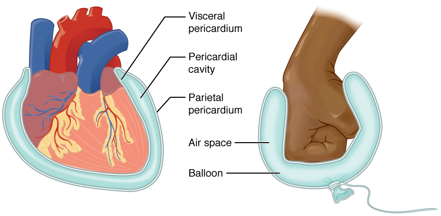
The pericardium (: pericardia), also called pericardial sac, is a double-walled sac containing the heart and the roots of the great vessels. It has two layers, an outer layer made of strong inelastic connective tissue (fibrous pericardium), and an inner layer made of serous membrane (serous pericardium). It encloses the pericardial cavity, which contains pericardial fluid, and defines the middle mediastinum. It separates the heart from interference of other structures, protects it against infection and blunt trauma, and lubricates the heart's movements. The English name originates from the Ancient Greek prefix ''peri-'' (περί) 'around' and the suffix ''-cardion'' (κάρδιον) 'heart'. Anatomy The pericardium is a tough fibroelastic sac which covers the heart from all sides except at the cardiac root (where the great vessels join the heart) and the bottom (where only the serous pericardium exists to cover the upper surface of the central tendon of diaphragm). ... [...More Info...] [...Related Items...] OR: [Wikipedia] [Google] [Baidu] |
Mesoderm
The mesoderm is the middle layer of the three germ layers that develops during gastrulation in the very early development of the embryo of most animals. The outer layer is the ectoderm, and the inner layer is the endoderm.Langman's Medical Embryology, 11th edition. 2010. The mesoderm forms mesenchyme, mesothelium and coelomocytes. Mesothelium lines coeloms. Mesoderm forms the muscles in a process known as myogenesis, septa (cross-wise partitions) and mesenteries (length-wise partitions); and forms part of the gonads (the rest being the gametes). Myogenesis is specifically a function of mesenchyme. The mesoderm differentiates from the rest of the embryo through intercellular signaling, after which the mesoderm is polarized by an organizing center. The position of the organizing center is in turn determined by the regions in which beta-catenin is protected from degradation by GSK-3. Beta-catenin acts as a co-factor that alters the activity of the transcription facto ... [...More Info...] [...Related Items...] OR: [Wikipedia] [Google] [Baidu] |
Serosa
The serous membrane (or serosa) is a smooth epithelial membrane of mesothelium lining the contents and inner walls of body cavities, which secrete serous fluid to allow lubricated sliding movements between opposing surfaces. The serous membrane that covers internal organs (viscera) is called ''visceral'', while the one that covers the cavity wall is called ''parietal''. For instance the parietal peritoneum is attached to the abdominal wall and the pelvic walls. The visceral peritoneum is wrapped around the visceral organs. For the heart, the layers of the serous membrane are called parietal and visceral pericardium. For the lungs they are called parietal and visceral pleura. The visceral serosa of the uterus is called the perimetrium. The potential space between two opposing serosal surfaces is mostly empty except for the small amount of serous fluid. The Latin anatomical name is tunica serosa. Serous membranes line and enclose several body cavities, also known as s ... [...More Info...] [...Related Items...] OR: [Wikipedia] [Google] [Baidu] |
Peritoneal
The peritoneum is the serous membrane forming the lining of the abdominal cavity or coelom in amniotes and some invertebrates, such as annelids. It covers most of the intra-abdominal (or coelomic) organs, and is composed of a layer of mesothelium supported by a thin layer of connective tissue. This peritoneal lining of the cavity supports many of the abdominal organs and serves as a conduit for their blood vessels, lymphatic vessels, and nerves. The abdominal cavity (the space bounded by the vertebrae, abdominal muscles, diaphragm, and pelvic floor) is different from the intraperitoneal space (located within the abdominal cavity but wrapped in peritoneum). The structures within the intraperitoneal space are called "intraperitoneal" (e.g., the stomach and intestines), the structures in the abdominal cavity that are located behind the intraperitoneal space are called "retroperitoneal" (e.g., the kidneys), and those structures below the intraperitoneal space are called "subperit ... [...More Info...] [...Related Items...] OR: [Wikipedia] [Google] [Baidu] |
Pleura
The pleurae (: pleura) are the two flattened closed sacs filled with pleural fluid, each ensheathing each lung and lining their surrounding tissues, locally appearing as two opposing layers of serous membrane separating the lungs from the mediastinum, the inside surfaces of the surrounding chest walls and the diaphragm. Although wrapped onto itself resulting in an apparent double layer, each lung is surrounded by a single, continuous pleural membrane. The portion of the pleura that covers the surface of each lung is often called the visceral pleura. This can lead to some confusion, as the lung is not the only visceral organ covered by the pleura. The pleura typically dips between the lobes of the lung as ''fissures'', and is formed by the invagination of lung buds into each thoracic sac during embryonic development. The portion of the pleura seen as the outer layer covers the chest wall, the diaphragm and the mediastinum and is often also misleadingly called the parieta ... [...More Info...] [...Related Items...] OR: [Wikipedia] [Google] [Baidu] |
Mesentery
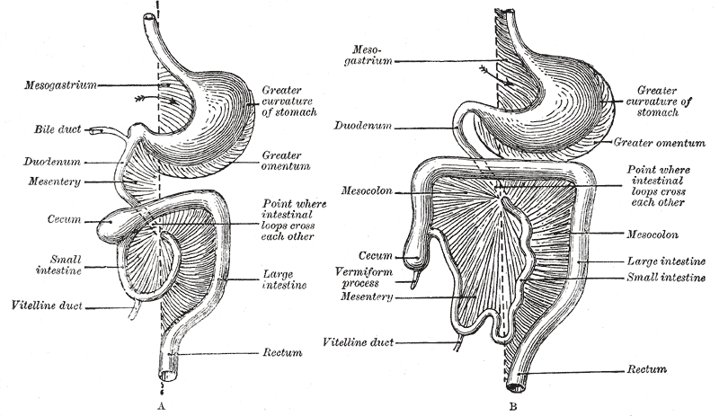
In human anatomy, the mesentery is an Organ (anatomy), organ that attaches the intestines to the posterior abdominal wall, consisting of a double fold of the peritoneum. It helps (among other functions) in storing Adipose tissue, fat and allowing blood vessels, lymphatics, and nerves to supply the intestines. The (the part of the mesentery that attaches the colon to the abdominal wall) was formerly thought to be a fragmented structure, with all named parts—the ascending, transverse, descending, and sigmoid mesocolons, the mesoappendix, and the mesorectum—separately terminating their insertion into the posterior abdominal wall. However, in 2012, new microscopy, microscopic and electron microscope, electron microscopic histology, examinations showed the mesocolon to be a single structure derived from the duodenojejunal flexure and extending to the distal mesorectal layer. Thus the mesentery is an internal organ. Structure The mesentery of the small intestine arises from th ... [...More Info...] [...Related Items...] OR: [Wikipedia] [Google] [Baidu] |
Body Cavities
A body cavity is any space or compartment, or potential space, in an animal body. Cavities accommodate organs and other structures; cavities as potential spaces contain fluid. The two largest human body cavities are the ventral body cavity, and the dorsal body cavity. In the dorsal body cavity the brain and spinal cord are located. The membranes that surround the central nervous system organs (the brain and the spinal cord, in the cranial and spinal cavities) are the three meninges. The differently lined spaces contain different types of fluid. In the meninges for example the fluid is cerebrospinal fluid; in the abdominal cavity the fluid contained in the peritoneum is a serous fluid. In amniotes and some invertebrates the peritoneum lines their largest body cavity called the coelom. Mammals Mammalian embryos develop two body cavities: the intraembryonic coelom and the extraembryonic coelom (or chorionic cavity). The intraembryonic coelom is lined by somatic and ... [...More Info...] [...Related Items...] OR: [Wikipedia] [Google] [Baidu] |
Perimetrium
The perimetrium (or serous coat of uterus) is the outer serosal layer of the uterus, derived from the peritoneum overlying the uterine fundus, and can be considered a visceral peritoneum. It consists of a superficial layer of mesothelium, and a thin layer of loose connective tissue beneath it. Anteriorly, the perimetrium covers the fundus and upper body of the uterus until it meets the superoposterior surface of the adjacent urinary bladder, resulting in a concave fold of peritoneum called the '' vesicouterine pouch''. Posteriorly, the perimetrium covers the entire surface of the uterus deep down to the cervix, where it then folds back onto the adjacent rectum to form the ''rectouterine pouch The rectouterine pouch (rectovaginal pouch, pouch of Douglas or cul-de-sac) is the extension of the peritoneum into the space between the posterior wall of the uterus and the rectum in the human female. Structure In women, the rectouterine pouch ...'', the lowest gutter of the peritone ... [...More Info...] [...Related Items...] OR: [Wikipedia] [Google] [Baidu] |
Simple Squamous Epithelium
A simple squamous epithelium, also known as pavement epithelium and tessellated epithelium, is a single layer of flattened, polygonal cells in contact with the basal lamina (one of the two layers of the basement membrane) of the epithelium. This type of epithelium is often permeable and occurs where small molecules need to pass quickly through membranes via filtration or diffusion. Simple squamous epithelia are found in endothelium (lining of blood and lymph capillaries), mesothelium (coelomic epithelium/peritoneum), alveoli of lungs, glomeruli, and other tissues where rapid diffusion is required.AAMC - 2015 MCAT Question Pack Explanations Within the cardiovascular system such as lining capillaries or the inside of the heart, simple squamous epithelium is specifically called the endothelium The endothelium (: endothelia) is a single layer of squamous endothelial cells that line the interior surface of blood vessels and lymphatic vessels. The endothelium forms an interface bet ... [...More Info...] [...Related Items...] OR: [Wikipedia] [Google] [Baidu] |
Visceral
In a multicellular organism, an organ is a collection of Tissue (biology), tissues joined in a structural unit to serve a common function. In the biological organization, hierarchy of life, an organ lies between Tissue (biology), tissue and an organ system. Tissues are formed from same type Cell (biology), cells to act together in a function. Tissues of different types combine to form an organ which has a specific function. The Gastrointestinal tract, intestinal wall for example is formed by epithelial tissue and smooth muscle tissue. Two or more organs working together in the execution of a specific body function form an organ system, also called a biological system or body system. An organ's tissues can be broadly categorized as parenchyma, the functional tissue, and stroma (tissue), stroma, the structural tissue with supportive, connective, or ancillary functions. For example, the gland's tissue that makes the hormones is the parenchyma, whereas the stroma includes the nerve t ... [...More Info...] [...Related Items...] OR: [Wikipedia] [Google] [Baidu] |


