|
Enamel Rod
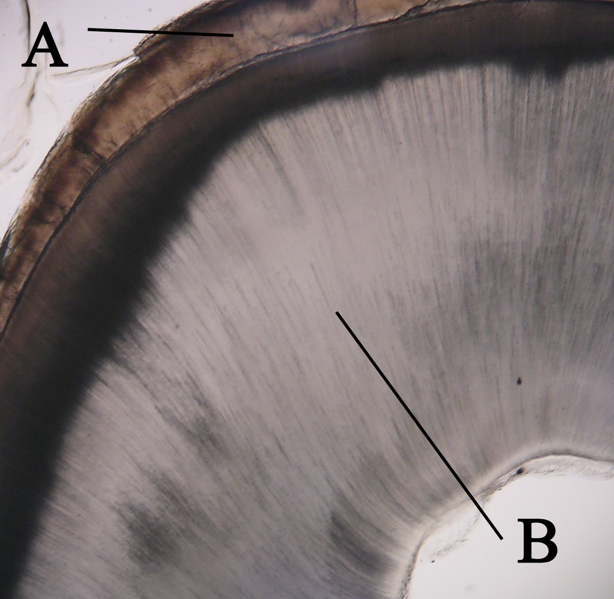
An enamel prism, or enamel rod, is the basic unit of tooth enamel. Measuring 3-6 μm in diameter in primates, enamel prism are tightly packed hydroxyapatite crystals structures. The hydroxyapatite crystals are hexagonal in shape, providing rigidity to the prism and strengthening the enamel. In cross-section, it is best compared to a complex “keyhole” or a “fish-like” shape. The head, which is called the prism core, is oriented toward the tooth’s crown; The tail, which is called the prism sheath, is oriented toward the tooth cervical margin /sup>. The prism core has tightly packed hydroxyapatite crystals. On the other hand, the prism sheath has its crystals less tightly packed and has more space for organic components. These prism structures can usually be visualised within ground sections and/or with the use of a scanning electron microscope on enamel that has been acid etched ">/sup>. The number of enamel prisms range approximately from 5 million to 12 million in ... [...More Info...] [...Related Items...] OR: [Wikipedia] [Google] [Baidu] |
Tooth Enamel
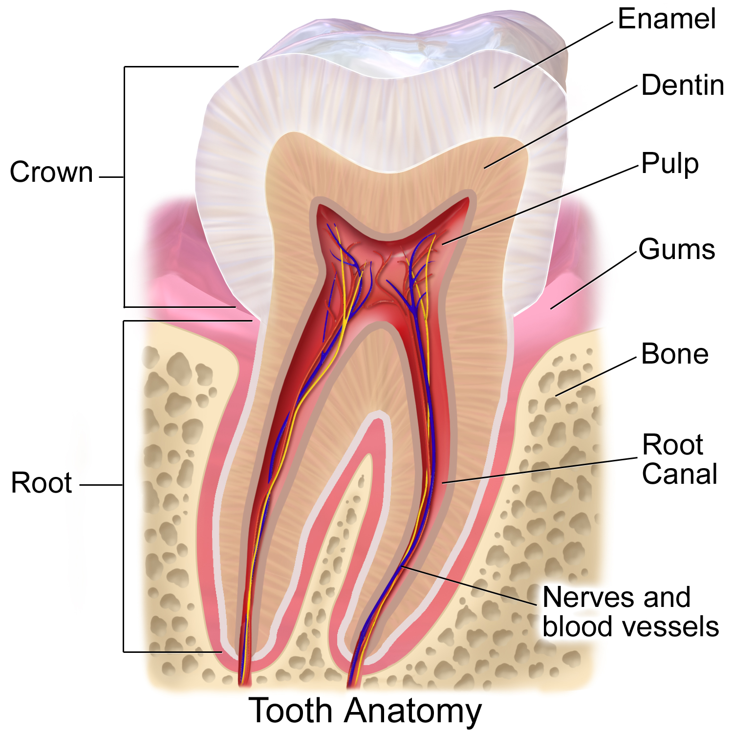
Tooth enamel is one of the four major Tissue (biology), tissues that make up the tooth in humans and many animals, including some species of fish. It makes up the normally visible part of the tooth, covering the Crown (tooth), crown. The other major tissues are dentin, cementum, and Pulp (tooth), dental pulp. It is a very hard, white to off-white, highly mineralised substance that acts as a barrier to protect the tooth but can become susceptible to degradation, especially by acids from food and drink. In rare circumstances enamel fails to form, leaving the underlying dentin exposed on the surface. Features Enamel is the hardest substance in the human body and contains the highest percentage of minerals (at 96%),Ross ''et al.'', p. 485 with water and organic material composing the rest.Ten Cate's Oral Histology, Nancy, Elsevier, pp. 70–94 The primary mineral is hydroxyapatite, which is a crystalline calcium phosphate. Enamel is formed on the tooth while the tooth develops wit ... [...More Info...] [...Related Items...] OR: [Wikipedia] [Google] [Baidu] |
Hydroxyapatite
Hydroxyapatite (International Mineralogical Association, IMA name: hydroxylapatite) (Hap, HAp, or HA) is a naturally occurring mineral form of calcium apatite with the Chemical formula, formula , often written to denote that the Crystal structure, crystal unit cell comprises two entities. It is the Hydroxy group, hydroxyl endmember of the complex apatite, apatite group. The ion can be replaced by fluorine, fluoride or chlorine, chloride, producing fluorapatite or chlorapatite. It crystallizes in the hexagonal (crystal system), hexagonal crystal system. Pure hydroxyapatite powder is white. Naturally occurring apatites can, however, also have brown, yellow, or green colorations, comparable to the discolorations of dental fluorosis. Up to 50% by volume and 70% by weight of human bone is a modified form of hydroxyapatite, known as bone mineral. Carbonated calcium-deficient hydroxyapatite is the main mineral of which dental enamel and dentin are composed. Hydroxyapatite crystals a ... [...More Info...] [...Related Items...] OR: [Wikipedia] [Google] [Baidu] |
Tooth’s Crown
In dentistry, the crown is the visible part of the tooth above the gingival margin and is an essential component of dental anatomy. Covered by enamel, the crown plays a crucial role in cutting, tearing, and grinding food. Its shape and structure vary depending on the type and function of the tooth (incisors, canines, premolars, or molars), and differ between primary dentition and permanent dentition. The crown also contributes to facial aesthetics, speech, and oral health. Anatomical crown vs clinical crown The anatomical crown refers to the portion of the tooth covered by enamel, regardless of whether it is visible. The clinical crown is the part of the tooth that is visible in the mouth. In a healthy young adult, the gums typically follow the contour where enamel meets the root, so the clinical and anatomical crowns are similar in size. However, with age or periodontal disease, this may change. Terminology of tooth surfaces To describe the location and orientation of t ... [...More Info...] [...Related Items...] OR: [Wikipedia] [Google] [Baidu] |
Tooth Cervical Margin
The cervical margins of teeth are the surfaces where the crown and root meet, and is also referred to as the tooth's neck or cervical line. Anatomy The cervical margin, also known as the cervical line or neck of the tooth, represents the boundary between the enamel covering the crown and the cementum covering the root. The cementum typically overlaps the enamel, although in some cases, it may meet edge-to-edge. The cervical region includes the residual tooth structure between the gingival margin and the bone crest, encompassing the supragingival tooth area (STA) and gingival sulcus. Periodontal consideration Biological width The biological width is a crucial factor in maintaining periodontal health. It refers to the soft tissue dimensions coronal to the alveolar bone, consisting of junctional epithelium and supracrestal connective tissue attachment. However, by violating the biological width during restorative procedures can lead to periodontal breakdown, inflammat ... [...More Info...] [...Related Items...] OR: [Wikipedia] [Google] [Baidu] |
Enamel Prism
An enamel prism, or enamel rod, is the basic unit of tooth enamel. Measuring 3-6 μm in diameter in primates, enamel prism are tightly packed hydroxyapatite crystals structures. The hydroxyapatite crystals are hexagonal in shape, providing rigidity to the prism and strengthening the enamel. In cross-section, it is best compared to a complex “keyhole” or a “fish-like” shape. The head, which is called the prism core, is oriented toward the tooth’s crown; The tail, which is called the prism sheath, is oriented toward the tooth cervical margin /sup>. The prism core has tightly packed hydroxyapatite crystals. On the other hand, the prism sheath has its crystals less tightly packed and has more space for organic components. These prism structures can usually be visualised within ground sections and/or with the use of a scanning electron microscope on enamel that has been acid etched ">/sup>. The number of enamel prisms range approximately from 5 million to 12 million in t ... [...More Info...] [...Related Items...] OR: [Wikipedia] [Google] [Baidu] |
Scanning Electron Microscope
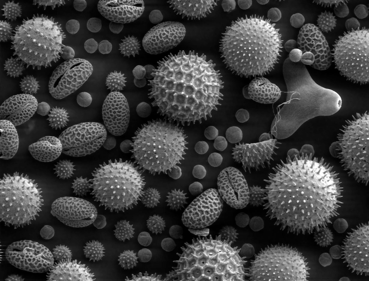
A scanning electron microscope (SEM) is a type of electron microscope that produces images of a sample by scanning the surface with a focused beam of electrons. The electrons interact with atoms in the sample, producing various signals that contain information about the surface topography and composition. The electron beam is scanned in a raster scan pattern, and the position of the beam is combined with the intensity of the detected signal to produce an image. In the most common SEM mode, secondary electrons emitted by atoms excited by the electron beam are detected using a secondary electron detector ( Everhart–Thornley detector). The number of secondary electrons that can be detected, and thus the signal intensity, depends, among other things, on specimen topography. Some SEMs can achieve resolutions better than 1 nanometer. Specimens are observed in high vacuum in a conventional SEM, or in low vacuum or wet conditions in a variable pressure or environmental SEM, an ... [...More Info...] [...Related Items...] OR: [Wikipedia] [Google] [Baidu] |
Mandibular Incisor (other)
{{disambiguation ...
Mandibular incisor may refer to: * Mandibular central incisor * Mandibular lateral incisor The mandibular lateral incisor is the tooth located distally (away from the midline of the face) from both mandibular central incisors of the mouth and mesially (toward the midline of the face) from both mandibular canines. As with all incisors, ... [...More Info...] [...Related Items...] OR: [Wikipedia] [Google] [Baidu] |
Maxillary Molar
The molars or molar teeth are large, flat teeth at the back of the mouth. They are more developed in mammals. They are used primarily to grind food during chewing. The name ''molar'' derives from Latin, ''molaris dens'', meaning "millstone tooth", from ''mola'', millstone and ''dens'', tooth. Molars show a great deal of diversity in size and shape across the mammal groups. The third molar of humans is sometimes vestigial. Human anatomy In humans, the molar teeth have either four or five cusps. Adult humans have 12 molars, in four groups of three at the back of the mouth. The third, rearmost molar in each group is called a wisdom tooth. It is the last tooth to appear, breaking through the front of the gum at about the age of 20, although this varies among individuals and populations, and in many cases the tooth is missing. The human mouth contains upper (maxillary) and lower (mandibular) molars. They are: maxillary first molar, maxillary second molar, maxillary third molar, man ... [...More Info...] [...Related Items...] OR: [Wikipedia] [Google] [Baidu] |
Primary Dentition
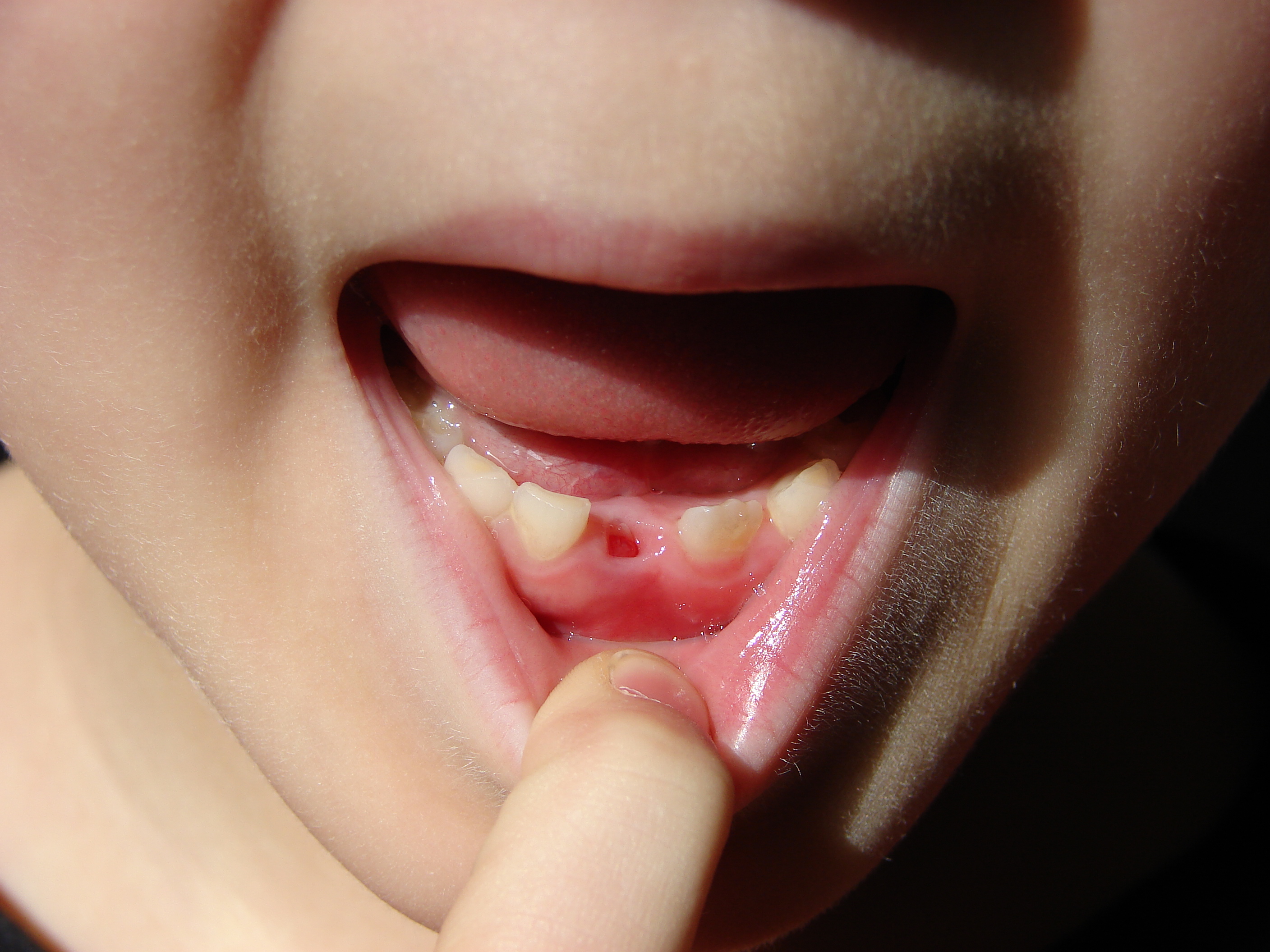
Deciduous teeth or primary teeth, also informally known as baby teeth, milk teeth, or temporary teeth,Fehrenbach, MJ and Popowics, T. (2026). ''Illustrated Dental Embryology, Histology, and Anatomy'', 6th edition, Elsevier, page 287–296. are the first set of teeth in the growth and development of humans and other diphyodonts, which include most mammals but not elephants, kangaroos, or manatees, which are polyphyodonts. Deciduous teeth develop during the embryonic stage of development and erupt (break through the gums and become visible in the mouth) during infancy. They are usually lost and replaced by permanent teeth, but in the absence of their permanent replacements, they can remain functional for many years into adulthood. Development Formation Primary teeth start to form during the embryonic phase of human life. The development of primary teeth starts at the sixth week of tooth development as the dental lamina. This process starts at the midline and then spreads ba ... [...More Info...] [...Related Items...] OR: [Wikipedia] [Google] [Baidu] |
Ameloblasts
Ameloblasts are cells present only during tooth development that deposit tooth enamel, which is the hard outermost layer of the tooth forming the surface of the crown. Structure Each ameloblast is a columnar cell approximately 4 micrometers in diameter, 40 micrometers in length and is hexagonal in cross section. The secretory end of the ameloblast ends in a six-sided pyramid-like projection known as the Tomes' process. The angulation of the Tomes' process is significant in the orientation of enamel rods, the basic unit of tooth enamel. Distal terminal bars are junctional complexes that separate the Tomes' processes from ameloblast proper. Development Ameloblasts are derived from oral epithelium tissue of ectodermal origin. Their differentiation from preameloblasts (whose origin is from inner enamel epithelium) is a result of signaling from the ectomesenchymal cells of the dental papilla. Initially the preameloblasts will differentiate into presecretory ameloblasts and then ... [...More Info...] [...Related Items...] OR: [Wikipedia] [Google] [Baidu] |
Dentin
Dentin ( ) (American English) or dentine ( or ) (British English) () is a calcified tissue (biology), tissue of the body and, along with tooth enamel, enamel, cementum, and pulp (tooth), pulp, is one of the four major components of teeth. It is usually covered by enamel on the crown and cementum on the root and surrounds the entire pulp. By volume, 45% of dentin consists of the mineral hydroxyapatite, 33% is organic material, and 22% is water. Yellow in appearance, it greatly affects the color of a tooth due to the translucency of enamel. Dentin, which is less mineralized and less brittle than enamel, is necessary for the support of enamel. Dentin rates approximately 3 on the Mohs scale of mineral hardness. There are two main characteristics which distinguish dentin from enamel: firstly, dentin forms throughout life; secondly, dentin is sensitive and can become hypersensitive to changes in temperature due to the sensory function of odontoblasts, especially when enamel recedes an ... [...More Info...] [...Related Items...] OR: [Wikipedia] [Google] [Baidu] |
Masticatory
Chewing or mastication is the process by which food is crushed and ground by the teeth. It is the first step in the process of digestion, allowing a greater surface area for digestive enzymes to break down the foods. During the mastication process, the food is positioned by the cheek and tongue between the teeth for grinding. The muscles of mastication move the jaws to bring the teeth into intermittent contact, repeatedly occluding and opening. As chewing continues, the food is made softer and warmer, and the enzymes in saliva begin to break down carbohydrates in the food. After chewing, the food (now called a bolus) is swallowed. It enters the esophagus and via peristalsis continues on to the stomach, where the next step of digestion occurs. Increasing the number of chews per bite stimulates the production of digestive enzymes and peptides and has been shown to increase diet-induced thermogenesis (DIT) by activating the sympathetic nervous system. Studies suggest that thorough ... [...More Info...] [...Related Items...] OR: [Wikipedia] [Google] [Baidu] |


