|
Ameloblasts
Ameloblasts are cells present only during tooth development that deposit tooth enamel, which is the hard outermost layer of the tooth forming the surface of the crown. Structure Each ameloblast is a columnar cell approximately 4 micrometers in diameter, 40 micrometers in length and is hexagonal in cross section. The secretory end of the ameloblast ends in a six-sided pyramid-like projection known as the Tomes' process. The angulation of the Tomes' process is significant in the orientation of enamel rods, the basic unit of tooth enamel. Distal terminal bars are junctional complexes that separate the Tomes' processes from ameloblast proper. Development Ameloblasts are derived from oral epithelium tissue of ectodermal origin. Their differentiation from preameloblasts (whose origin is from inner enamel epithelium) is a result of signaling from the ectomesenchymal cells of the dental papilla. Initially the preameloblasts will differentiate into presecretory ameloblasts and then ... [...More Info...] [...Related Items...] OR: [Wikipedia] [Google] [Baidu] |
Ameloblast Life Cycle & Amelogenesis
Ameloblasts are Cell (biology), cells present only during tooth development that deposit tooth enamel, which is the hard outermost layer of the tooth forming the surface of the crown. Structure Each ameloblast is a columnar cell approximately 4 micrometers in diameter, 40 micrometers in length and is hexagonal in cross section. The secretory end of the ameloblast ends in a six-sided pyramid-like projection known as the Tomes' process. The angulation of the Tomes' process is significant in the orientation of enamel rods, the basic unit of tooth enamel. Distal terminal bars are junctional complexes that separate the Tomes' processes from ameloblast proper. Development Ameloblasts are derived from oral epithelium tissue of ectodermal origin. Their differentiation from preameloblasts (whose origin is from inner enamel epithelium) is a result of signaling from the Ectomesenchyme, ectomesenchymal cells of the dental papilla. Initially the preameloblasts will differentiate into presecr ... [...More Info...] [...Related Items...] OR: [Wikipedia] [Google] [Baidu] |
Tooth Enamel
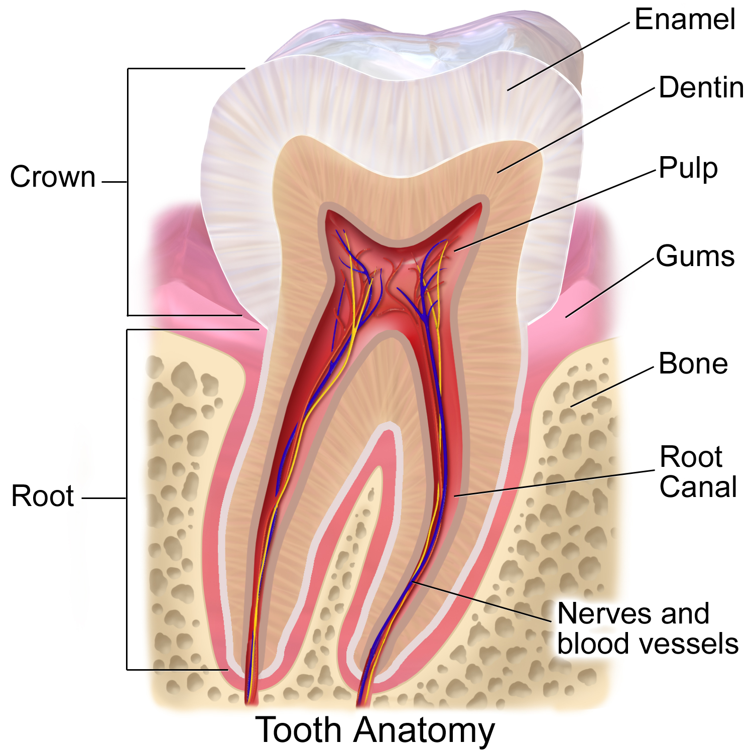
Tooth enamel is one of the four major Tissue (biology), tissues that make up the tooth in humans and many animals, including some species of fish. It makes up the normally visible part of the tooth, covering the Crown (tooth), crown. The other major tissues are dentin, cementum, and Pulp (tooth), dental pulp. It is a very hard, white to off-white, highly mineralised substance that acts as a barrier to protect the tooth but can become susceptible to degradation, especially by acids from food and drink. In rare circumstances enamel fails to form, leaving the underlying dentin exposed on the surface. Features Enamel is the hardest substance in the human body and contains the highest percentage of minerals (at 96%),Ross ''et al.'', p. 485 with water and organic material composing the rest.Ten Cate's Oral Histology, Nancy, Elsevier, pp. 70–94 The primary mineral is hydroxyapatite, which is a crystalline calcium phosphate. Enamel is formed on the tooth while the tooth develops wit ... [...More Info...] [...Related Items...] OR: [Wikipedia] [Google] [Baidu] |
Human Tooth Development
Tooth development or odontogenesis is the complex process by which teeth form from embryonic cells, grow, and erupt into the mouth. For human teeth to have a healthy oral environment, all parts of the tooth must develop during appropriate stages of fetal development. Primary (baby) teeth start to form between the sixth and eighth week of prenatal development, and permanent teeth begin to form in the twentieth week.Ten Cate's Oral Histology, Nanci, Elsevier, 2013, pages 70-94 If teeth do not start to develop at or near these times, they will not develop at all, resulting in hypodontia or anodontia. A significant amount of research has focused on determining the processes that initiate tooth development. It is widely accepted that there is a factor within the tissues of the first pharyngeal arch that is necessary for the development of teeth. Overview The tooth germ is an aggregation of cells that eventually forms a tooth.University of Texas Medical Branch. These cells are ... [...More Info...] [...Related Items...] OR: [Wikipedia] [Google] [Baidu] |
Amelogenesis
Amelogenesis is the process of forming tooth enamel, the hard, protective outer layer of teeth. This process begins during tooth development after the initial formation of dentin ( dentinogenesis), the layer beneath the enamel. The inner enamel epithelium (IEE), a layer of cells within the developing tooth, plays a crucial role by signaling the differentiation of specialized cells called ameloblasts, which then secrete the proteins and minerals that make up enamel. The formation of dentin is essential for amelogenesis to occur, and a reciprocal signaling process between the developing dentin and the IEE-derived ameloblasts ensures the proper coordination of these two crucial stages of tooth development. Stages Amelogenesis is considered to have three stages. The first stage is known as the inductive stage, the second is the secretory stage, and the third stage is known as the maturation stage. During the inductive stage, ameloblast differentiation from IEE is initiated. Prote ... [...More Info...] [...Related Items...] OR: [Wikipedia] [Google] [Baidu] |
Ameloblastoma
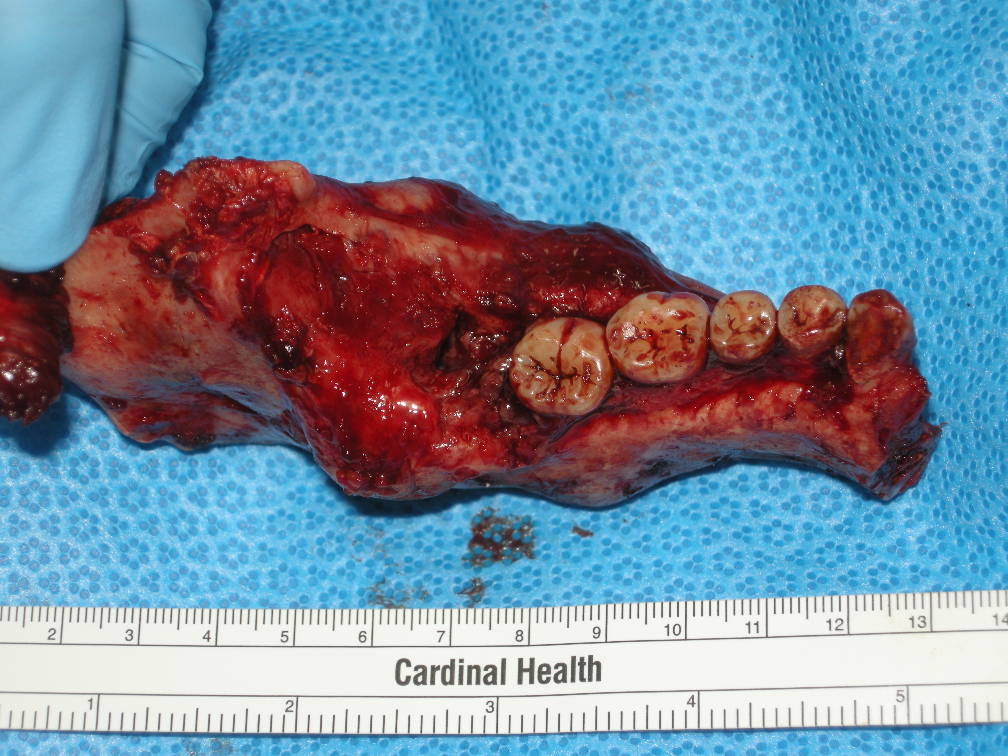
Ameloblastoma is a rare, benign or cancerous tumor of odontogenic epithelium ( ameloblasts, or outside portion, of the teeth during development) much more commonly appearing in the lower jaw than the upper jaw. It was recognized in 1827 by Cusack. This type of odontogenic neoplasm was designated as an '' adamantinoma'' in 1885 by the French physician Louis-Charles Malassez. It was finally renamed to the modern name ''ameloblastoma'' in 1930 by Ivey and Churchill. While these tumors are rarely malignant or metastatic (that is, they rarely spread to other parts of the body), and progress slowly, the resulting lesions can cause severe abnormalities of the face and jaw leading to severe disfiguration. Additionally, as abnormal cell growth easily infiltrates and destroys surrounding bony tissues, wide surgical excision is required to treat this disorder. If an aggressive tumor is left untreated, it can obstruct the nasal and oral airways making it impossible to breathe without oro ... [...More Info...] [...Related Items...] OR: [Wikipedia] [Google] [Baidu] |
Enamelin
Enamelin is an enamel matrix protein (EMPs), that in humans is encoded by the ''ENAM'' gene In biology, the word gene has two meanings. The Mendelian gene is a basic unit of heredity. The molecular gene is a sequence of nucleotides in DNA that is transcribed to produce a functional RNA. There are two types of molecular genes: protei .... It is part of the non- amelogenins, which comprise 10% of the total enamel matrix proteins. It is one of the key proteins thought to be involved in amelogenesis (enamel development). The formation of enamel's intricate architecture is thought to be rigorously controlled in ameloblasts through interactions of various organic matrix protein molecules that include: enamelin, amelogenin, ameloblastin, tuftelin, dentine sialophosphoprotein, and a variety of enzymes. Enamelin is the largest protein (~168kDa) in the enamel matrix of developing teeth and is the least abundant (encompasses approximately 1-5%) of total enamel matrix proteins. It ... [...More Info...] [...Related Items...] OR: [Wikipedia] [Google] [Baidu] |
Striae Of Retzius
The striae of Retzius are incremental growth lines or bands seen in tooth enamel. They represent the incremental pattern of enamel, the successive apposition of different layers of enamel during crown formation. There are 3 types of incremental lines: Daily incremental lines (cross striation), striae of Retzius and neonatal lines. Appearance When viewed microscopically in cross-section, they appear as concentric rings. In a longitudinal section, they appear as a series of dark bands. The presence of the dark lines is similar to the annual rings on a tree. They are named after Swedish anatomist Anders Retzius. In the longitudinal section of a tooth, these lines appear near the dentin. They bend obliquely near the cervical region. They curve occlusally near the cuspal regions or the incisal regions. Produced during the second stage of enamel calcification, also known as the maturation stage, ameloblasts produce matrix and enamel at the rate of 4 micrometers per day; however e ... [...More Info...] [...Related Items...] OR: [Wikipedia] [Google] [Baidu] |
Dental Fluorosis
Dental fluorosis is a common disorder, characterized by Enamel hypocalcification, hypocalcification of tooth enamel caused by ingestion of excessive fluoride during enamel formation. Dental fluorosis appears as a range of visual changes in enamel causing degrees of Tooth discoloration#Intrinsic discoloration, intrinsic tooth discoloration, and, in some cases, physical damage to the teeth. The severity of the condition is dependent on the dose, duration, and age of the individual during the exposure. The "very mild" (and most common) form of fluorosis, is characterized by small, opaque, "paper white" areas scattered irregularly over the tooth, covering less than 25% of the tooth surface. In the "mild" form of the disease, these mottled patches can involve up to half of the surface area of the teeth. When fluorosis is moderate, all of the surfaces of the teeth are mottled and teeth may be ground down and brown stains frequently "disfigure" the teeth. Severe fluorosis is character ... [...More Info...] [...Related Items...] OR: [Wikipedia] [Google] [Baidu] |
Tomes' Processes
Tomes's processes (also called Tomes processes) are a histologic landmark identified on an ameloblast, cells involved in the production of tooth enamel. During the synthesis of enamel, the ameloblast moves away from the enamel, forming a projection surrounded by the developing enamel. Tomes's processes are those projections and give the ameloblast a "picket-fence" appearance under a microscope. They are located on the secretory, basal, end of the ameloblast. Terminal bar apparatuses connect the Tomes's processes. Tonofilaments separate the developing enamel from the enamel organ. Gap junctions synchronize cell activation. The body of the cell between the processes first deposits enamel, which will become the periphery of the enamel prisms, then the Tomes's process will infill the main body of the enamel prism. More than one ameloblast contributes to a single prism. Tomes's processes are distinctly different from Tomes's fibers, which are odontoblastic processes that occupy ... [...More Info...] [...Related Items...] OR: [Wikipedia] [Google] [Baidu] |
Odontoblasts
In vertebrates, an odontoblast is a Cell (biology), cell of neural crest origin that is part of the outer surface of the pulp (tooth), dental pulp, and whose biological function is dentinogenesis, which is the formation of dentin, the substance beneath the tooth enamel on the crown and the cementum on the root. Structure Odontoblasts are large columnar cells, whose cell bodies are arranged along the interface between dentin and pulp, from the crown to the cervix to the Root apex (dental), root apex in a mature tooth. The cell is rich in endoplasmic reticulum and Golgi complex, especially during primary dentin formation, which allows it to have a high secretory capacity; it first forms the collagenous matrix to form predentin, then mineral levels to form the mature dentin. Odontoblasts form approximately 4 μm of predentin daily during tooth development.Ten Cate's Oral Histology, Nanci, Elsevier, 2013, page 170 During secretion after differentiation from the outer cells of the dent ... [...More Info...] [...Related Items...] OR: [Wikipedia] [Google] [Baidu] |
Dental Papilla
In embryology and prenatal development, the dental papilla is a condensation of ectomesenchymal cells called odontoblasts, seen in histologic sections of a developing tooth. It lies below a cellular aggregation known as the enamel organ. The dental papilla appears after 8–10 weeks of intra uteral life. The dental papilla gives rise to the dentin and pulp of a tooth. The enamel organ, dental papilla, and dental follicle together forms one unit, called the tooth germ. This is of importance because all the tissues of a tooth and its supporting structures form from these distinct cellular aggregations. Similar to dental follicle, the dental papilla has a very rich blood supply and provides nutrition to the enamel organ. Embryology Formation of dental papilla occurs in the cap stage of odontogenesis. Cap stage The cap stage is the second stage of tooth development and occurs during the ninth or tenth week of prenatal development. Unequal proliferation of the tooth bu ... [...More Info...] [...Related Items...] OR: [Wikipedia] [Google] [Baidu] |


