|
Drawer Test
The drawer test is used in the initial clinical assessment of suspected rupture of the cruciate ligaments in the knee. The patient should be supine with the hips flexed to 45 degrees, the knees flexed to 90 degrees and the feet flat on table. The examiner positions himself by sitting on the examination table in front of the involved knee and grasping the tibia just below the joint line of the knee. The thumbs are placed along the joint line on either side of the patellar tendon. The tibia is then drawn forward anteriorly. An increased amount of anterior tibial translation compared with the opposite limb or lack of a firm end-point may indicate either a sprain of the anteromedial bundle or complete tear of the ACL. If the tibia pulls forward or backward more than normal, the test is considered positive. Excessive displacement of the tibia anteriorly suggests that the anterior cruciate ligament is injured, whereas excessive posterior displacement of the tibia may indicate injury of ... [...More Info...] [...Related Items...] OR: [Wikipedia] [Google] [Baidu] |
Knee
In humans and other primates, the knee joins the thigh with the human leg, leg and consists of two joints: one between the femur and tibia (tibiofemoral joint), and one between the femur and patella (patellofemoral joint). It is the largest joint in the human body. The knee is a modified hinge joint, which permits flexion and extension (kinesiology), extension as well as slight internal and external rotation. The knee is vulnerable to injury and to the development of osteoarthritis. It is often termed a ''compound joint'' having tibiofemoral and patellofemoral components. (The fibular collateral ligament is often considered with tibiofemoral components.) Structure The knee is a modified hinge joint, a type of synovial joint, which is composed of three functional compartments: the patellofemoral articulation, consisting of the patella, or "kneecap", and the patellar groove on the front of the femur through which it slides; and the medial and lateral tibiofemoral articulation ... [...More Info...] [...Related Items...] OR: [Wikipedia] [Google] [Baidu] |
Supine Position
The supine position ( or ) means lying horizontally with the face and torso facing up, as opposed to the prone position, which is face down. When used in surgical procedures, it grants access to the peritoneal, thoracic and pericardial regions; as well as the head, neck and extremities. Using anatomical terms of location, the dorsal side is down, and the ventral side is up, when supine. Semi-supine In scientific literature "semi-supine" commonly refers to positions where the upper body is tilted (at 45° or variations) and not completely horizontal. Relation to sudden infant death syndrome The decline in death due to sudden infant death syndrome (SIDS) is said to be attributable to having babies sleep in the supine position. The realization that infants sleeping face down, or in a prone position, had an increased mortality rate re-emerged into medical awareness at the end of the 1980s when two researchers, Susan Beal in Australia and Gus De Jonge in the Netherlands, in ... [...More Info...] [...Related Items...] OR: [Wikipedia] [Google] [Baidu] |
Anterior Cruciate Ligament
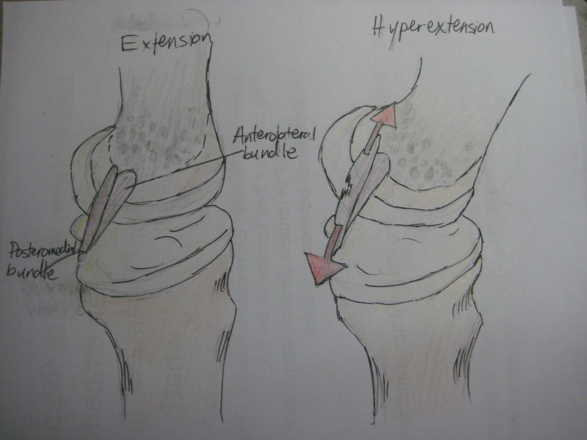
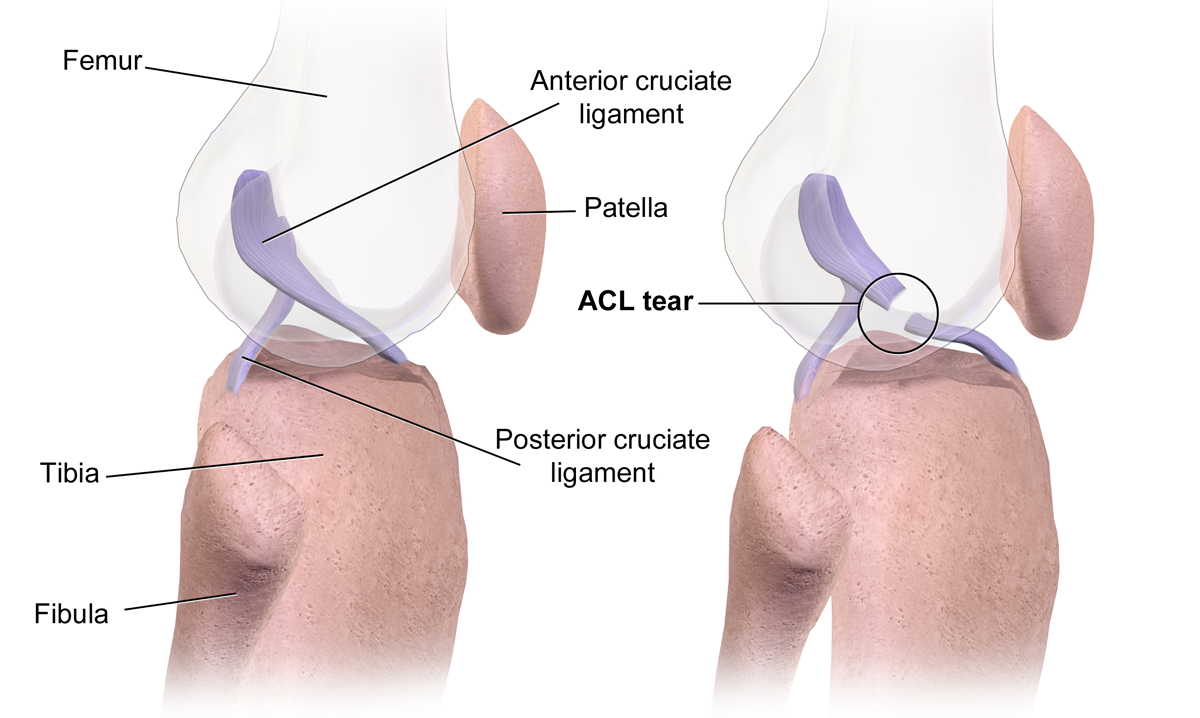
The anterior cruciate ligament (ACL) is one of a pair of cruciate ligaments (the other being the posterior cruciate ligament) in the human knee. The two ligaments are also called "cruciform" ligaments, as they are arranged in a crossed formation. In the quadruped stifle joint (analogous to the knee), based on its anatomical position, it is also referred to as the cranial cruciate ligament. The term cruciate translates to cross. This name is fitting because the ACL crosses the posterior cruciate ligament to form an “X”. It is composed of strong, fibrous material and assists in controlling excessive motion. This is done by limiting mobility of the joint. The anterior cruciate ligament is one of the four main ligaments of the knee, providing 85% of the restraining force to anterior tibial displacement at 30 and 90° of knee flexion. The ACL is the most injured ligament of the four located in the knee. Structure The ACL originates from deep within the notch of the distal femu ... [...More Info...] [...Related Items...] OR: [Wikipedia] [Google] [Baidu] |
Posterior Cruciate Ligament
The posterior cruciate ligament (PCL) is a ligament in each knee of humans and various other animals. It works as a counterpart to the anterior cruciate ligament (ACL). It connects the posterior intercondylar area of the tibia to the medial condyle of the femur. This configuration allows the PCL to resist forces pushing the tibia posteriorly relative to the femur. The PCL and ACL are intracapsular ligaments because they lie deep within the knee joint. They are both isolated from the fluid-filled synovial cavity, with the synovial membrane wrapped around them. The PCL gets its name by attaching to the posterior portion of the tibia. The PCL, ACL, MCL, and LCL are the four main ligaments of the knee in primates. Structure The PCL is located within the knee joint where it stabilizes the articulating bones, particularly the femur and the tibia, during movement. It originates from the lateral edge of the medial femoral condyle and the roof of the intercondyle notch then stretches ... [...More Info...] [...Related Items...] OR: [Wikipedia] [Google] [Baidu] |
Anterior Cruciate Ligament Injury
An anterior cruciate ligament injury occurs when the anterior cruciate ligament (ACL) is either stretched, partially torn, or completely torn. The most common injury is a complete tear. Symptoms include pain, an audible cracking sound during injury, instability of the knee, and joint swelling. Swelling generally appears within a couple of hours. In approximately 50% of cases, other structures of the knee such as surrounding ligaments, cartilage, or meniscus are damaged. The underlying mechanism often involves a rapid change in direction, sudden stop, landing after a jump, or direct contact to the knee. It is more common in athletes, particularly those who participate in alpine skiing, football (soccer), netball, American football, or basketball. Diagnosis is typically made by physical examination and is sometimes supported by magnetic resonance imaging (MRI). Physical examination will often show tenderness around the knee joint, reduced range of motion of the knee, and inc ... [...More Info...] [...Related Items...] OR: [Wikipedia] [Google] [Baidu] |


