|
Charles Alfred Ballance
Sir Charles Alfred Ballance (30 August 1856 – 9 February 1936) was an English surgeon who specialized in the fields of otology and neurotology. Biography Charles Alfred Ballance was the eldest son of Charles and Caroline Ballance (née Pollard). His three brothers all entered the medical profession, the youngest of whom, Sir Hamilton Ashley Ballance, was also a distinguished surgeon. Charles studied at St Thomas' Hospital in London, where he passed his finals in 1881 and became a Master of Surgery the following year. He was appointed Aural Surgeon there in 1888, becoming assistant surgeon in 1891, surgeon in 1900 and consulting surgeon in 1919. For much of his professional life he was associated with St Thomas's and National Hospital, Queen Square in London, where he was appointed consulting surgeon in 1908, and was also made Chief Surgeon of the Metropolitan Police in 1912. During the First World War he worked in Malta, organising and supervising military hospitals with C ... [...More Info...] [...Related Items...] OR: [Wikipedia] [Google] [Baidu] |
Upper Clapton
Clapton is a district of East London, England, in the London Borough of Hackney. Clapton is divided into Upper Clapton, in the north, and Lower Clapton to the south. Clapton railway station lies north-east of Charing Cross. Geography and origins The hamlet of Clapton emerged in the manor and Ancient Parish of Hackney. Origins The hamlet of Clapton was, from 1339 (when first recorded) until the 18th century normally rendered as Clopton, meaning the "farm on the hill". The Old English ''clop'' - "lump" or "hill" - presumably denoted the high ground which rises from the River Lea. Clapton grew up as a linear hamlet along the road subsequently known as Lower and Upper Clapton Road. As the area became urbanised, the extent of the area called Clapton eventually increased to encompass most of the north-eastern quarter of Hackney. Scope Because Clapton has never been an administrative unit, it has never had any defined boundaries, though the E5 postcode area (established in 1917) h ... [...More Info...] [...Related Items...] OR: [Wikipedia] [Google] [Baidu] |
Victor Horsley
Sir Victor Alexander Haden Horsley (14 April 1857 – 16 July 1916) was a British scientist and professor. He was born in Kensington, London. Educated at Cranbrook School, Kent, he studied medicine at University College London and in Berlin, Germany (1881) and, in the same year, started his career as a house surgeon and registrar at the University College Hospital. From 1884 to 1890, Horsley was Professor-Superintendent of the Brown Institute. In 1886, he was appointed as Assistant Professor of Surgery at the National Hospital for Paralysis and Epilepsy, and as a Professor of Pathology (1887–1896) and Professor of Clinical Surgery (1899–1902) at University College London. He was a supporter of women's suffrage and was an opponent of tobacco and alcohol. Personal life Victor Horsley was born in Kensington, London, the son of Rosamund Haden and John Callcott Horsley, R.A. His given names, Victor Alexander, were given to him by Queen Victoria. In 1883, he became engaged t ... [...More Info...] [...Related Items...] OR: [Wikipedia] [Google] [Baidu] |
Cranial Nerve
Cranial nerves are the nerves that emerge directly from the brain (including the brainstem), of which there are conventionally considered twelve pairs. Cranial nerves relay information between the brain and parts of the body, primarily to and from regions of the head and neck, including the special senses of vision, taste, smell, and hearing. The cranial nerves emerge from the central nervous system above the level of the first vertebra of the vertebral column. Each cranial nerve is paired and is present on both sides. There are conventionally twelve pairs of cranial nerves, which are described with Roman numerals I–XII. Some considered there to be thirteen pairs of cranial nerves, including cranial nerve zero. The numbering of the cranial nerves is based on the order in which they emerge from the brain and brainstem, from front to back. The terminal nerves (0), olfactory nerves (I) and optic nerves (II) emerge from the cerebrum, and the remaining ten pairs arise from ... [...More Info...] [...Related Items...] OR: [Wikipedia] [Google] [Baidu] |
Vestibulocochlear Nerve
The vestibulocochlear nerve or auditory vestibular nerve, also known as the eighth cranial nerve, cranial nerve VIII, or simply CN VIII, is a cranial nerve that transmits sound and equilibrium (balance) information from the inner ear to the brain. Through olivocochlear fibers, it also transmits motor and modulatory information from the superior olivary complex in the brainstem to the cochlea. Structure The vestibulocochlear nerve consists mostly of bipolar neurons and splits into two large divisions: the cochlear nerve and the vestibular nerve. Cranial nerve 8, the vestibulocochlear nerve, goes to the middle portion of the brainstem called the pons (which then is largely composed of fibers going to the cerebellum). The 8th cranial nerve runs between the base of the pons and medulla oblongata (the lower portion of the brainstem). This junction between the pons, medulla, and cerebellum that contains the 8th nerve is called the cerebellopontine angle. The vestibulocochlea ... [...More Info...] [...Related Items...] OR: [Wikipedia] [Google] [Baidu] |
Jugular Vein
The jugular veins are veins that take deoxygenated blood from the head back to the heart via the superior vena cava. The internal jugular vein descends next to the internal carotid artery and continues posteriorly to the sternocleidomastoid muscle. Structure and Function There are two sets of jugular veins: external and internal. The left and right external jugular veins drain into the subclavian veins. The internal jugular veins join with the subclavian veins more medially to form the brachiocephalic veins. Finally, the left and right brachiocephalic veins join to form the superior vena cava, which delivers deoxygenated blood to the right atrium of the heart. The Jugular veins help carry blood from the heart to and from the brain. An average human brain weighs about 3 pounds, and gets about 15%-20% of the blood that the heart pumps out. It is important for the brain to get enough blood for many reasons. Th ... [...More Info...] [...Related Items...] OR: [Wikipedia] [Google] [Baidu] |
Ligature (medicine)
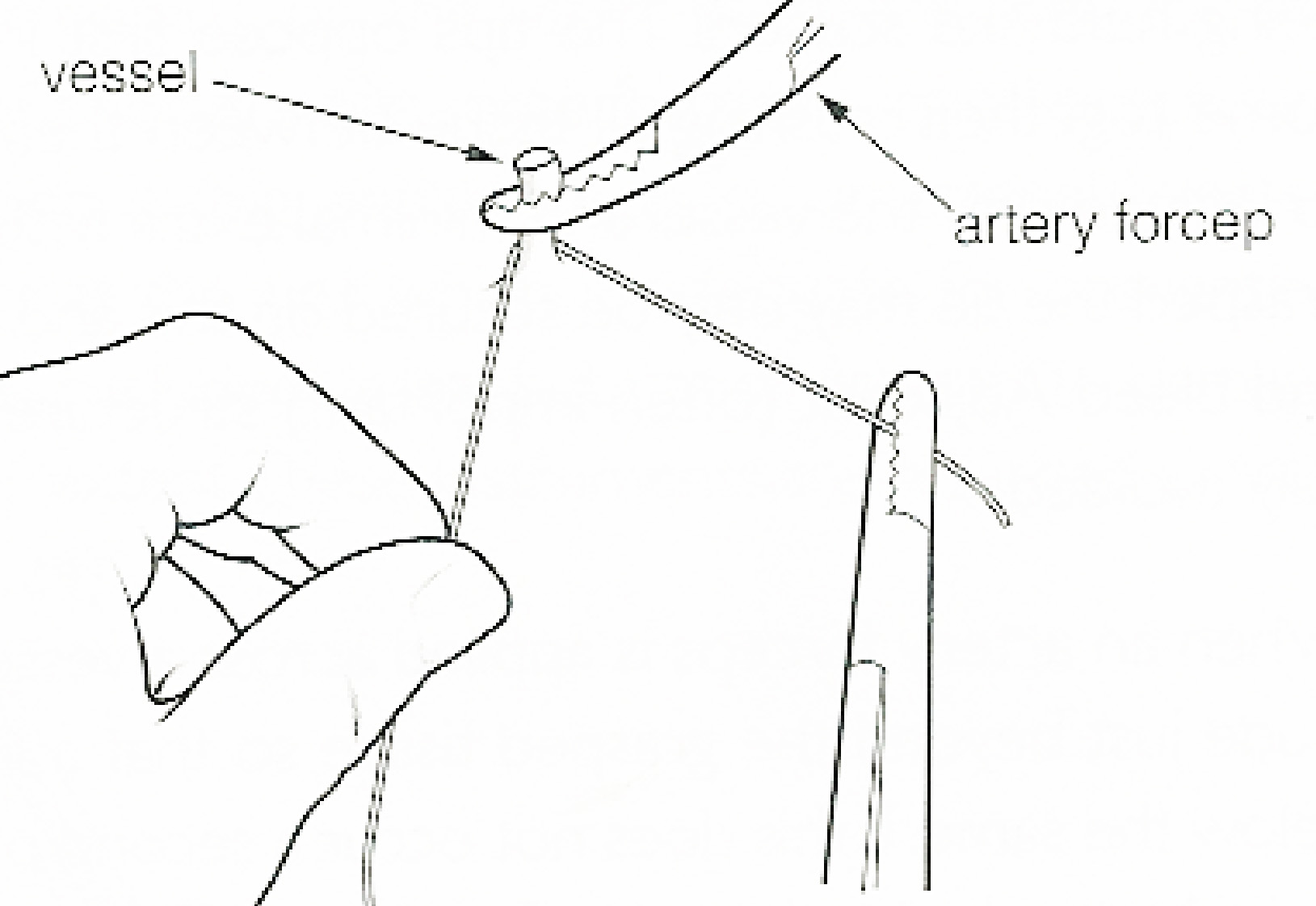
In surgery or medical procedure, a ligature consists of a piece of thread ( suture) tied around an anatomical structure, usually a blood vessel or another hollow structure (e.g. urethra) to shut it off. History The principle of ligation is attributed to Hippocrates and Galen. In ancient Rome, ligatures were used to treat hemorrhoids. The concept of a ligature was reintroduced some 1,500 years later by Ambroise Paré, and finally it found its modern use in 1870–80, made popular by Jules-Émile Péan. Procedure With a blood vessel the surgeon will clamp the vessel perpendicular to the axis of the artery or vein with a hemostat, then secure it by ligating it; i.e. using a piece of suture around it before dividing the structure and releasing the hemostat. It is different from a tourniquet in that the tourniquet will not be secured by knots and it can therefore be released/tightened at will. Ligature is one of the remedies to treat skin tag, or acrochorda. It is done by tying s ... [...More Info...] [...Related Items...] OR: [Wikipedia] [Google] [Baidu] |
Mastoidectomy
A mastoidectomy is a procedure performed to remove the mastoid air cells, air bubbles in the skull, near the inner ears. This can be done as part of treatment for mastoiditis, chronic suppurative otitis media or cholesteatoma. In addition, it is sometimes performed as part of other procedures (cochlear implant) or for access to the middle ear. There are classically 5 different types of mastoidectomy: ;* Radical : Removal of posterior and superior canal wall, meatoplasty and exteriorisation of middle ear. ;* Canal wall down : Removal of posterior and superior canal wall, meatoplasty. Tympanic membrane In the anatomy of humans and various other tetrapods, the eardrum, also called the tympanic membrane or myringa, is a thin, cone-shaped membrane that separates the external ear from the middle ear. Its function is to transmit sound from the ai ... left in place. ;* Canal wall up : Posterior and superior canal wall are kept intact. A facial recess approach is taken. ;* Cortical : ... [...More Info...] [...Related Items...] OR: [Wikipedia] [Google] [Baidu] |
Tumor
A neoplasm () is a type of abnormal and excessive growth of tissue. The process that occurs to form or produce a neoplasm is called neoplasia. The growth of a neoplasm is uncoordinated with that of the normal surrounding tissue, and persists in growing abnormally, even if the original trigger is removed. This abnormal growth usually forms a mass, when it may be called a tumor. ICD-10 classifies neoplasms into four main groups: benign neoplasms, in situ neoplasms, malignant neoplasms, and neoplasms of uncertain or unknown behavior. Malignant neoplasms are also simply known as cancers and are the focus of oncology. Prior to the abnormal growth of tissue, as neoplasia, cells often undergo an abnormal pattern of growth, such as metaplasia or dysplasia. However, metaplasia or dysplasia does not always progress to neoplasia and can occur in other conditions as well. The word is from Ancient Greek 'new' and 'formation, creation'. Types A neoplasm can be benign, potenti ... [...More Info...] [...Related Items...] OR: [Wikipedia] [Google] [Baidu] |
Cerebellopontine Angle
The cerebellopontine angle (CPA) ( la, angulus cerebellopontinus) is located between the cerebellum and the pons. The cerebellopontine angle is the site of the cerebellopontine angle cistern one of the subarachnoid cisterns that contains cerebrospinal fluid, arachnoid tissue, cranial nerves, and associated vessels. The cerebellopontine angle is also the site of a set of neurological disorders known as the cerebellopontine angle syndrome. Structure The cerebellopontine angle is formed by the cerebellopontine fissure. This fissure is made when the cerebellum folds over to the pons, creating a sharply defined angle between them. The angle formed in turn creates a subarachnoid cistern, the cerebellopontine angle cistern. The pia mater follows the outline of the fissure and the arachnoid mater continues across the divide so that the subarachnoid space is dilated at this area, forming the cerebellopontine angle cistern. The anterior inferior cerebellar artery (AICA) is the princ ... [...More Info...] [...Related Items...] OR: [Wikipedia] [Google] [Baidu] |
Anastomosis
An anastomosis (, plural anastomoses) is a connection or opening between two things (especially cavities or passages) that are normally diverging or branching, such as between blood vessels, leaf veins, or streams. Such a connection may be normal (such as the foramen ovale in a fetus's heart) or abnormal (such as the patent foramen ovale in an adult's heart); it may be acquired (such as an arteriovenous fistula) or innate (such as the arteriovenous shunt of a metarteriole); and it may be natural (such as the aforementioned examples) or artificial (such as a surgical anastomosis). The reestablishment of an anastomosis that had become blocked is called a reanastomosis. Anastomoses that are abnormal, whether congenital or acquired, are often called fistulas. The term is used in medicine, biology, mycology, geology, and geography. Etymology Anastomosis: medical or Modern Latin, from Greek ἀναστόμωσις, anastomosis, "outlet, opening", Gr ana- "up, on, upon", stoma "mouth", ... [...More Info...] [...Related Items...] OR: [Wikipedia] [Google] [Baidu] |
Spinal Accessory Nerve
The accessory nerve, also known as the eleventh cranial nerve, cranial nerve XI, or simply CN XI, is a cranial nerve that supplies the sternocleidomastoid and trapezius muscles. It is classified as the eleventh of twelve pairs of cranial nerves because part of it was formerly believed to originate in the brain. The sternocleidomastoid muscle tilts and rotates the head, whereas the trapezius muscle, connecting to the scapula, acts to shrug the shoulder. Traditional descriptions of the accessory nerve divide it into a spinal part and a cranial part. The cranial component rapidly joins the vagus nerve, and there is ongoing debate about whether the cranial part should be considered part of the accessory nerve proper. Consequently, the term "accessory nerve" usually refers only to nerve supplying the sternocleidomastoid and trapezius muscles, also called the spinal accessory nerve. Strength testing of these muscles can be measured during a neurological examination to assess functio ... [...More Info...] [...Related Items...] OR: [Wikipedia] [Google] [Baidu] |


