|
Carotid
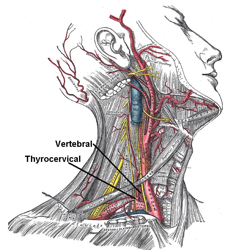
In anatomy, the left and right common carotid arteries (carotids) () are arteries that supply the head and neck with oxygenated blood; they divide in the neck to form the external and internal carotid arteries. Structure The common carotid arteries are present on the left and right sides of the body. These arteries originate from different arteries but follow symmetrical courses. The right common carotid originates in the neck from the brachiocephalic trunk; the left from the aortic arch in the thorax. These split into the external and internal carotid arteries at the upper border of the thyroid cartilage, at around the level of the fourth cervical vertebra. The left common carotid artery can be thought of as having two parts: a thoracic (chest) part and a cervical (neck) part. The right common carotid originates in or close to the neck and contains only a small thoracic portion. There are studies in the bioengineering literature that have looked into characterizing the geo ... [...More Info...] [...Related Items...] OR: [Wikipedia] [Google] [Baidu] |
Internal Carotid Artery
The internal carotid artery is an artery in the neck which supplies the anterior cerebral artery, anterior and middle cerebral artery, middle cerebral circulation. In human anatomy, the internal and external carotid artery, external carotid arise from the common carotid artery, where it bifurcates at cervical vertebrae C3 or C4. The internal carotid artery supplies the brain, including the eyes, while the external carotid nourishes other portions of the head, such as the face, scalp, skull, and meninges. Classification Terminologia Anatomica in 1998 subdivided the artery into four parts: "cervical", "petrous", "cavernous", and "cerebral". In clinical settings, however, usually the classification system of the internal carotid artery follows the 1996 recommendations by Bouthillier, describing seven anatomical segments of the internal carotid artery, each with a corresponding alphanumeric identifier: C1 cervical; C2 petrous; C3 lacerum; C4 cavernous; C5 clinoid; C6 ophthalmic; ... [...More Info...] [...Related Items...] OR: [Wikipedia] [Google] [Baidu] |
External Carotid Artery
The external carotid artery is the major artery of the head and upper neck. It arises from the common carotid artery. It terminates by splitting into the superficial temporal and maxillary artery within the parotid gland. Structure Origin The external carotid artery arises from the common carotid artery just inferior to the upper border of the thyroid cartilage. At its origin, this artery is closer to the skin and more medial than the internal carotid, and is situated within the carotid triangle. Course and fate It curves to pass anterosuperiorly before inclining posterior-ward to reach the space posterior the neck of the mandible, where it divides into the superficial temporal and maxillary artery within the parotid gland. It rapidly diminishes in size as it travels up the neck, owing to the number and large size of its branches. Relations At the origin, external carotid artery is more medial than internal carotid artery. When external carotid artery ascends the ... [...More Info...] [...Related Items...] OR: [Wikipedia] [Google] [Baidu] |
Subclavian Artery
In human anatomy, the subclavian arteries are paired major arteries of the upper thorax, below the clavicle. They receive blood from the aortic arch. The left subclavian artery supplies blood to the left arm and the right subclavian artery supplies blood to the right arm, with some branches supplying the head and thorax. On the left side of the body, the subclavian comes directly off the aortic arch, while on the right side it arises from the relatively short brachiocephalic artery when it bifurcates into the subclavian and the right common carotid artery. The usual branches of the subclavian on both sides of the body are the vertebral artery, the internal thoracic artery, the thyrocervical trunk, the costocervical trunk and the dorsal scapular artery, which may branch off the transverse cervical artery, which is a branch of the thyrocervical trunk. The subclavian becomes the axillary artery at the lateral border of the first rib. Structure From its origin, the subclavian art ... [...More Info...] [...Related Items...] OR: [Wikipedia] [Google] [Baidu] |
Sternohyoid
The sternohyoid muscle is a bilaterally paired, long, thin, narrow strap muscle of the anterior neck. It is one of the infrahyoid muscles. It is innervated by the ansa cervicalis. It acts to depress the hyoid bone. The sternohyoid muscle is a flat muscle located on both sides of the neck, part of the infrahyoid muscle group. It originates from the medial edge of the clavicle, sternoclavicular ligament, and posterior side of the manubrium, and ascends to attach to the body of the hyoid bone. The sternohyoid muscle, along with other infrahyoid muscles, functions to depress the hyoid bone, which is important for activities such as speaking, chewing, and swallowing. Additionally, this muscle group contributes to the protection of the trachea, esophagus, blood vessels, and thyroid gland. The sternohyoid muscle also plays a minor role in head movements. Structure The sternohyoid muscle is one of the paired strap muscles of the infrahyoid muscles. The muscle is directed superome ... [...More Info...] [...Related Items...] OR: [Wikipedia] [Google] [Baidu] |
Pleura
The pleurae (: pleura) are the two flattened closed sacs filled with pleural fluid, each ensheathing each lung and lining their surrounding tissues, locally appearing as two opposing layers of serous membrane separating the lungs from the mediastinum, the inside surfaces of the surrounding chest walls and the diaphragm. Although wrapped onto itself resulting in an apparent double layer, each lung is surrounded by a single, continuous pleural membrane. The portion of the pleura that covers the surface of each lung is often called the visceral pleura. This can lead to some confusion, as the lung is not the only visceral organ covered by the pleura. The pleura typically dips between the lobes of the lung as ''fissures'', and is formed by the invagination of lung buds into each thoracic sac during embryonic development. The portion of the pleura seen as the outer layer covers the chest wall, the diaphragm and the mediastinum and is often also misleadingly called the parieta ... [...More Info...] [...Related Items...] OR: [Wikipedia] [Google] [Baidu] |
Lung
The lungs are the primary Organ (biology), organs of the respiratory system in many animals, including humans. In mammals and most other tetrapods, two lungs are located near the Vertebral column, backbone on either side of the heart. Their function in the respiratory system is to extract oxygen from the atmosphere and transfer it into the bloodstream, and to release carbon dioxide from the bloodstream into the atmosphere, in a process of gas exchange. Respiration is driven by different muscular systems in different species. Mammals, reptiles and birds use their musculoskeletal systems to support and foster breathing. In early tetrapods, air was driven into the lungs by the pharyngeal muscles via buccal pumping, a mechanism still seen in amphibians. In humans, the primary muscle that drives breathing is the Thoracic diaphragm, diaphragm. The lungs also provide airflow that makes Animal communication#Auditory, vocalisation including speech possible. Humans have two lungs, a ri ... [...More Info...] [...Related Items...] OR: [Wikipedia] [Google] [Baidu] |
Brachiocephalic Vein
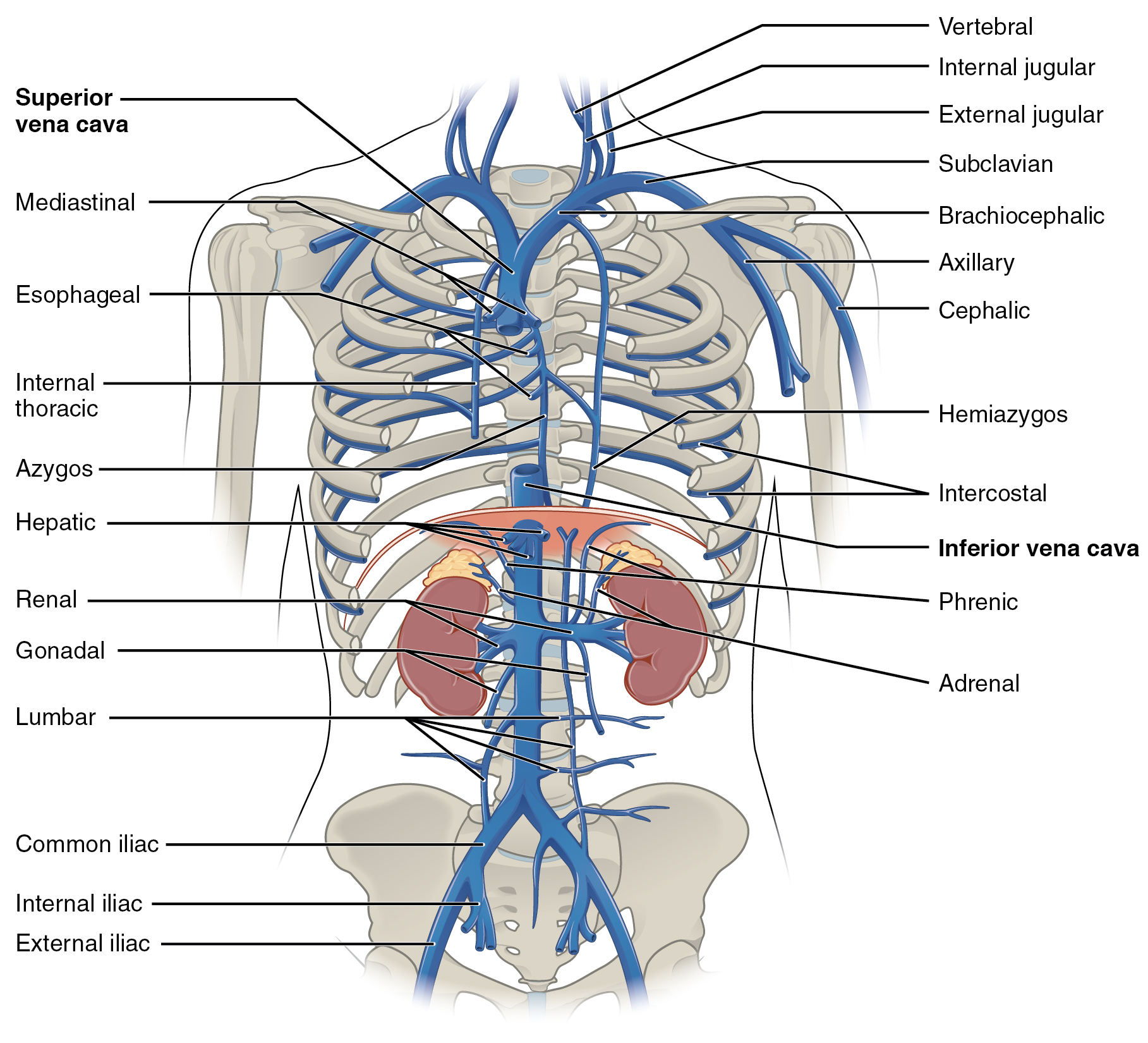
The left and right brachiocephalic veins (previously called innominate veins) are major veins in the Thorax, upper chest, formed by the union of the ipsilateral internal jugular vein and subclavian vein (the so-called venous angle) behind the sternoclavicular joint. The left brachiocephalic vein is more than twice the length of the right brachiocephalic vein. These veins merge to form the superior vena cava, a great vessel, posterior to the junction of the first costal cartilage with the Manubrium, manubrium of the sternum. The brachiocephalic veins are the major veins returning blood to the superior vena cava. Left and right veins Left brachiocephalic vein The left brachiocephalic vein is about 6cm, more than twice the length of the right brachiocephalic vein. and is formed by the confluence of the left subclavian vein, subclavian and left internal jugular veins. In addition the left vein receives drainage from the following tributaries: * The left vertebral vein, internal thor ... [...More Info...] [...Related Items...] OR: [Wikipedia] [Google] [Baidu] |
Thymus
The thymus (: thymuses or thymi) is a specialized primary lymphoid organ of the immune system. Within the thymus, T cells mature. T cells are critical to the adaptive immune system, where the body adapts to specific foreign invaders. The thymus is located in the upper front part of the chest, in the anterior superior mediastinum, behind the sternum, and in front of the heart. It is made up of two lobes, each consisting of a central medulla and an outer cortex, surrounded by a capsule. The thymus is made up of immature T cells called thymocytes, as well as lining cells called epithelial cells which help the thymocytes develop. T cells that successfully develop react appropriately with Major histocompatibility complex, MHC immune receptors of the body (called ''positive selection'') and not against proteins of the body (called ''negative selection''). The thymus is the largest and most active during the neonatal and pre-adolescent periods. By the early teens, the Thymic involuti ... [...More Info...] [...Related Items...] OR: [Wikipedia] [Google] [Baidu] |
Vertebrate Trachea
The trachea (: tracheae or tracheas), also known as the windpipe, is a cartilaginous tube that connects the larynx to the bronchi of the lungs, allowing the passage of air, and so is present in almost all animals' lungs. The trachea extends from the larynx and branches into the two primary bronchi. At the top of the trachea, the cricoid cartilage attaches it to the larynx. The trachea is formed by a number of horseshoe-shaped rings, joined together vertically by overlying ligaments, and by the trachealis muscle at their ends. The epiglottis closes the opening to the larynx during swallowing. The trachea begins to form in the second month of embryo development, becoming longer and more fixed in its position over time. Its epithelium is lined with column-shaped cells that have hair-like extensions called cilia, with scattered goblet cells that produce protective mucins. The trachea can be affected by inflammation or infection, usually as a result of a viral illness affectin ... [...More Info...] [...Related Items...] OR: [Wikipedia] [Google] [Baidu] |
Recurrent Laryngeal Nerve
The recurrent laryngeal nerve (RLN), also known as nervus recurrens, is a branch of the vagus nerve ( cranial nerve X) that supplies all the intrinsic muscles of the larynx, with the exception of the cricothyroid muscles. There are two recurrent laryngeal nerves, right and left. The right and left nerves are not symmetrical, with the left nerve looping under the aortic arch, and the right nerve looping under the right subclavian artery, then traveling upwards. They both travel alongside the trachea. Additionally, the nerves are among the few nerves that follow a ''recurrent'' course, moving in the opposite direction to the nerve they branch from, a fact from which they gain their name. The recurrent laryngeal nerves supply sensation to the larynx below the vocal cords, give cardiac branches to the deep cardiac plexus, and branch to the trachea, esophagus and the inferior constrictor muscles. The posterior cricoarytenoid muscles, the only muscles that can open the vocal fo ... [...More Info...] [...Related Items...] OR: [Wikipedia] [Google] [Baidu] |
Esophagus
The esophagus (American English), oesophagus (British English), or œsophagus (Œ, archaic spelling) (American and British English spelling differences#ae and oe, see spelling difference) all ; : ((o)e)(œ)sophagi or ((o)e)(œ)sophaguses), colloquially known also as the food pipe, food tube, or gullet, is an Organ (anatomy), organ in vertebrates through which food passes, aided by Peristalsis, peristaltic contractions, from the Human pharynx, pharynx to the stomach. The esophagus is a :wiktionary:fibromuscular, fibromuscular tube, about long in adults, that travels behind the trachea and human heart, heart, passes through the Thoracic diaphragm, diaphragm, and empties into the uppermost region of the stomach. During swallowing, the epiglottis tilts backwards to prevent food from going down the larynx and lungs. The word ''esophagus'' is from Ancient Greek οἰσοφάγος (oisophágos), from οἴσω (oísō), future form of φέρω (phérō, "I carry") + ἔφαγον ( ... [...More Info...] [...Related Items...] OR: [Wikipedia] [Google] [Baidu] |


