|
Broad Ligament
The broad ligament of the uterus is the wide fold of peritoneum that connects the sides of the uterus to the walls and floor of the pelvis. Structure Subdivisions Contents The contents of the broad ligament include the following: * Reproductive ** uterine tubes (or fallopian tube) ** ovary (some sources consider the ovary to be on the broad ligament, but not in it.) * vessels ** ovarian artery (in the suspensory ligament) ** uterine artery (in reality, travels in the cardinal ligament) * ligaments ** ovarian ligament ** round ligament of uterus ** suspensory ligament of the ovary (Some sources consider it a part of the broad ligament, while other sources just consider it a "termination" of the ligament.) Relations The peritoneum surrounds the uterus like a flat sheet that folds over its fundus, covering it anteriorly and posteriorly; on the sides of the uterus, this sheet of peritoneum comes in direct contact with itself, forming the double layer of peritoneum known as t ... [...More Info...] [...Related Items...] OR: [Wikipedia] [Google] [Baidu] |
Uterus
The uterus (from Latin ''uterus'', : uteri or uteruses) or womb () is the hollow organ, organ in the reproductive system of most female mammals, including humans, that accommodates the embryonic development, embryonic and prenatal development, fetal development of one or more Fertilized egg, fertilized eggs until birth. The uterus is a hormone-responsive sex organ that contains uterine gland, glands in its endometrium, lining that secrete uterine milk for embryonic nourishment. (The term ''uterus'' is also applied to analogous structures in some non-mammalian animals.) In humans, the lower end of the uterus is a narrow part known as the Uterine isthmus, isthmus that connects to the cervix, the anterior gateway leading to the vagina. The upper end, the body of the uterus, is connected to the fallopian tubes at the uterine horns; the rounded part, the fundus, is above the openings to the fallopian tubes. The connection of the uterine cavity with a fallopian tube is called the utero ... [...More Info...] [...Related Items...] OR: [Wikipedia] [Google] [Baidu] |
Round Ligament Of Uterus
The round ligament of the uterus is a ligament that connects the uterus to the labia majora. It originates at the junction of the uterus and uterine tube. It passes through the inguinal canal to insert at the labium majus. The two round ligaments of uterus develop from the gubernaculum; they are the female homologue of the male gubernaculum testis. Structure The round ligament of the uterus originates at the uterine horns, in the parametrium. The round ligament exits the pelvis via the deep inguinal ring. It passes through the inguinal canal to reach the labium majus, inserting into the fibro-fatty substance of the labium majus. Blood supply The round ligament is supplied by the artery of the round ligament of uterus, also known as ''Sampson's artery''. Development The round ligament develops from the gubernaculum which attaches the gonad to the labioscrotal swellings in the embryo. Function The round ligament of uterus acts to hold the uterus anterior-ward to in ... [...More Info...] [...Related Items...] OR: [Wikipedia] [Google] [Baidu] |
Parametrium
The parametrium is the fibrous and fatty connective tissue that surrounds the uterus. This tissue separates the supravaginal portion of the cervix from the bladder. The parametrium lies in front of the cervix and extends laterally between the layers of the broad ligaments. It connects the uterus to other tissues in the pelvis. It is different from the perimetrium, which is the outermost layer of the uterus. The uterine artery and ovarian ligament are located in the parametrium. An associated form of pelvic inflammatory disease Pelvic inflammatory disease (PID), also known as pelvic inflammatory disorder, is an infection of the upper part of the female reproductive system, mainly the uterus, fallopian tubes, and ovaries, and inside of the pelvis. Often, there may be no ... is inflammation of the parametrium known as parametritis. References * External links Mammal female reproductive system {{genitourinary-stub ... [...More Info...] [...Related Items...] OR: [Wikipedia] [Google] [Baidu] |
Pelvic Diaphragm
The pelvic floor or pelvic diaphragm is an anatomical location in the human body which has an important role in urinary and anal continence, sexual function, and support of the pelvic organs. The pelvic floor includes muscles, both skeletal and smooth, ligaments, and fascia and separates between the pelvic cavity from above, and the perineum from below. It is formed by the levator ani muscle and coccygeus muscle, and associated connective tissue. The pelvic floor has two hiatuses (gaps): (anteriorly) the urogenital hiatus through which urethra and vagina pass, and (posteriorly) the rectal hiatus through which the anal canal passes. Structure Definition Some sources do not consider "pelvic floor" and "pelvic diaphragm" to be identical, with the "diaphragm" consisting of only the levator ani and coccygeus, while the "floor" also includes the perineal membrane and deep perineal pouch. However, other sources include the fascia as part of the diaphragm. In practice, the two ... [...More Info...] [...Related Items...] OR: [Wikipedia] [Google] [Baidu] |
Cardinal Ligament
The cardinal ligament (also transverse cervical ligament, lateral cervical ligament, or Mackenrodt's ligament) is a major ligament of the uterus formed as a thickening of connective tissue of the base of the broad ligament of the uterus. It extends laterally (on either side) from the cervix and vaginal fornix to attach onto the lateral wall of the pelvis. The female ureter, uterine artery, and inferior hypogastric (nervous) plexus course within the cardinal ligament. The cardinal ligament supports the uterus. Structure The cardinal ligament is a paired structure on the lateral side of the uterus. It originates from the lateral part of the cervix. Attachments It attaches the cervix to the lateral pelvic wall by its attachment to the obturator fascia of the obturator internus muscle. It attaches to the uterosacral ligament. Relations It is continuous externally with the fibrous tissue surrounding the pelvic blood vessels. Function The cardinal ligament supports the uter ... [...More Info...] [...Related Items...] OR: [Wikipedia] [Google] [Baidu] |
Epoophoron
The epoophoron or epoöphoron (also called organ of RosenmüllerJ. C. Rosenmüller. De ovariis embryonum et foetuum humanorum. 1802. or the parovarium; : epoophora) is a remnant of the mesonephric duct that can be found next to the ovary and fallopian tube. Anatomy It may contain 10–15 transverse small ducts or tubules that lead to the Gartner's duct (also longitudinal duct of epoophoron) that represents the caudal remnant of the mesonephric ducts and passes through the broad ligament and the lateral wall of the cervix and vagina. The epoophoron is a homologue to the epididymis in the male. While the epoophoron is located in the lateral portion of the mesosalpinx and mesovarium, the paroophoron (residual remnant of that part of the mesonephric duct that forms the paradidymis in the male) lies more medially in the mesosalpinx. Histology It has a unique histological profile. Clinical significance Clinically the organ may give rise to a local paraovarian cyst or adenoma A ... [...More Info...] [...Related Items...] OR: [Wikipedia] [Google] [Baidu] |
Mesentery
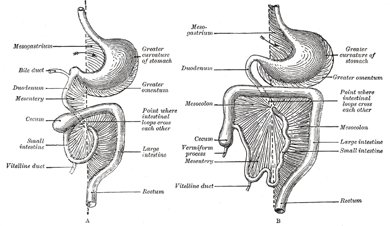
In human anatomy, the mesentery is an Organ (anatomy), organ that attaches the intestines to the posterior abdominal wall, consisting of a double fold of the peritoneum. It helps (among other functions) in storing Adipose tissue, fat and allowing blood vessels, lymphatics, and nerves to supply the intestines. The (the part of the mesentery that attaches the colon to the abdominal wall) was formerly thought to be a fragmented structure, with all named parts—the ascending, transverse, descending, and sigmoid mesocolons, the mesoappendix, and the mesorectum—separately terminating their insertion into the posterior abdominal wall. However, in 2012, new microscopy, microscopic and electron microscope, electron microscopic histology, examinations showed the mesocolon to be a single structure derived from the duodenojejunal flexure and extending to the distal mesorectal layer. Thus the mesentery is an internal organ. Structure The mesentery of the small intestine arises from th ... [...More Info...] [...Related Items...] OR: [Wikipedia] [Google] [Baidu] |
Mesosalpinx
The mesosalpinx is part of the lining of the abdominal cavity in higher vertebrates, specifically the portion of the broad ligament that stretches from the ovary to the level of the fallopian tube The fallopian tubes, also known as uterine tubes, oviducts or salpinges (: salpinx), are paired tubular sex organs in the human female body that stretch from the Ovary, ovaries to the uterus. The fallopian tubes are part of the female reproduct .... See also * Mesometrium * Mesovarium * Salpinx in anatomy References External links * (, ) Mammal female reproductive system {{ligament-stub ... [...More Info...] [...Related Items...] OR: [Wikipedia] [Google] [Baidu] |
Uterine Tube
The fallopian tubes, also known as uterine tubes, oviducts or salpinges (: salpinx), are paired tubular sex organs in the human female body that stretch from the ovaries to the uterus. The fallopian tubes are part of the female reproductive system. In other vertebrates, they are only called oviducts. Each tube is a muscular hollow organ that is on average between in length, with an external diameter of . It has four described parts: the intramural part, isthmus, ampulla, and infundibulum with associated fimbriae. Each tube has two openings: a proximal opening nearest to the uterus, and a distal opening nearest to the ovary. The fallopian tubes are held in place by the mesosalpinx, a part of the broad ligament mesentery that wraps around the tubes. Another part of the broad ligament, the mesovarium suspends the ovaries in place. An egg cell is transported from an ovary to a fallopian tube where it may be fertilized in the ampulla of the tube. The fallopian tubes are lined with ... [...More Info...] [...Related Items...] OR: [Wikipedia] [Google] [Baidu] |
Fundus (uterus)
The uterus (from Latin ''uterus'', : uteri or uteruses) or womb () is the organ in the reproductive system of most female mammals, including humans, that accommodates the embryonic and fetal development of one or more fertilized eggs until birth. The uterus is a hormone-responsive sex organ that contains glands in its lining that secrete uterine milk for embryonic nourishment. (The term ''uterus'' is also applied to analogous structures in some non-mammalian animals.) In humans, the lower end of the uterus is a narrow part known as the isthmus that connects to the cervix, the anterior gateway leading to the vagina. The upper end, the body of the uterus, is connected to the fallopian tubes at the uterine horns; the rounded part, the fundus, is above the openings to the fallopian tubes. The connection of the uterine cavity with a fallopian tube is called the uterotubal junction. The fertilized egg is carried to the uterus along the fallopian tube. It will have divided on its ... [...More Info...] [...Related Items...] OR: [Wikipedia] [Google] [Baidu] |
Suspensory Ligament Of The Ovary
The suspensory ligament of the ovary, also infundibulopelvic ligament (commonly abbreviated IP ligament or simply IP), is a fold of peritoneum that extends out from the ovary to the wall of the pelvis. Some sources consider it a part of the broad ligament of uterus while other sources just consider it a "termination" of the ligament. It is not considered a true ligament in that it does not physically support any anatomical structures; however it is an important landmark and it houses the ovarian vessels. The suspensory ligament is directed upward over the iliac vessels (other), iliac vessels. Structure It contains the ovarian artery, ovarian vein, ovarian plexus, ovarian nerve plexus, at eMedicine Dictionary and lymphatic vessels. ... [...More Info...] [...Related Items...] OR: [Wikipedia] [Google] [Baidu] |
Ovarian Ligament
The ovarian ligament (also called the utero-ovarian ligament or proper ovarian ligament) is a fibrous ligament that connects the ovary to the lateral surface of the uterus. Structure The ovarian ligament is composed of muscular and fibrous tissue; it extends from the uterine extremity of the ovary to the lateral aspect of the uterus, just below the point where the uterine tube and uterus meet. The ligament runs in the broad ligament of the uterus, which is a fold of peritoneum rather than a fibrous ligament. Specifically, it is located in the parametrium. Development Embryologically, each ovary (which forms from the gonadal ridge) is connected to a band of mesoderm, the gubernaculum. This strip of mesoderm remains in connection with the ovary throughout its development, and eventually spans this distance by attachment within the labia majora. During the latter parts of urogenital development, the gubernaculum forms a long fibrous band of connective tissue stretching from th ... [...More Info...] [...Related Items...] OR: [Wikipedia] [Google] [Baidu] |


