|
Cardinal Ligament
The cardinal ligament (also transverse cervical ligament, lateral cervical ligament, or Mackenrodt's ligament) is a major ligament of the uterus formed as a thickening of connective tissue of the base of the broad ligament of the uterus. It extends laterally (on either side) from the cervix and vaginal fornix to attach onto the lateral wall of the pelvis. The female ureter, uterine artery, and inferior hypogastric (nervous) plexus course within the cardinal ligament. The cardinal ligament supports the uterus. Structure The cardinal ligament is a paired structure on the lateral side of the uterus. It originates from the lateral part of the cervix. Attachments It attaches the cervix to the lateral pelvic wall by its attachment to the obturator fascia of the obturator internus muscle. It attaches to the uterosacral ligament. Relations It is continuous externally with the fibrous tissue surrounding the pelvic blood vessels. Function The cardinal ligament supports the uter ... [...More Info...] [...Related Items...] OR: [Wikipedia] [Google] [Baidu] |
Major Ligament Of The Uterus
The uterus (from Latin ''uterus'', : uteri or uteruses) or womb () is the organ in the reproductive system of most female mammals, including humans, that accommodates the embryonic and fetal development of one or more fertilized eggs until birth. The uterus is a hormone-responsive sex organ that contains glands in its lining that secrete uterine milk for embryonic nourishment. (The term ''uterus'' is also applied to analogous structures in some non-mammalian animals.) In humans, the lower end of the uterus is a narrow part known as the isthmus that connects to the cervix, the anterior gateway leading to the vagina. The upper end, the body of the uterus, is connected to the fallopian tubes at the uterine horns; the rounded part, the fundus, is above the openings to the fallopian tubes. The connection of the uterine cavity with a fallopian tube is called the uterotubal junction. The fertilized egg is carried to the uterus along the fallopian tube. It will have divided on its jour ... [...More Info...] [...Related Items...] OR: [Wikipedia] [Google] [Baidu] |
Broad Ligament Of The Uterus
The broad ligament of the uterus is the wide fold of peritoneum that connects the sides of the uterus to the walls and floor of the pelvis. Structure Subdivisions Contents The contents of the broad ligament include the following: * Reproductive ** uterine tubes (or fallopian tube) ** ovary (some sources consider the ovary to be on the broad ligament, but not in it.) * vessels ** ovarian artery (in the suspensory ligament) ** uterine artery (in reality, travels in the cardinal ligament) * ligaments ** ovarian ligament ** round ligament of uterus ** suspensory ligament of the ovary (Some sources consider it a part of the broad ligament, while other sources just consider it a "termination" of the ligament.) Relations The peritoneum surrounds the uterus like a flat sheet that folds over its fundus (uterus), fundus, covering it anteriorly and posteriorly; on the sides of the uterus, this sheet of peritoneum comes in direct contact with itself, forming the double layer of perito ... [...More Info...] [...Related Items...] OR: [Wikipedia] [Google] [Baidu] |
Uterine Artery
The uterine artery is an artery that supplies blood to the uterus in females. Structure The uterine artery usually arises from the anterior division of the internal iliac artery. It travels to the uterus, crossing the ureter anteriorly, to the uterus by traveling in the cardinal ligament. It travels through the parametrium of the inferior broad ligament of the uterus. It commonly anastomoses (connects with) the ovarian artery. The uterine artery is the major blood supply to the uterus and enlarges significantly during pregnancy. Branches and organs supplied * round ligament of the uterus * ovary ("ovarian branches") * uterus ( arcuate vessels) * vagina ( Vaginal branches of uterine artery) * uterine tube ("tubal branch") Anatomical variants Uterine artery can arise from the first branch of inferior gluteal artery. It can also arise as the 2nd or 3rd branch from the inferior gluteal artery. On the other hand, uterine artery can be first branch from internal iliac artery bef ... [...More Info...] [...Related Items...] OR: [Wikipedia] [Google] [Baidu] |
Inferior Hypogastric Plexus
Inferior may refer to: * Inferiority complex * An anatomical term of location * Inferior angle of the scapula, in the human skeleton * ''Inferior'' (book), by Angela Saini * '' The Inferior'', a 2007 novel by Peadar Ó Guilín * Inferior good: economics term for goods that consumers buy less of as they become wealthier (vs "normal goods" where they buy more) See also * Junior (other) {{disambiguation ... [...More Info...] [...Related Items...] OR: [Wikipedia] [Google] [Baidu] |
Uterus
The uterus (from Latin ''uterus'', : uteri or uteruses) or womb () is the hollow organ, organ in the reproductive system of most female mammals, including humans, that accommodates the embryonic development, embryonic and prenatal development, fetal development of one or more Fertilized egg, fertilized eggs until birth. The uterus is a hormone-responsive sex organ that contains uterine gland, glands in its endometrium, lining that secrete uterine milk for embryonic nourishment. (The term ''uterus'' is also applied to analogous structures in some non-mammalian animals.) In humans, the lower end of the uterus is a narrow part known as the Uterine isthmus, isthmus that connects to the cervix, the anterior gateway leading to the vagina. The upper end, the body of the uterus, is connected to the fallopian tubes at the uterine horns; the rounded part, the fundus, is above the openings to the fallopian tubes. The connection of the uterine cavity with a fallopian tube is called the utero ... [...More Info...] [...Related Items...] OR: [Wikipedia] [Google] [Baidu] |
Cervix
The cervix (: cervices) or cervix uteri is a dynamic fibromuscular sexual organ of the female reproductive system that connects the vagina with the uterine cavity. The human female cervix has been documented anatomically since at least the time of Hippocrates, over 2,000 years ago. The cervix is approximately 4 cm long with a diameter of approximately 3 cm and tends to be described as a cylindrical shape, although the front and back walls of the cervix are contiguous. The size of the cervix changes throughout a woman's life cycle. For example, women in the fertile years of their reproductive cycle tend to have larger cervixes than postmenopausal women; likewise, women who have produced offspring have a larger cervix than those who have not. In relation to the vagina, the part of the cervix that opens to the uterus is called the ''internal os'' and the opening of the cervix in the vagina is called the ''external os''. Between them is a conduit commonly called the cervic ... [...More Info...] [...Related Items...] OR: [Wikipedia] [Google] [Baidu] |
Obturator Fascia
The obturator fascia, or fascia of the internal obturator muscle, covers the pelvic surface of that muscle and is attached around the margin of its origin. Above, it is loosely connected to the back part of the arcuate line, and here it is continuous with the iliac fascia. In front of this, as it follows the line of origin of the internal obturator, it gradually separates from the iliac fascia and the continuity between the two is retained only through the periosteum. It arches beneath the obturator vessels and nerve, completing the obturator canal, and at the front of the pelvis is attached to the back of the superior ramus of the pubis. Below, the obturator fascia is attached to the falciform process of the sacrotuberous ligament and to the pubic arch, where it becomes continuous with the superior fascia of the urogenital diaphragm. Behind, it is prolonged into the gluteal region. The internal pudendal vessels and pudendal nerve cross the pelvic surface of the intern ... [...More Info...] [...Related Items...] OR: [Wikipedia] [Google] [Baidu] |
Obturator Internus Muscle
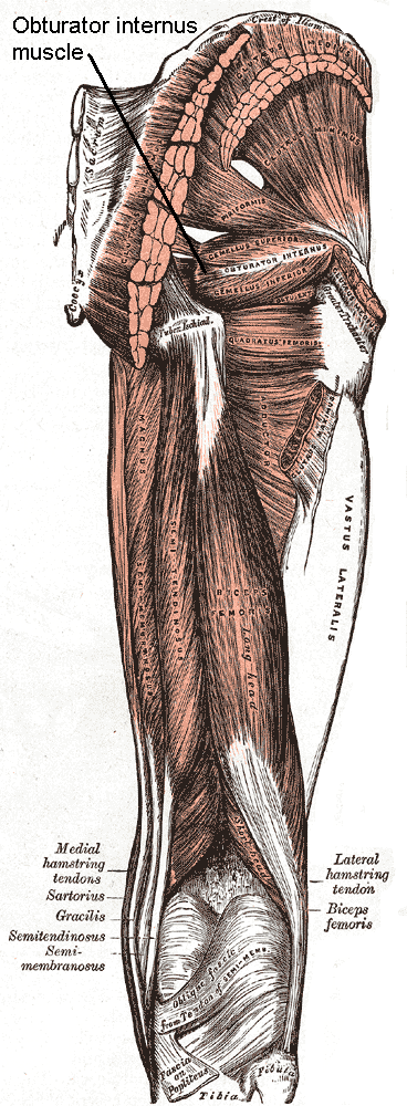
The internal obturator muscle or obturator internus muscle originates on the medial surface of the obturator membrane, the ischium near the membrane, and the rim of the pubis. It exits the pelvic cavity through the lesser sciatic foramen. The internal obturator is situated partly within the lesser pelvis, and partly at the back of the hip-joint. It functions to help laterally rotate femur with hip extension and abduct femur with hip flexion, as well as to steady the femoral head in the acetabulum. Structure Origin The internal obturator muscle arises from the inner surface of the antero-lateral wall of the pelvis. It surrounds the obturator foramen. It is attached to the inferior pubic ramus and ischium, and at the side to the inner surface of the hip bone below and behind the pelvic brim. It reaches from the upper part of the greater sciatic foramen above and behind to the obturator foramen below and in front. It also arises from the pelvic surface of the obturator mem ... [...More Info...] [...Related Items...] OR: [Wikipedia] [Google] [Baidu] |
Uterosacral Ligament
The uterosacral ligaments (or rectouterine ligaments) are major ligaments of uterus that extend posterior-ward from the cervix to attach onto the (anterior aspect of the) sacrum The sacrum (: sacra or sacrums), in human anatomy, is a triangular bone at the base of the spine that forms by the fusing of the sacral vertebrae (S1S5) between ages 18 and 30. The sacrum situates at the upper, back part of the pelvic cavity, .... Structure Microanatomy/histology The uterosacral ligaments consist of fibrous connective tissue, and smooth muscle tissue. Relations The uterosacral ligaments pass inferior to the peritoneum. They embrace the rectouterine pouch, and rectum. The pelvic splanchnic nerves run on top of the ligament. Function The uterosacral ligaments pull the cervix posterior-ward, counteracting the anterior-ward pull exerted by the round ligament of uterus upon the fundus of the uterus, thus maintaining anteversion of the body of the uterus. Clinical signi ... [...More Info...] [...Related Items...] OR: [Wikipedia] [Google] [Baidu] |
Hysterectomy
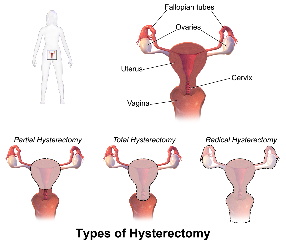
Hysterectomy is the surgical removal of the uterus and cervix. Supracervical hysterectomy refers to removal of the uterus while the cervix is spared. These procedures may also involve removal of the ovaries (oophorectomy), fallopian tubes ( salpingectomy), and other surrounding structures. The term “partial” or “total” hysterectomy are lay-terms that incorrectly describe the addition or omission of oophorectomy at the time of hysterectomy. These procedures are usually performed by a gynecologist. Removal of the uterus is a form of sterilization, rendering the patient unable to bear children (as does removal of ovaries and fallopian tubes) and has surgical risks as well as long-term effects, so the surgery is normally recommended only when other treatment options are not available or have failed. It is the second most commonly performed gynecological surgical procedure, after cesarean section, in the United States. Nearly 68 percent were performed for conditions such as ... [...More Info...] [...Related Items...] OR: [Wikipedia] [Google] [Baidu] |
Pelvic Splanchnic Nerves
Pelvic splanchnic nerves or nervi erigentes are splanchnic nerves that arise from sacral spinal nerves S2, S3, S4 to provide parasympathetic innervation to the organs of the pelvic cavity. Structure The pelvic splanchnic nerves arise from the anterior rami of the sacral spinal nerves S2, S3, and S4, and enter the sacral plexus. They travel to their side's corresponding inferior hypogastric plexus, located bilaterally on the walls of the rectum. They contain both preganglionic parasympathetic fibers as well as visceral afferent fibers. Visceral afferent fibers go to spinal cord following pathway of pelvic splanchnic nerve fibers. The parasympathetic nervous system is referred to as the ''craniosacral outflow''; the pelvic splanchnic nerves are the ''sacral'' component. They are in the same region as the sacral splanchnic nerves, which arise from the sympathetic trunk and provide sympathetic efferent fibers. Function The pelvic splanchnic nerves contribute to the inner ... [...More Info...] [...Related Items...] OR: [Wikipedia] [Google] [Baidu] |
Mammal Female Reproductive System
Most mammals are viviparous, giving birth to live young. However, the five species of monotreme, the platypuses and the echidnas, lay eggs. The monotremes have a sex determination system different from that of most other mammals. In particular, the sex chromosomes of a platypus are more like those of a chicken than those of a therian mammal. The mammary glands of mammals are specialized to produce milk, a liquid used by newborns as their primary source of nutrition. The monotremes branched early from other mammals and do not have the teats seen in most mammals, but they do have mammary glands. The young lick the milk from a mammary patch on the mother's belly. Viviparous mammals are in the subclass Theria; those living today are in the Marsupialia and Placentalia infraclasses. A marsupial has a short gestation period, typically shorter than its estrous cycle, and gives birth to an underdeveloped (altricial) newborn that then undergoes further development; in many species, this ... [...More Info...] [...Related Items...] OR: [Wikipedia] [Google] [Baidu] |


