|
Brenner Tumor
Brenner tumours are an uncommon subtype of the Surface epithelial-stromal tumor, surface epithelial-stromal tumour group of Ovarian cancer, ovarian neoplasms. The majority are benign, but some can be malignant. They are most frequently found incidentally on pelvic examination or at laparotomy. Brenner tumours very rarely can occur in other locations, including the testes. Presentation On gross pathology, pathological examination, they are solid, sharply circumscribed and pale yellow-tan in colour. 90% are unilateral (arising in one ovary, the other is unaffected). The tumours can vary in size from less than to . Borderline and malignant Brenner tumours are possible but each are rare. Diagnosis Histology, Histologically, there are nests of transitional epithelial (urothelial) cells with longitudinal nuclear grooves (coffee bean nuclei) lying in abundant fibrous stroma. The coffee bean nuclei are the nuclear grooves exceptionally pathognomonic to the sex cord stromal tumour, ... [...More Info...] [...Related Items...] OR: [Wikipedia] [Google] [Baidu] |
Gross Examination
Gross processing, "grossing" or "gross pathology" is the process by which pathology specimens undergo examination with the bare eye to obtain diagnosis, diagnostic information, as well as cutting and tissue sampling in order to prepare material for subsequent histopathology, microscopic ''examination.'' Responsibility Gross examination of surgical pathology, surgical specimens is typically performed by a pathology, pathologist, or by a pathologists' assistant working within a pathology practice. Individuals trained in these fields are often able to gather diagnostically critical information in this stage of processing, including the stage and margin status of surgically removed tumors. Steps The initial step in any examination of a clinical specimen is confirmation of the identity of the patient and the anatomy, anatomical site from which the specimen was obtained. Sufficient clinical data should be communicated by the clinical team to the pathology team in order to guide the app ... [...More Info...] [...Related Items...] OR: [Wikipedia] [Google] [Baidu] |
Transitional Epithelial
Transitional epithelium is a type of stratified epithelium. Transitional epithelium is a type of tissue that changes shape in response to stretching (stretchable epithelium). The transitional epithelium usually appears cuboidal when relaxed and squamous when stretched. This tissue consists of multiple layers of epithelial cells which can contract and expand in order to adapt to the degree of distension needed. Transitional epithelium lines the organs of the urinary system and is known here as urothelium (: urothelia). The bladder, for example, has a need for great distension. Structure The appearance of transitional epithelium differs according to its cell layer. Cells of the basal layer are cuboidal (cube-shaped), or columnar (column-shaped), while the cells of the superficial layer vary in appearance depending on the degree of distension. These cells appear to be cuboidal with a domed apex when the organ or the tube in which they reside is not stretched. When the organ or tube ... [...More Info...] [...Related Items...] OR: [Wikipedia] [Google] [Baidu] |
General Electric
General Electric Company (GE) was an American Multinational corporation, multinational Conglomerate (company), conglomerate founded in 1892, incorporated in the New York (state), state of New York and headquartered in Boston. Over the years, the company had multiple divisions, including GE Aerospace, aerospace, GE Power, energy, GE HealthCare, healthcare, lighting, locomotives, appliances, and GE Capital, finance. In 2020, GE ranked among the Fortune 500, ''Fortune'' 500 as the 33rd largest firm in the United States by gross revenue. In 2023, the company was ranked 64th in the Forbes Global 2000, ''Forbes'' Global 2000. In 2011, GE ranked among the Fortune 20 as the 14th most profitable company, but later very severely underperformed the market (by about 75%) as its profitability collapsed. Two employees of GE—Irving Langmuir (1932) and Ivar Giaever (1973)—have been awarded the Nobel Prize. From 1986 until 2013, GE was the owner of the NBC television network through its ... [...More Info...] [...Related Items...] OR: [Wikipedia] [Google] [Baidu] |
H&E Stain
Hematoxylin and eosin stain ( or haematoxylin and eosin stain or hematoxylin–eosin stain; often abbreviated as H&E stain or HE stain) is one of the principal tissue stains used in histology. It is the most widely used stain in medical diagnosis and is often the ''gold standard.'' For example, when a pathologist looks at a biopsy of a suspected cancer, the histological section is likely to be stained with H&E. H&E is the combination of two histological stains: hematoxylin and eosin. The hematoxylin stains cell nuclei a purplish blue, and eosin stains the extracellular matrix and cytoplasm pink, with other structures taking on different shades, hues, and combinations of these colors. Hence a pathologist can easily differentiate between the nuclear and cytoplasmic parts of a cell, and additionally, the overall patterns of coloration from the stain show the general layout and distribution of cells and provides a general overview of a tissue sample's structure. Thus, patte ... [...More Info...] [...Related Items...] OR: [Wikipedia] [Google] [Baidu] |
Walthard Cell Nest
Walthard cell rests, sometimes called Walthard cell nests, are a benign cluster of epithelial cells most commonly found in the connective tissue of the fallopian tubes, but also seen in the mesovarium, mesosalpinx and ovarian hilus. Appearance They appear as white/yellow cysts or nodules that can reach a size of 2 millimeters. They typically have elliptical nuclei with a long groove (along the major axis) – so-called "coffee bean" nuclei. Pathology It has been suggested that these cell rests are the histogenetic origins of Brenner tumors, due to the histological similarity of the epithelium of Walthard cell rests and Brenner tumors to the urothelium of the lower urinary tract. Also, it has been proposed that Brenner tumors and Walthard cell rests signify urothelial differentiation within the female genital tract. Eponym They are named after Swiss gynecologist Max Walthard (1867–1933), who provided a comprehensive description of them in 1903. Additional images Image:Bren ... [...More Info...] [...Related Items...] OR: [Wikipedia] [Google] [Baidu] |
Micrograph
A micrograph is an image, captured photographically or digitally, taken through a microscope or similar device to show a magnify, magnified image of an object. This is opposed to a macrograph or photomacrograph, an image which is also taken on a microscope but is only slightly magnified, usually less than 10 times. Micrography is the practice or art of using microscopes to make photographs. A photographic micrograph is a photomicrograph, and one taken with an electron microscope is an electron micrograph. A micrograph contains extensive details of microstructure. A wealth of information can be obtained from a simple micrograph like behavior of the material under different conditions, the phases found in the system, failure analysis, grain size estimation, elemental analysis and so on. Micrographs are widely used in all fields of microscopy. Types Photomicrograph A light micrograph or photomicrograph is a micrograph prepared using an optical microscope, a process referred to ... [...More Info...] [...Related Items...] OR: [Wikipedia] [Google] [Baidu] |
Robert Meyer (pathologist)
Robert Meyer (11 January 1864, in Hanover – 12 December 1947, in Minneapolis) was a German pathologist. He studied medicine at the universities of Leipzig, Heidelberg and Strassburg, receiving his doctorate at the latter institution in 1889. From 1890 to 1894 he was a medical practitioner in the community of Dedeleben, and afterwards worked as assistant to gynecologist Johann Veit in Berlin.Menghin - Pötel / edited by Rudolf Vierhaus Deutsche Biographische Enzyklopaedie at From 1909 to 1911 he was he ... [...More Info...] [...Related Items...] OR: [Wikipedia] [Google] [Baidu] |
Fritz Brenner (pathologist)
Fritz Brenner (16 December 1877, in Osthofen – 26 December 1969, in Johannesburg) was a German physician and pathologist. He studied medicine at the universities of Strasbourg, Freiburg and Heidelberg, receiving his doctorate at the latter institution in 1904. Following graduation, he worked as an assistant under Eugen Albrecht at the Senckenberg Institute of Pathology in Frankfurt am Main. In 1910 he relocated to German South-West Africa, where he worked as a doctor at the seaport of Swakopmund. Later on, he was a physician in Windhoek (from 1922) and Johannesburg (from 1935). January/February 2007 - Volume 13 - Issue 1 > Powell's Pearls: Fritz Brenner, MD (1877–1969)Female Pelvic Medicine & Reconstructive Surgery In 1907 he described a unique ovarian tumour in an article titled ''Das Oophoroma folliculare'', published in the journal ''Frankfurter Zeitschrift für Pathologie''. In 1932 Berlin pathologist Robert Meyer (1864–1947) coined the term "Brenner tumour Bre ... [...More Info...] [...Related Items...] OR: [Wikipedia] [Google] [Baidu] |
Reinke Crystals
Reinke crystals are rod-like cytoplasmic inclusions which can be found in Leydig cells of the testes. Occurring only in adult humans and wild bush rats, their function is unknown. Ovarian stromal tumors having a predominant pattern of fibroma or thecoma but also containing cells typical of steroid hormone-secreting cells were reported. Some of the tumors were classified as luteinized thecomas because the steroid cells resembled lutein cells and lacked crystalloids of Reinke. Others were classified as stromal Leydig cell tumors as seen in tumors of the testes because Reinke crystalloids were identified in the steroid cells. The stromal Leydig tumors occurred at an average age of 61 years and were associated with ovarian hyperandrogenism which led to virilization in some cases, endometrial hyperplasia in other cases, and endometrial hyperplasia with carcinoma in the rest of the cases. Luteinized thecomas and stromal Leydig cell tumors are indistinguishable except for the presence of c ... [...More Info...] [...Related Items...] OR: [Wikipedia] [Google] [Baidu] |
Leydig Cell Tumour
Leydig cell tumour, also Leydig cell tumor (US spelling), (testicular) interstitial cell tumour and (testicular) interstitial cell tumor (US spelling), is a member of the sex cord-stromal tumour group of ovarian and testicular cancers. It arises from Leydig cells. While the tumour can occur at any age, it occurs most often in young adults. However, in women it tends to happen after menopapuse. A Sertoli–Leydig cell tumour is a combination of a Leydig cell tumour and a Sertoli cell tumour from Sertoli cells. Presentation The majority of Leydig cell tumors are found in males, usually at 5–10 years of age or in middle adulthood (30–60 years). Children typically present with precocious puberty. In men, testicular swelling is the most common presenting feature. Other symptoms depend on age and the type of tumour. If it is secreting androgens the tumour is usually asymptomatic, but can cause precocious puberty in pre-pubertal boys. If the tumour secretes oestrogens it can cause ... [...More Info...] [...Related Items...] OR: [Wikipedia] [Google] [Baidu] |
Call–Exner Bodies
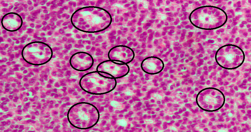
Call–Exner bodies, giving a follicle-like appearance, are small eosinophilic fluid-filled punched out spaces between granulosa cells. The granulosa cells are usually arranged haphazardly around the space. They are pathognomonic for granulosa cell tumors. Histologically, these tumors consists of monotonous islands of granulosa cells with "coffee-bean" nuclei. That same nuclear groove appearance noted in Brenner tumour, an epithelial-stromal ovarian tumor distinguishable by nests of transitional epithelial cells (urothelial) with longitudinal nuclear grooves (coffee bean nuclei) in abundant fibrous stroma. They are composed of membrane-packaged secretion of granulosa cells and have relations to the formation of liquor folliculi which are seen among closely arranged granulosa cells. They are named for Emma Louise Call (1847–1937), an American physician, and Sigmund Exner (1846–1926), an Austrian physiologist.Emma Louise Call and Sigmund Exner: ''Zur Kenntniss des Graafschen ... [...More Info...] [...Related Items...] OR: [Wikipedia] [Google] [Baidu] |


