|
Blebbistatin
Blebbistatin is a myosin inhibitor mostly specific for myosin II. It is widely used in research to inhibit heart muscle myosin, non-muscle myosin II, and skeletal muscle myosin. Blebbistatin has been especially useful in optical mapping of the heart, and its recent use in cardiac muscle cell cultures has improved cell survival time. However, its adverse characteristics e.g. its cytotoxicity and blue-light instability or low solubility in water often make its application challenging. Recently its applicability was improved by chemical design and its derivatives overcome the limitations of blebbistatin. E.g. para-nitroblebbistatin and para-aminoblebbistatin are photostable, and they are neither cytotoxic nor fluorescent. Mode of action and biological effects Blebbistatin inhibits myosin ATPase activity and this way acto-myosin based motility. It binds halfway between the nucleotide binding pocket and the actin binding cleft of myosin, predominantly in an actin detached conformat ... [...More Info...] [...Related Items...] OR: [Wikipedia] [Google] [Baidu] |
Bleb (cell Biology)
In cell biology, a bleb is a bulge of the plasma membrane of a cell, characterized by a spherical, bulky morphology. It is characterized by the decoupling of the cytoskeleton from the plasma membrane, degrading the internal structure of the cell, allowing the flexibility required for the cell to separate into individual bulges or pockets of the intercellular matrix. Most commonly, blebs are seen in apoptosis (programmed cell death) but are also seen in other non-apoptotic functions. ''Blebbing'', or ''zeiosis'', is the formation of blebs. Formation Initiation and expansion Bleb growth is driven by intracellular pressure generated in the cytoplasm when the actin cortex undergoes actomyosin contractions. The disruption of the membrane-actin cortex interactions are dependent on the activity of myosin-ATPase Bleb initiation is affected by three main factors: high intracellular pressure, decreased amounts of cortex-membrane linker proteins, and deterioration of the actin cortex. T ... [...More Info...] [...Related Items...] OR: [Wikipedia] [Google] [Baidu] |
Para-Aminoblebbistatin
''para''-Aminoblebbistatin is a water-soluble, non-fluorescent, photostable myosin II inhibitor, developed from blebbistatin. Among the several blebbistatin derivatives it is one of the most promising for research applications. Furthermore, it has a favourable overall profile considering inhibitory properties and ADME ADME is an abbreviation in pharmacokinetics and pharmacology for " absorption, distribution, metabolism, and excretion", and describes the disposition of a pharmaceutical compound within an organism. The four criteria all influence the drug l ... calculations. Myosin specificity References {{DEFAULTSORT:Aminoblebbistatin, para- Anilines Pyrroloquinolines Acyloins Tertiary alcohols ... [...More Info...] [...Related Items...] OR: [Wikipedia] [Google] [Baidu] |
Para-aminoblebbistatin 2D Structure
''para''-Aminoblebbistatin is a water-soluble, non-fluorescent, photostable myosin II inhibitor, developed from blebbistatin. Among the several blebbistatin derivatives it is one of the most promising for research applications. Furthermore, it has a favourable overall profile considering inhibitory properties and ADME ADME is an abbreviation in pharmacokinetics and pharmacology for " absorption, distribution, metabolism, and excretion", and describes the disposition of a pharmaceutical compound within an organism. The four criteria all influence the drug le ... calculations. Myosin specificity References {{DEFAULTSORT:Aminoblebbistatin, para- Anilines Pyrroloquinolines Acyloins Tertiary alcohols ... [...More Info...] [...Related Items...] OR: [Wikipedia] [Google] [Baidu] |
Myosin
Myosins () are a superfamily of motor proteins best known for their roles in muscle contraction and in a wide range of other motility processes in eukaryotes. They are ATP-dependent and responsible for actin-based motility. The first myosin (M2) to be discovered was in 1864 by Wilhelm Kühne. Kühne had extracted a viscous protein from skeletal muscle that he held responsible for keeping the tension state in muscle. He called this protein ''myosin''. The term has been extended to include a group of similar ATPases found in the cells of both striated muscle tissue and smooth muscle tissue. Following the discovery in 1973 of enzymes with myosin-like function in ''Acanthamoeba castellanii'', a global range of divergent myosin genes have been discovered throughout the realm of eukaryotes. Although myosin was originally thought to be restricted to muscle cells (hence '' myo-''(s) + '' -in''), there is no single "myosin"; rather it is a very large superfamily of genes whose prote ... [...More Info...] [...Related Items...] OR: [Wikipedia] [Google] [Baidu] |
Dictyostelium Discoideum
''Dictyostelium discoideum'' is a species of soil-dwelling amoeba belonging to the phylum Amoebozoa, infraphylum Mycetozoa. Commonly referred to as slime mold, ''D. discoideum'' is a eukaryote that transitions from a collection of unicellular amoebae into a multicellular slug and then into a fruiting body within its lifetime. Its unique asexual lifecycle consists of four stages: vegetative, aggregation, migration, and culmination. The lifecycle of ''D. discoideum'' is relatively short, which allows for timely viewing of all stages. The cells involved in the lifecycle undergo movement, chemical signaling, and development, which are applicable to human cancer research. The simplicity of its lifecycle makes ''D. discoideum'' a valuable model organism to study genetic, cellular, and biochemical processes in other organisms. Natural habitat and diet In the wild, ''D. discoideum'' can be found in soil and moist leaf litter. Its primary diet consists of bacteria, such as '' Escheric ... [...More Info...] [...Related Items...] OR: [Wikipedia] [Google] [Baidu] |
Structure–activity Relationship
The structure–activity relationship (SAR) is the relationship between the chemical structure of a molecule and its biological activity. This idea was first presented by Crum-Brown and Fraser in 1865. The analysis of SAR enables the determination of the chemical group responsible for evoking a target biological effect in the organism. This allows modification of the effect or the potency of a bioactive compound (typically a drug) by changing its chemical structure. Medicinal chemists use the techniques of chemical synthesis to insert new chemical groups into the biomedical compound and test the modifications for their biological effects. This method was refined to build mathematical relationships between the chemical structure and the biological activity, known as quantitative structure–activity relationships (QSAR). A related term is structure affinity relationship (SAFIR). Structure-biodegradability relationship The large number of synthetic organic chemicals currently in p ... [...More Info...] [...Related Items...] OR: [Wikipedia] [Google] [Baidu] |
Two-photon Excitation Microscopy
Two-photon excitation microscopy (TPEF or 2PEF) is a fluorescence imaging technique that allows imaging of living tissue up to about one millimeter in thickness, with 0.64 μm lateral and 3.35 μm axial spatial resolution. Unlike traditional fluorescence microscopy, in which the excitation wavelength is shorter than the emission wavelength, two-photon excitation requires simultaneous excitation by two photons with longer wavelength than the emitted light. Two-photon excitation microscopy typically uses near-infrared (NIR) excitation light which can also excite fluorescent dyes. However, for each excitation, two photons of NIR light are absorbed. Using infrared light minimizes scattering in the tissue. Due to the multiphoton absorption, the background signal is strongly suppressed. Both effects lead to an increased penetration depth for this technique. Two-photon excitation can be a superior alternative to confocal microscopy due to its deeper tissue penetration, efficient light d ... [...More Info...] [...Related Items...] OR: [Wikipedia] [Google] [Baidu] |
Photoaffinity Labeling
Photoaffinity labeling is a chemoproteomics technique used to attach "labels" to the active site of a large molecule, especially a protein. The "label" attaches to the molecule loosely and reversibly, and has an inactive site which can be converted using photolysis into a highly reactive form, which causes the label to bind more permanently to the large molecule via a covalent bond. The technique was first described in the 1970s. Molecules that have been used as labels in this process are often analogs of complex molecules, in which certain functional groups are replaced with a photoreactive group, such as an azide, a diazirine or a benzophenone Benzophenone is the organic compound with the formula (C6H5)2CO, generally abbreviated Ph2CO. It is a white solid that is soluble in organic solvents. Benzophenone is a widely used building block in organic chemistry, being the parent diarylke .... References {{Reflist Molecular biology techniques ... [...More Info...] [...Related Items...] OR: [Wikipedia] [Google] [Baidu] |
Cnidaria
Cnidaria () is a phylum under kingdom Animalia containing over 11,000 species of aquatic animals found both in Fresh water, freshwater and Marine habitats, marine environments, predominantly the latter. Their distinguishing feature is cnidocytes, specialized cells that they use mainly for capturing prey. Their bodies consist of mesoglea, a non-living jelly-like substance, sandwiched between two layers of epithelium that are mostly one cell (biology), cell thick. Cnidarians mostly have two basic body forms: swimming Medusa (biology), medusae and Sessility (motility), sessile polyp (zoology), polyps, both of which are Symmetry (biology)#Radial symmetry, radially symmetrical with mouths surrounded by tentacles that bear cnidocytes. Both forms have a single Body orifice, orifice and body cavity that are used for digestion and respiration (physiology), respiration. Many cnidarian species produce Colony (biology), colonies that are single organisms composed of medusa-like or polyp (z ... [...More Info...] [...Related Items...] OR: [Wikipedia] [Google] [Baidu] |
Caenorhabditis Elegans
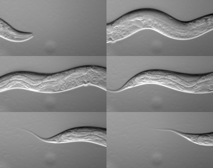
''Caenorhabditis elegans'' () is a free-living transparent nematode about 1 mm in length that lives in temperate soil environments. It is the type species of its genus. The name is a blend of the Greek ''caeno-'' (recent), ''rhabditis'' (rod-like) and Latin ''elegans'' (elegant). In 1900, Maupas initially named it ''Rhabditides elegans.'' Osche placed it in the subgenus ''Caenorhabditis'' in 1952, and in 1955, Dougherty raised ''Caenorhabditis'' to the status of genus. ''C. elegans'' is an unsegmented pseudocoelomate and lacks respiratory or circulatory systems. Most of these nematodes are hermaphrodites and a few are males. Males have specialised tails for mating that include spicules. In 1963, Sydney Brenner proposed research into ''C. elegans,'' primarily in the area of neuronal development. In 1974, he began research into the molecular and developmental biology of ''C. elegans'', which has since been extensively used as a model organism. It was the first multice ... [...More Info...] [...Related Items...] OR: [Wikipedia] [Google] [Baidu] |


