|
Biliary Endoscopic Sphincterotomy
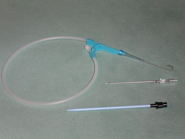
Biliary endoscopic sphincterotomy is a procedure where the sphincter of Oddi and the segment of the common bile duct where it enters the duodenum are cannulated and then cut with a sphincterotome, a device that includes a wire which cuts with an electric current (electrocautery). This procedure was developed in both Germany and Japan and was first published in each nation in 1974. It has become a very common technique, useful for treatment of a wide variety of conditions of the biliary system such as the evacuation of gallstones within the bile duct ( choledocholithiasis), biliary or papillary strictures, sphincter of Oddi dysfunction, bile leaks, and others. In addition, it is commonly performed during an endoscopic retrograde cholangiopancreatography (ERCP), and it may be used for facilitating diagnostic procedures such as transpapillary bile duct biopsy, papillary tumor biopsy, and insertion of a cholangioscope. Medical Uses Therapeutic Extraction of choledocholithiasi ... [...More Info...] [...Related Items...] OR: [Wikipedia] [Google] [Baidu] |
Gallstone
A gallstone is a stone formed within the gallbladder from precipitated bile components. The term cholelithiasis may refer to the presence of gallstones or to any disease caused by gallstones, and choledocholithiasis refers to the presence of migrated gallstones within bile ducts. Most people with gallstones (about 80%) are asymptomatic. However, when a gallstone obstructs the bile duct and causes acute cholestasis, a reflexive smooth muscle spasm often occurs, resulting in an intense cramp-like visceral pain in the right upper part of the abdomen known as a biliary colic (or "gallbladder attack"). This happens in 1–4% of those with gallstones each year. Complications from gallstones may include inflammation of the gallbladder ( cholecystitis), inflammation of the pancreas ( pancreatitis), obstructive jaundice, and infection in bile ducts ( cholangitis). Symptoms of these complications may include pain that lasts longer than five hours, fever, yellowish skin, vomiting, ... [...More Info...] [...Related Items...] OR: [Wikipedia] [Google] [Baidu] |
Choledochocele
Choledochal cysts (a.k.a. bile duct cyst) are congenital conditions involving cystic dilatation of bile ducts. They are uncommon in western countries but not as rare in East Asian nations like Japan and China. Signs and symptoms Most patients have symptoms in the first year of life. It is rare for symptoms to be undetected until adulthood, and usually adults have associated complications. The classic triad of intermittent abdominal pain, jaundice, and a right upper quadrant abdominal mass is found only in minority of patients. In infants, choledochal cysts usually lead to obstruction of the bile ducts and retention of bile. This leads to jaundice and an enlarged liver. If the obstruction is not relieved, permanent damage may occur to the liver - scarring and cirrhosis - with the signs of portal hypertension (obstruction to the flow of blood through the liver) and ascites (fluid accumulation in the abdomen). There is an increased risk of cancer in the wall of the cyst. In older ... [...More Info...] [...Related Items...] OR: [Wikipedia] [Google] [Baidu] |
Septum
In biology, a septum (Latin for ''something that encloses''; plural septa) is a wall, dividing a cavity or structure into smaller ones. A cavity or structure divided in this way may be referred to as septate. Examples Human anatomy * Interatrial septum, the wall of tissue that is a sectional part of the left and right atria of the heart * Interventricular septum, the wall separating the left and right ventricles of the heart * Lingual septum, a vertical layer of fibrous tissue that separates the halves of the tongue. * Nasal septum: the cartilage wall separating the nostrils of the nose * Alveolar septum: the thin wall which separates the alveoli from each other in the lungs * Orbital septum, a palpebral ligament in the upper and lower eyelids * Septum pellucidum or septum lucidum, a thin structure separating two fluid pockets in the brain * Uterine septum, a malformation of the uterus * Vaginal septum, a lateral or transverse partition inside the vagina * Interm ... [...More Info...] [...Related Items...] OR: [Wikipedia] [Google] [Baidu] |
Main Pancreatic Duct
The pancreatic duct, or duct of Wirsung (also, the major pancreatic duct due to the existence of an accessory pancreatic duct), is a duct joining the pancreas to the common bile duct. This supplies it with pancreatic juice from the exocrine pancreas, which aids in digestion. Structure The pancreatic duct joins the common bile duct just prior to the ampulla of Vater, after which both ducts perforate the medial side of the second portion of the duodenum at the major duodenal papilla. There are many anatomical variants reported, but these are quite rare. Accessory pancreatic duct Most people have just one pancreatic duct. However, some have an additional accessory pancreatic duct, also called the Duct of Santorini. An accessory pancreatic duct can be functional or non-functional. It may open separately into the second part of the duodenum,Moore KL, Dalley AF. 2006. Clinically Oriented Anatomy. 5th Ed. Lippincott Williams & Wilkins. p 287.1. Mchonde GJ, Gesase AP. Termination ... [...More Info...] [...Related Items...] OR: [Wikipedia] [Google] [Baidu] |
Cholangiogram
Cholangiography is the imaging of the bile duct (also known as the biliary tree) by x-rays and an injection of contrast medium. __TOC__ Types There are at least four types of cholangiography: # Percutaneous transhepatic cholangiography (PTC): Examination of liver The liver is a major organ only found in vertebrates which performs many essential biological functions such as detoxification of the organism, and the synthesis of proteins and biochemicals necessary for digestion and growth. In humans, it ... and bile ducts by x-rays. This is accomplished by the insertion of a thin needle into the liver carrying a contrast medium to help to see blockage in liver and bile ducts. # Endoscopic retrograde cholangiopancreatography (ERCP). Although this is a form of imaging, it is both diagnostic and therapeutic, and is often classified with surgeries rather than with imaging. # Primary cholangiography (or ''perioperative''): Done in the operation room during a biliary drainage in ... [...More Info...] [...Related Items...] OR: [Wikipedia] [Google] [Baidu] |
Billroth II
Billroth II, more formally Billroth's operation II, is an operation in which a partial gastrectomy (removal of the stomach) is performed and the cut end of the stomach is closed. The greater curvature of the stomach (not involved with the previous closure of the stomach) is then connected to the first part of the jejunum in end-to-side anastomosis. The Billroth II always follows resection of the lower part of the stomach ( antrum). The surgical procedure is called a partial gastrectomy and gastrojejunostomy. The Billroth II is often indicated in refractory peptic ulcer disease and gastric adenocarcinoma. Robinson, JO. The History of Gastric Surgery. Postgraduate Medical Journal. Dec 1960, p 706-713. Over the years, the Billroth II operation has been colloquially referred to as any partial removal of the stomach with an end to side connection to the stomach as shown in the picture; however, technically, this picture is a modification of Billroth's operation called a parti ... [...More Info...] [...Related Items...] OR: [Wikipedia] [Google] [Baidu] |
Major Duodenal Papilla
Major (commandant in certain jurisdictions) is a military rank of commissioned officer status, with corresponding ranks existing in many military forces throughout the world. When used unhyphenated and in conjunction with no other indicators, major is one rank above captain, and one rank below lieutenant colonel. It is considered the most junior of the field officer ranks. Background Majors are typically assigned as specialised executive or operations officers for battalion-sized units of 300 to 1,200 soldiers while in some nations, like Germany, majors are often in command of a company. When used in hyphenated or combined fashion, the term can also imply seniority at other levels of rank, including ''general-major'' or ''major general'', denoting a low-level general officer, and ''sergeant major'', denoting the most senior non-commissioned officer (NCO) of a military unit. The term ''major'' can also be used with a hyphen to denote the leader of a military band such as ... [...More Info...] [...Related Items...] OR: [Wikipedia] [Google] [Baidu] |
Teflon
Polytetrafluoroethylene (PTFE) is a synthetic fluoropolymer of tetrafluoroethylene that has numerous applications. It is one of the best-known and widely applied PFAS. The commonly known brand name of PTFE-based composition is Teflon by Chemours, a spin-off from DuPont, which originally discovered the compound in 1938. Polytetrafluoroethylene is a fluorocarbon solid, as it is a high- molecular-weight polymer consisting wholly of carbon and fluorine. PTFE is hydrophobic: neither water nor water-containing substances wet PTFE, as fluorocarbons exhibit only small London dispersion forces due to the low electric polarizability of fluorine. PTFE has one of the lowest coefficients of friction of any solid. Polytetrafluoroethylene is used as a non-stick coating for pans and other cookware. It is non-reactive, partly because of the strength of carbon–fluorine bonds, so it is often used in containers and pipework for reactive and corrosive chemicals. Where used as a lubrican ... [...More Info...] [...Related Items...] OR: [Wikipedia] [Google] [Baidu] |
Guide Wires
The Seldinger technique, also known as Seldinger wire technique, is a medical procedure to obtain safe access to blood vessels and other hollow organs. It is named after Sven Ivar Seldinger (1921–1998), a Swedish radiologist who introduced the procedure in 1953. Uses The Seldinger technique is used for angiography, insertion of chest drains and central venous catheters, insertion of PEG tubes using the push technique, insertion of the leads for an artificial pacemaker or implantable cardioverter-defibrillator, and numerous other interventional medical procedures. Complications The initial puncture is with a sharp instrument, and this may lead to hemorrhage or perforation of the organ in question. Infection is a possible complication, and hence asepsis is practiced during most Seldinger procedures. Loss of the guidewire into the cavity or blood vessel is a significant and generally preventable complication. Description The desired vessel or cavity is punctured with a shar ... [...More Info...] [...Related Items...] OR: [Wikipedia] [Google] [Baidu] |
Catheter
In medicine, a catheter (/ˈkæθətər/) is a thin tube made from medical grade materials serving a broad range of functions. Catheters are medical devices that can be inserted in the body to treat diseases or perform a surgical procedure. Catheters are manufactured for specific applications, such as cardiovascular, urological, gastrointestinal, neurovascular and ophthalmic procedures. The process of inserting a catheter is ''catheterization''. In most uses, a catheter is a thin, flexible tube (''soft'' catheter) though catheters are available in varying levels of stiffness depending on the application. A catheter left inside the body, either temporarily or permanently, may be referred to as an "indwelling catheter" (for example, a peripherally inserted central catheter). A permanently inserted catheter may be referred to as a "permcath" (originally a trademark). Catheters can be inserted into a body cavity, duct, or vessel, brain, skin or adipose tissue. Functionally, they a ... [...More Info...] [...Related Items...] OR: [Wikipedia] [Google] [Baidu] |
Thrombosis
Thrombosis (from Ancient Greek "clotting") is the formation of a blood clot inside a blood vessel, obstructing the flow of blood through the circulatory system. When a blood vessel (a vein or an artery) is injured, the body uses platelets (thrombocytes) and fibrin to form a blood clot to prevent blood loss. Even when a blood vessel is not injured, blood clots may form in the body under certain conditions. A clot, or a piece of the clot, that breaks free and begins to travel around the body is known as an embolus. Thrombosis may occur in veins ( venous thrombosis) or in arteries ( arterial thrombosis). Venous thrombosis (sometimes called DVT, deep vein thrombosis) leads to a blood clot in the affected part of the body, while arterial thrombosis (and, rarely, severe venous thrombosis) affects the blood supply and leads to damage of the tissue supplied by that artery (ischemia and necrosis). A piece of either an arterial or a venous thrombus can break off as an embolus, which c ... [...More Info...] [...Related Items...] OR: [Wikipedia] [Google] [Baidu] |
Hemorrhage
Bleeding, hemorrhage, haemorrhage or blood loss, is blood escaping from the circulatory system from damaged blood vessels. Bleeding can occur internally, or externally either through a natural opening such as the mouth, nose, ear, urethra, vagina or anus, or through a puncture in the skin. Hypovolemia is a massive decrease in blood volume, and death by excessive loss of blood is referred to as exsanguination. Typically, a healthy person can endure a loss of 10–15% of the total blood volume without serious medical difficulties (by comparison, blood donation typically takes 8–10% of the donor's blood volume). The stopping or controlling of bleeding is called hemostasis and is an important part of both first aid and surgery. Types * Upper head ** Intracranial hemorrhage – bleeding in the skull. ** Cerebral hemorrhage – a type of intracranial hemorrhage, bleeding within the brain tissue itself. ** Intracerebral hemorrhage – bleeding in the brain caused by the ruptur ... [...More Info...] [...Related Items...] OR: [Wikipedia] [Google] [Baidu] |


