|
Athletic Pubalgia
Athletic pubalgia, also called sports hernia, core injury, hockey hernia, hockey groin, Gilmore's groin, or groin disruption, is a medical condition of the pubic joint affecting athletes. It is a syndrome characterized by chronic groin pain in athletes and a dilated superficial ring of the inguinal canal. Football and ice hockey players are affected most frequently. Both recreational and professional athletes may be affected. Presentation Symptoms include pain during sports movements, particularly hip extension, and twisting and turning. This pain usually radiates to the adductor muscle region and even the testicles, although it is often difficult for the patient to pin-point the exact location. Following sporting activity the person with athletic pubalgia will be stiff and sore. The day after a match, getting out of bed or a car will be difficult. Any exertion that increases intra-abdominal pressure, such as coughing, sneezing, or sporting activity can cause pain. In the ear ... [...More Info...] [...Related Items...] OR: [Wikipedia] [Google] [Baidu] |
Pubic Symphysis
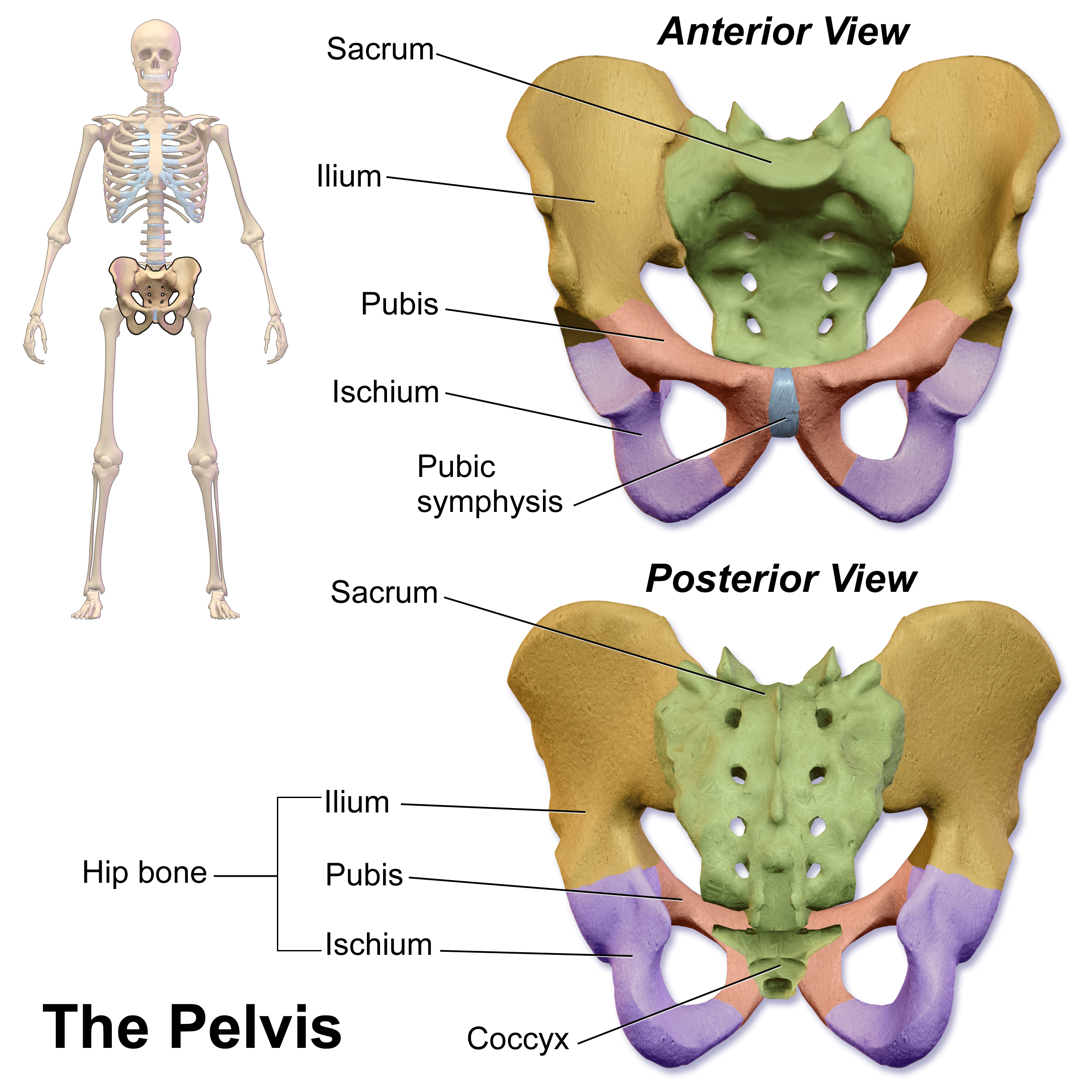
The pubic symphysis (: symphyses) is a secondary cartilaginous joint between the left and right superior rami of the pubis of the hip bones. It is in front of and below the urinary bladder. In males, the suspensory ligament of the penis attaches to the pubic symphysis. In females, the pubic symphysis is attached to the suspensory ligament of the clitoris. In most adults, it can be moved roughly 2 mm and with 1 degree rotation. This increases for women at the time of childbirth. The name comes from the Greek word ''symphysis'', meaning 'growing together'. Structure The pubic symphysis is a nonsynovial amphiarthrodial joint. The width of the pubic symphysis at the front is 3–5 mm greater than its width at the back. This joint is connected by fibrocartilage and may contain a fluid-filled cavity; the center is avascular, possibly due to the nature of the compressive forces passing through this joint, which may lead to harmful vascular disease. The ends of both pubi ... [...More Info...] [...Related Items...] OR: [Wikipedia] [Google] [Baidu] |
Pubic Tubercle
The pubic tubercle is a prominent tubercle on the superior ramus of the pubis bone of the pelvis. Structure The pubic tubercle is a prominent forward-projecting tubercle on the upper border of the medial portion of the superior ramus of the pubis bone. The inguinal ligament attaches to it. Part of the abdominal external oblique muscle inserts onto it. The inferior epigastric artery passes between the pubic tubercle and the anterior superior iliac spine. The pubic spine is a rough ridge that extends from the pubic tubercle to the upper border of the pubic symphysis. Clinical significance The pubic tubercle may be palpated. It serves as a landmark for local anaesthetic A local anesthetic (LA) is a medication that causes absence of all sense, sensation (including pain) in a specific body part without loss of consciousness, providing local anesthesia, as opposed to a general anesthetic, which eliminates all sen ... of the genital branch of the genitofemoral nerve, whi ... [...More Info...] [...Related Items...] OR: [Wikipedia] [Google] [Baidu] |
Incidence (epidemiology)
In epidemiology Epidemiology is the study and analysis of the distribution (who, when, and where), patterns and Risk factor (epidemiology), determinants of health and disease conditions in a defined population, and application of this knowledge to prevent dise ..., incidence reflects the number of new cases of a given medical condition in a population within a specified period of time. Incidence proportion Incidence proportion (IP), also known as cumulative incidence, is defined as the probability that a particular event, such as occurrence of a particular disease, has occurred in a specified period: Incidence = \frac For example, if a population contains 1,000 persons and 28 develop a condition from the time the disease first occurred until two years later, the cumulative incidence is 28 cases per 1,000 persons, i.e. 2.8%. Incidence rate The incidence rate can be calculated by dividing the number of subjects developing a disease by the total time at risk from all patie ... [...More Info...] [...Related Items...] OR: [Wikipedia] [Google] [Baidu] |
Stretching
Stretching is a form of physical exercise in which a specific muscle or tendon (or muscle group) is deliberately expanded and flexed in order to improve the muscle's felt elasticity and achieve comfortable muscle tone. The result is a feeling of increased muscle control, flexibility, and range of motion. Stretching is also used therapeutically to alleviate cramps and to improve function in daily activities by increasing range of motion. In its most basic form, stretching is a natural and instinctive activity; it is performed by humans and many other animals. It can be accompanied by yawning. Stretching often occurs instinctively after waking from sleep, after long periods of inactivity, or after exiting confined spaces and areas. In addition to vertebrates (e.g. mammals and birds), spiders have also been found to exhibit stretching. Increasing flexibility through stretching is one of the basic tenets of physical fitness. It is common for athletes to stretch before (for ... [...More Info...] [...Related Items...] OR: [Wikipedia] [Google] [Baidu] |
Genitofemoral Nerve
The genitofemoral nerve is a mixed branch of the lumbar plexus derived from anterior rami of lumbar nerves L1–L2. It splits into a genital branch and a femoral branch. It provides sensory innervation to the upper anterior thigh, as well as the skin of the anterior scrotum in males and mons pubis in females. It also provides motor innervation to the cremaster muscle (via its genital branch). Structure Origin The genitofemoral nerve is a branch of the lumbar plexus. It is derived from the anterior rami of lumbar nerves L1–L2. It coalesces within the substances of the psoas major muscle. Course It passes downwards, pierces the psoas major and emerges from its anterior surface. The nerve divides into two branches, the genital branch and the lumboinguinal nerve also known as the femoral branch, both of which then continue downwards and medially to the inguinal and femoral canal respectively. Branches Genital branch The genital branch continues downward on the s ... [...More Info...] [...Related Items...] OR: [Wikipedia] [Google] [Baidu] |
Ilioinguinal Nerve
The ilioinguinal nerve is a branch of the first lumbar nerve (L1). It separates from the first lumbar nerve along with the larger iliohypogastric nerve. It emerges from the lateral border of the psoas major just inferior to the iliohypogastric, and passes obliquely across the quadratus lumborum and iliacus. The ilioinguinal nerve then perforates the transversus abdominis near the anterior part of the iliac crest, and communicates with the iliohypogastric nerve between the transversus and the internal oblique muscle. It then pierces the internal oblique muscle, distributing filaments to it, and then accompanies the spermatic cord (in males) or the round ligament of uterus (in females) through the superficial inguinal ring. Its fibres are then distributed to the skin of the upper and medial part of the thigh, and to the following locations in the male and female: * In the male (" anterior scrotal nerve"): to the skin over the root of the penis and upper part of the scrotum. ... [...More Info...] [...Related Items...] OR: [Wikipedia] [Google] [Baidu] |
Abdominal Internal Oblique Muscle
The abdominal internal oblique muscle, also internal oblique muscle or interior oblique, is an abdominal muscle in the abdominal wall that lies below the external oblique muscle and just above the transverse abdominal muscle. Structure Its fibers run perpendicular to the external oblique muscle, beginning in the thoracolumbar fascia of the lower back, the anterior 2/3 of the iliac crest (upper part of hip bone) and the lateral half of the inguinal ligament. The muscle fibers run from these points superomedially (up and towards midline) to the muscle's insertions on the inferior borders of the 10th through 12th ribs and the linea alba. In males, the cremaster muscle is also attached to the internal oblique. Nerve supply The internal oblique is supplied by the lower intercostal nerves, as well as the iliohypogastric nerve and the ilioinguinal nerve. Function The internal oblique performs two major functions. Firstly as an accessory muscle of respiration, it acts as an ... [...More Info...] [...Related Items...] OR: [Wikipedia] [Google] [Baidu] |
Rectus Abdominis Muscle
The rectus abdominis muscle, () also known as the "abdominal muscle" or simply better known as the "abs", is a pair of segmented skeletal muscle on the ventral aspect of a person's abdomen. The paired muscle is separated at the midline by a band of dense connective tissue called the linea alba, and the connective tissue defining each lateral margin of the rectus abdominus is the linea semilunaris. The muscle extends from the pubic symphysis, pubic crest and pubic tubercle inferiorly, to the xiphoid process and costal cartilages of the 5th–7th ribs superiorly. The rectus abdominis muscle is contained in the rectus sheath, which consists of the aponeuroses of the lateral abdominal muscles. Each rectus abdominus is traversed by bands of connective tissue called the tendinous intersections, which interrupt it into distinct muscle bellies. Structure The rectus abdominis is a very long flat muscle, which extends along the whole length of the front of the abdomen, and is s ... [...More Info...] [...Related Items...] OR: [Wikipedia] [Google] [Baidu] |
Inguinal Ligament
The inguinal ligament (), also known as Poupart's ligament or groin ligament, is a band running from the pubic tubercle to the anterior superior iliac spine. It forms the base of the inguinal canal through which an indirect inguinal hernia may develop. Structure The inguinal ligament runs from the anterior superior iliac crest of the ilium to the pubic tubercle of the pubic bone. It is formed by the external abdominal oblique aponeurosis and is continuous with the fascia lata of the thigh. There is some dispute over the attachments. Structures that pass deep to the inguinal ligament include: * Psoas major, iliacus, pectineus * Femoral nerve, artery, and vein * Lateral cutaneous nerve of thigh *Lymphatics Function The ligament serves to contain soft tissues as they course anteriorly from the trunk to the lower extremity. This structure demarcates the superior border of the femoral triangle. It demarcates the inferior border of the inguinal triangle. The midpoint ... [...More Info...] [...Related Items...] OR: [Wikipedia] [Google] [Baidu] |
Conjoint Tendon
also known as superior tendon of abdominal cavity. The conjoint tendon (previously known as the inguinal aponeurotic falx) is a sheath of connective tissue formed from the lower part of the common aponeurosis of the abdominal internal oblique muscle and the transversus abdominis muscle, joining the muscle to the pelvis. It forms the medial part of the posterior wall of the inguinal canal. Structure The conjoint tendon is formed from the lower part of the common aponeurosis of the abdominal internal oblique muscle and the transversus abdominis muscle. It inserts into the pubic crest and the pectineal line immediately behind the superficial inguinal ring. It is usually conjoint with the tendon of the internal oblique muscle, but they may be separate as well. It forms the medial part of the posterior wall of the inguinal canal. Clinical significance The conjoint tendon serves to protect what would otherwise be a weak point in the abdominal wall. A weakening of the conjoint ... [...More Info...] [...Related Items...] OR: [Wikipedia] [Google] [Baidu] |
Inguinodynia
Post herniorrhaphy pain syndrome, or inguinodynia is pain or discomfort lasting greater than 3 months after surgery of inguinal hernia. Randomized trials of laparoscopic vs open inguinal hernia repair have demonstrated similar recurrence rates with the use of mesh and have identified that chronic groin pain (>10%) surpasses recurrence (<2%) and is an important measure of success. Chronic groin pain is potentially disabling with , parasthesia, , and |
Aponeurosis Of The Obliquus Externus Abdominis
The aponeurosis of the abdominal external oblique muscle is a thin but strong membranous structure, the fibers of which are directed downward and medially. It is joined with that of the opposite muscle along the middle line, and covers the whole of the front of the abdomen; above, it is covered by and gives origin to the lower fibers of the pectoralis major; below, its fibers are closely aggregated together, and extend obliquely across from the anterior superior iliac spine to the pubic tubercle and the pectineal line to form the inguinal ligament. In the middle line, it interlaces with the aponeurosis of the opposite muscle, forming the linea alba, which extends from the xiphoid process to the pubic symphysis. That portion of the aponeurosis which extends between the anterior superior iliac spine and the pubic tubercle is a thick band, folded inward, and continuous below with the fascia lata; it is called the inguinal ligament. The portion which is reflected from the inguina ... [...More Info...] [...Related Items...] OR: [Wikipedia] [Google] [Baidu] |

