|
Afferent Nerve Fiber
Afferent nerve fibers are axons (nerve fibers) of sensory neurons that carry sensory information from sensory receptors to the central nervous system. Many afferent projections ''arrive'' at a particular brain region. In the peripheral nervous system, afferent nerve fibers are part of the sensory nervous system and arise from outside of the central nervous system. Sensory and mixed nerves contain afferent fibers. Structure Afferent neurons are pseudounipolar neurons that have a single process leaving the cell body dividing into two branches: the long one towards the sensory organ, and the short one toward the central nervous system (e.g. spinal cord). These cells do have sensory afferent dendrites, similar to those typically inherent in neurons. They have a smooth and rounded cell body located in the ganglia of the peripheral nervous system. Just outside the spinal cord, thousands of afferent neuronal cell bodies are aggregated in a swelling in the dorsal root known a ... [...More Info...] [...Related Items...] OR: [Wikipedia] [Google] [Baidu] |
Nervous System
In biology, the nervous system is the complex system, highly complex part of an animal that coordinates its behavior, actions and sense, sensory information by transmitting action potential, signals to and from different parts of its body. The nervous system detects environmental changes that impact the body, then works in tandem with the endocrine system to respond to such events. Nervous tissue first arose in Ediacara biota, wormlike organisms about 550 to 600 million years ago. In Vertebrate, vertebrates, it consists of two main parts, the central nervous system (CNS) and the peripheral nervous system (PNS). The CNS consists of the brain and spinal cord. The PNS consists mainly of nerves, which are enclosed bundles of the long fibers, or axons, that connect the CNS to every other part of the body. Nerves that transmit signals from the brain are called motor nerves (efferent), while those nerves that transmit information from the body to the CNS are called sensory nerves (aff ... [...More Info...] [...Related Items...] OR: [Wikipedia] [Google] [Baidu] |
Intrafusal Muscle Fiber
Intrafusal muscle fibers are skeletal muscle fibers that serve as specialized sensory organs ( proprioceptors). They detect the amount and rate of change in length of a muscle.Casagrand, Janet (2008) ''Action and Movement: Spinal Control of Motor Units and Spinal Reflexes.'' University of Colorado, Boulder. They constitute the muscle spindle, and are innervated by both sensory (afferent) and motor (efferent) fibers. Intrafusal muscle fibers are not to be confused with extrafusal muscle fibers, which contract, generating skeletal movement and are innervated by alpha motor neurons. Structure Types There are two types of intrafusal muscle fibers: nuclear bag fibers and nuclear chain fibers. They bear two types of sensory ending, known as annulospiral and flower-spray endings. Both ends of these fibers contract, but the central region only stretches and does not contract. Intrafusal muscle fibers are walled off from the rest of the muscle by an outer connective tissue ... [...More Info...] [...Related Items...] OR: [Wikipedia] [Google] [Baidu] |
Special Somatic Afferent Fiber
Special somatic afferent fibers (SSA) are the afferent nerve fibers that carry information from the special senses of vision, hearing and balance. The cranial nerves containing SSA fibers are the optic nerve (CN II) and the vestibulocochlear nerve (CN VIII). See also * General somatic afferent fiber (GSA) * General visceral afferent fiber (GVA) * Special visceral afferent fiber Special visceral afferent fibers (SVA) are afferent fibers that develop in association with the gastrointestinal tract. They carry the special sense of taste (gustation). The cranial nerves containing SVA fibers are the facial nerve (VII), the glos ... (SVA) References External links Overview at mmi.mcgill.ca Neuroanatomy {{Neuroanatomy-stub ... [...More Info...] [...Related Items...] OR: [Wikipedia] [Google] [Baidu] |
General Visceral Afferent Fiber
The general visceral afferent (GVA) fibers conduct sensory impulses (usually pain or reflex sensations) from the internal organs, glands, and blood vessels to the central nervous system. They are considered to be part of the visceral nervous system, which is closely related to the autonomic nervous system, but 'visceral nervous system' and 'autonomic nervous system' are not direct synonyms and care should be taken when using these terms. Unlike the efferent fibers of the autonomic nervous system, the afferent fibers are not classified as either sympathetic or parasympathetic. GVA fibers create referred pain by activating general somatic afferent fibers where the two meet in the posterior grey column. The cranial nerves that contain GVA fibers include the glossopharyngeal nerve (CN IX) and the vagus nerve (CN X). Generally, they are insensitive to cutting, crushing or burning; however, excessive tension in smooth muscle and some pathological conditions produce visceral pain ( ... [...More Info...] [...Related Items...] OR: [Wikipedia] [Google] [Baidu] |
General Somatic Afferent Fiber
The general somatic afferent fibers (GSA or somatic sensory fibers) are afferent fibers that arise from neurons in sensory ganglia and are found in all the spinal nerves, except occasionally the first cervical. General somatic afferents conduct impulses of pain, touch and temperature from the surface of the body through the dorsal roots to the spinal cord The spinal cord is a long, thin, tubular structure made up of nervous tissue that extends from the medulla oblongata in the lower brainstem to the lumbar region of the vertebral column (backbone) of vertebrate animals. The center of the spinal c ..., and impulses of muscle sense, tendon sense and joint sense from the deeper structures. See also * Afferent nerve * General visceral afferent fiber (GVA) * Special somatic afferent fiber (SSA) * Special visceral afferent fiber (SVA) References Spinal cord {{Portal bar, Anatomy ... [...More Info...] [...Related Items...] OR: [Wikipedia] [Google] [Baidu] |
Parietal Lobe
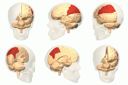
The parietal lobe is one of the four Lobes of the brain, major lobes of the cerebral cortex in the brain of mammals. The parietal lobe is positioned above the temporal lobe and behind the frontal lobe and central sulcus. The parietal lobe integrates sensory information among various sensory modality, modalities, including spatial sense and navigation (proprioception), the main sensory receptive area for the sense of touch in the somatosensory cortex which is just posterior to the central sulcus in the postcentral gyrus, and the two-streams hypothesis#Dorsal stream, dorsal stream of the visual system. The major sensory inputs from the skin (mechanoreceptor, touch, thermoreceptor, temperature, and nociceptor, pain receptors), relay through the thalamus to the parietal lobe. Several areas of the parietal lobe are important in language processing in the brain, language processing. The somatosensory cortex can be illustrated as a distorted figure – the cortical homunculus (Latin: "li ... [...More Info...] [...Related Items...] OR: [Wikipedia] [Google] [Baidu] |
Primary Somatosensory Cortex
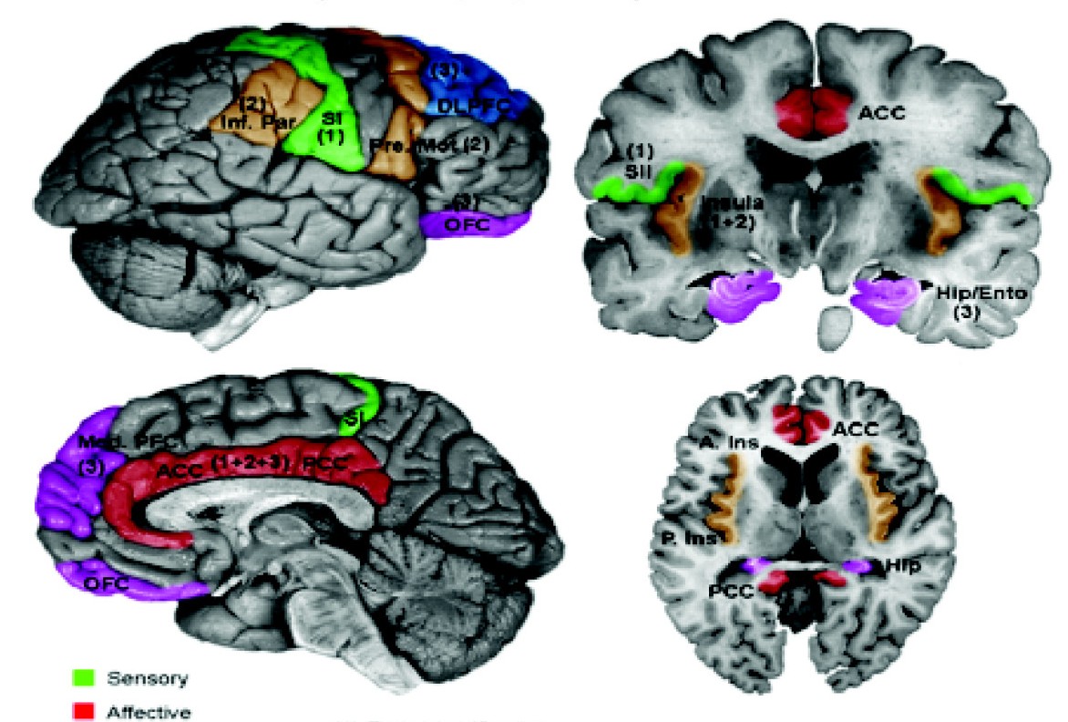
In neuroanatomy, the primary somatosensory cortex is located in the postcentral gyrus of the brain's parietal lobe, and is part of the somatosensory system. It was initially defined from surface stimulation studies of Wilder Penfield, and parallel surface potential studies of Bard, Woolsey, and Marshall. Although initially defined to be roughly the same as Brodmann areas 3, 1 and 2, more recent work by Kaas has suggested that for homogeny with other sensory fields only area 3 should be referred to as "primary somatosensory cortex", as it receives the bulk of the thalamocortical projections from the sensory input fields. At the primary somatosensory cortex, tactile representation is orderly arranged (in an inverted fashion) from the toe (at the top of the cerebral hemisphere) to mouth (at the bottom). However, some body parts may be controlled by partially overlapping regions of cortex. Each cerebral hemisphere of the primary somatosensory cortex only contains a tactile representatio ... [...More Info...] [...Related Items...] OR: [Wikipedia] [Google] [Baidu] |
Thalamus
The thalamus (: thalami; from Greek language, Greek Wikt:θάλαμος, θάλαμος, "chamber") is a large mass of gray matter on the lateral wall of the third ventricle forming the wikt:dorsal, dorsal part of the diencephalon (a division of the forebrain). Nerve fibers project out of the thalamus to the cerebral cortex in all directions, known as the thalamocortical radiations, allowing hub (network science), hub-like exchanges of information. It has several functions, such as the relaying of sensory neuron, sensory and motor neuron, motor signals to the cerebral cortex and the regulation of consciousness, sleep, and alertness. Anatomically, the thalami are paramedian symmetrical structures (left and right), within the vertebrate brain, situated between the cerebral cortex and the midbrain. It forms during embryonic development as the main product of the diencephalon, as first recognized by the Swiss embryologist and anatomist Wilhelm His Sr. in 1893. Anatomy The thalami ar ... [...More Info...] [...Related Items...] OR: [Wikipedia] [Google] [Baidu] |
Medial Lemniscus
The medial lemniscus, also known as Reil's band or Reil's ribbon (for German anatomist Johann Christian Reil), is a large ascending bundle of heavily myelinated axons that decussate in the brainstem The brainstem (or brain stem) is the posterior stalk-like part of the brain that connects the cerebrum with the spinal cord. In the human brain the brainstem is composed of the midbrain, the pons, and the medulla oblongata. The midbrain is conti ..., specifically in the medulla oblongata. The medial lemniscus is formed by the crossings of the internal arcuate fibers. The internal arcuate fibers are composed of axons of the gracile nucleus and the cuneate nucleus. The cell bodies of the nuclei lie contralaterally. The medial lemniscus is part of the somatosensory dorsal column–medial lemniscus pathway, which ascends in the spinal cord to the thalamus.Kamali A, Kramer LA, Butler IJ, Hasan KM. Diffusion tensor tractography of the somatosensory system in the human brains ... [...More Info...] [...Related Items...] OR: [Wikipedia] [Google] [Baidu] |
Medulla Oblongata
The medulla oblongata or simply medulla is a long stem-like structure which makes up the lower part of the brainstem. It is anterior and partially inferior to the cerebellum. It is a cone-shaped neuronal mass responsible for autonomic (involuntary) functions, ranging from vomiting to sneezing. The medulla contains the cardiovascular center, the respiratory center, vomiting and vasomotor centers, responsible for the autonomic functions of breathing, heart rate and blood pressure as well as the sleep–wake cycle. "Medulla" is from Latin, ‘pith or marrow’. And "oblongata" is from Latin, ‘lengthened or longish or elongated'. During embryonic development, the medulla oblongata develops from the myelencephalon. The myelencephalon is a secondary brain vesicle which forms during the maturation of the rhombencephalon, also referred to as the hindbrain. The bulb is an archaic term for the medulla oblongata. In modern clinical usage, the word bulbar (as in bulbar palsy) is r ... [...More Info...] [...Related Items...] OR: [Wikipedia] [Google] [Baidu] |
Decussates
Decussation is used in biological contexts to describe a crossing (due to the shape of the Roman numeral for ten, an uppercase 'X' (), ). In Latin anatomical terms, the form is used, e.g. . Similarly, the anatomical term chiasma is named after the Greek uppercase 'Χ' ( chi). Whereas a decussation refers to a crossing within the central nervous system, various kinds of crossings in the peripheral nervous system are called chiasma. Examples include: * In the brain, where nerve fibers obliquely cross from one lateral side of the brain to the other, that is to say they cross at a level other than their origin. See for examples decussation of pyramids and sensory decussation. In neuroanatomy, the term ''chiasma'' is reserved for crossing of- or within nerves such as in the optic chiasm. * In botanical leaf taxology, the word ''decussate'' describes an opposite pattern of leaves which has successive pairs at right angles to each other (i.e. rotated 90 degrees along the stem w ... [...More Info...] [...Related Items...] OR: [Wikipedia] [Google] [Baidu] |
Brainstem
The brainstem (or brain stem) is the posterior stalk-like part of the brain that connects the cerebrum with the spinal cord. In the human brain the brainstem is composed of the midbrain, the pons, and the medulla oblongata. The midbrain is continuous with the thalamus of the diencephalon through the tentorial notch, and sometimes the diencephalon is included in the brainstem. The brainstem is very small, making up around only 2.6 percent of the brain's total weight. It has the critical roles of regulating heart and respiratory system, respiratory function, helping to control heart rate and breathing rate. It also provides the main motor and sensory nerve supply to the face and neck via the cranial nerves. Ten pairs of cranial nerves come from the brainstem. Other roles include the regulation of the central nervous system and the body's sleep cycle. It is also of prime importance in the conveyance of motor and sensory pathways from the rest of the brain to the body, and from the b ... [...More Info...] [...Related Items...] OR: [Wikipedia] [Google] [Baidu] |


