|
Pilocytic Astrocytoma
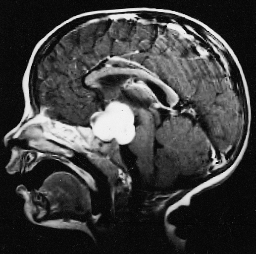
Pilocytic astrocytoma (and its variant pilomyxoid astrocytoma) is a brain tumor that occurs most commonly in children and young adults (in the first 20 years of life). They usually arise in the cerebellum, near the brainstem, in the hypothalamic region, or the optic chiasm, but they may occur in any area where astrocytes are present, including the cerebral hemispheres and the spinal cord. These tumors are usually slow growing and benign, corresponding to WHO malignancy grade 1. Signs and symptoms Children affected by pilocytic astrocytoma can present with different symptoms that might include failure to thrive (lack of appropriate weight gain/ weight loss), headache, nausea, vomiting, irritability, torticollis (tilt neck or wry neck), difficulty to coordinate movements, and visual complaints (including nystagmus). The complaints may vary depending on the location and size of the neoplasm. The most common symptoms are associated with increased intracranial pressure due to the size ... [...More Info...] [...Related Items...] OR: [Wikipedia] [Google] [Baidu] |
Micrograph
A micrograph or photomicrograph is a photograph or digital image taken through a microscope or similar device to show a magnified image of an object. This is opposed to a macrograph or photomacrograph, an image which is also taken on a microscope but is only slightly magnified, usually less than 10 times. Micrography is the practice or art of using microscopes to make photographs. A micrograph contains extensive details of microstructure. A wealth of information can be obtained from a simple micrograph like behavior of the material under different conditions, the phases found in the system, failure analysis, grain size estimation, elemental analysis and so on. Micrographs are widely used in all fields of microscopy. Types Photomicrograph A light micrograph or photomicrograph is a micrograph prepared using an optical microscope, a process referred to as ''photomicroscopy''. At a basic level, photomicroscopy may be performed simply by connecting a camera to a microscope ... [...More Info...] [...Related Items...] OR: [Wikipedia] [Google] [Baidu] |
BRAF (gene)
BRAF is a human gene that encodes a protein called B-Raf. The gene is also referred to as proto-oncogene B-Raf and v-Raf murine sarcoma viral oncogene homolog B, while the protein is more formally known as serine/threonine-protein kinase B-Raf. The B-Raf protein is involved in sending signals inside cells which are involved in directing cell growth. In 2002, it was shown to be mutated in some human cancers. Certain other inherited ''BRAF'' mutations cause birth defects. Drugs that treat cancers driven by ''BRAF'' mutations have been developed. Two of these drugs, vemurafenib and dabrafenib are approved by FDA for treatment of late-stage melanoma. Vemurafenib was the first approved drug to come out of fragment-based drug discovery. Function B-Raf is a member of the Raf kinase family of growth signal transduction protein kinases. This protein plays a role in regulating the MAP kinase/ ERKs signaling pathway, which affects cell division, differentiation, and secretion. ... [...More Info...] [...Related Items...] OR: [Wikipedia] [Google] [Baidu] |
Brain Tumor
A brain tumor occurs when abnormal cells form within the brain. There are two main types of tumors: malignant tumors and benign (non-cancerous) tumors. These can be further classified as primary tumors, which start within the brain, and secondary tumors, which most commonly have spread from tumors located outside the brain, known as brain metastasis tumors. All types of brain tumors may produce symptoms that vary depending on the size of the tumor and the part of the brain that is involved. Where symptoms exist, they may include headaches, seizures, problems with vision, vomiting and mental changes. Other symptoms may include difficulty walking, speaking, with sensations, or unconsciousness. The cause of most brain tumors is unknown. Uncommon risk factors include exposure to vinyl chloride, Epstein–Barr virus, ionizing radiation, and inherited syndromes such as neurofibromatosis, tuberous sclerosis, and von Hippel-Lindau Disease. Studies on Mobile phone radiation and h ... [...More Info...] [...Related Items...] OR: [Wikipedia] [Google] [Baidu] |
Jill S
Jill is an English feminine given name, a short form of the name Jillian (Gillian), which in turn originates as a Middle English variant of Juliana, the feminine form of the name Julian. People with the given name * Jill Astbury, Australian researcher into violence against women * Jill Balcon (1925–2009), British actress * Jill S. Barnholtz-Sloan, American biostatistician and data scientist * Jill Becker, American psychological researcher * Jill Biden (born 1951), American educator and the First Lady of the United States * Jill E. Brown (born 1950), African American aviator * Jill Carroll (born 1977), American journalist * Jill Clayburgh (1944–2010), American actress * Jill Costello (1987–2010), American athlete and lung cancer activist * Jill Craigie (1911–1999), British film director and writer * Jill Craybas (born 1974), American tennis player * Jill Dando (1961–1999), British television presenter * Jill Dickman, Republican member of the Nevada Assembly * J ... [...More Info...] [...Related Items...] OR: [Wikipedia] [Google] [Baidu] |
Oligodendroglioma
Oligodendrogliomas are a type of glioma that are believed to originate from the oligodendrocytes of the brain or from a glial precursor cell. They occur primarily in adults (9.4% of all primary brain and central nervous system tumors) but are also found in children (4% of all primary brain tumors). Signs and symptoms Oligodendroglioma arise mainly in the frontal lobe and in 50–80% of cases, the first symptom is the onset of seizure activity, without having any symptoms beforehand. Headaches combined with increased intracranial pressure are also a common symptom of oligodendroglioma. Depending on the location of the tumor, many different neurological deficits can be induced, including, but not limited to, visual loss, motor weakness and cognitive decline. A computed tomography (CT) or magnetic resonance imaging (MRI) scan is necessary to characterize tumor size, location, and hetero- or homogeneity. Final diagnosis relies on biopsy and histopathologic examination of the tumor mas ... [...More Info...] [...Related Items...] OR: [Wikipedia] [Google] [Baidu] |
Eosinophilic
Eosinophilic (Greek suffix -phil-, meaning ''loves eosin'') is the staining of tissues, cells, or organelles after they have been washed with eosin, a dye. Eosin is an acidic dye for staining cell cytoplasm, collagen, and muscle fibers. ''Eosinophilic'' describes the appearance of cells and structures seen in histological sections that take up the staining dye eosin. Such eosinophilic structures are, in general, composed of protein. Eosin is usually combined with a stain called hematoxylin to produce a hematoxylin- and eosin-stained section (also called an H&E stain, HE or H+E section). It is the most widely used histological stain for a medical diagnosis. When a pathologist examines a biopsy of a suspected cancer, they will stain the biopsy with H&E. Some structures seen inside cells are described as being eosinophilic; for example, Lewy and Mallory bodies. [...More Info...] [...Related Items...] OR: [Wikipedia] [Google] [Baidu] |
Rosenthal Fiber
A Rosenthal fiber is a thick, elongated, worm-like or "corkscrew" eosinophilic (pink) bundle that is found on staining of brain tissue in the presence of long-standing gliosis, occasional tumors, and some metabolic disorders. Associated conditions Its presence is associated with either pilocytic astrocytoma (more common) or Alexander's disease (a rare leukodystrophy). They are also seen in the context of fucosidosis. Pilocytic astrocytoma is the most common primitive tumor found in pediatrics. Composition The fibers are found in astrocytic processes and are thought to be clumped intermediate filament proteins, primarily glial fibrillary acidic protein Glial fibrillary acidic protein (GFAP) is a protein that is encoded by the ''GFAP'' gene in humans. It is a type III intermediate filament (IF) protein that is expressed by numerous cell types of the central nervous system (CNS), including astro .... Other reported constituents include alphaB crystallin, heat shock protein 2 ... [...More Info...] [...Related Items...] OR: [Wikipedia] [Google] [Baidu] |
Glial Fibrillary Acidic Protein
Glial fibrillary acidic protein (GFAP) is a protein that is encoded by the ''GFAP'' gene in humans. It is a type III intermediate filament (IF) protein that is expressed by numerous cell types of the central nervous system (CNS), including astrocytes and ependymal cells during development. GFAP has also been found to be expressed in glomeruli and peritubular fibroblasts taken from rat kidneys, Leydig cells of the testis in both hamsters and humans, human keratinocytes, human osteocytes and chondrocytes and stellate cells of the pancreas and liver in rats. GFAP is closely related to the other three non-epithelial type III IF family members, vimentin, desmin and peripherin, which are all involved in the structure and function of the cell’s cytoskeleton. GFAP is thought to help to maintain astrocyte mechanical strength as well as the shape of cells, but its exact function remains poorly understood, despite the number of studies using it as a cell marker. The protein was ... [...More Info...] [...Related Items...] OR: [Wikipedia] [Google] [Baidu] |
Cystic
A cyst is a closed sac, having a distinct envelope and division compared with the nearby tissue. Hence, it is a cluster of cells that have grouped together to form a sac (like the manner in which water molecules group together to form a bubble); however, the distinguishing aspect of a cyst is that the cells forming the "shell" of such a sac are distinctly abnormal (in both appearance and behaviour) when compared with all surrounding cells for that given location. A cyst may contain air, fluids, or semi-solid material. A collection of pus is called an abscess, not a cyst. Once formed, a cyst may resolve on its own. When a cyst fails to resolve, it may need to be removed surgically, but that would depend upon its type and location. Cancer-related cysts are formed as a defense mechanism for the body following the development of mutations that lead to an uncontrolled cellular division. Once that mutation has occurred, the affected cells divide incessantly and become cancerous, form ... [...More Info...] [...Related Items...] OR: [Wikipedia] [Google] [Baidu] |
MRI Scan
Magnetic resonance imaging (MRI) is a medical imaging technique used in radiology to form pictures of the anatomy and the physiological processes of the body. MRI scanners use strong magnetic fields, magnetic field gradients, and radio waves to generate images of the organs in the body. MRI does not involve X-rays or the use of ionizing radiation, which distinguishes it from CT and PET scans. MRI is a medical application of nuclear magnetic resonance (NMR) which can also be used for imaging in other NMR applications, such as NMR spectroscopy. MRI is widely used in hospitals and clinics for medical diagnosis, staging and follow-up of disease. Compared to CT, MRI provides better contrast in images of soft-tissues, e.g. in the brain or abdomen. However, it may be perceived as less comfortable by patients, due to the usually longer and louder measurements with the subject in a long, confining tube, though "Open" MRI designs mostly relieve this. Additionally, implants and other ... [...More Info...] [...Related Items...] OR: [Wikipedia] [Google] [Baidu] |
T1-weighted Coronal MRI Image Post Contrast Tectal Plate Glioma
Magnetic resonance imaging (MRI) is a medical imaging technique used in radiology to form pictures of the anatomy and the physiological processes of the body. MRI scanners use strong magnetic fields, magnetic field gradients, and radio waves to generate images of the organs in the body. MRI does not involve X-rays or the use of ionizing radiation, which distinguishes it from CT and PET scans. MRI is a medical application of nuclear magnetic resonance (NMR) which can also be used for imaging in other NMR applications, such as NMR spectroscopy. MRI is widely used in hospitals and clinics for medical diagnosis, staging and follow-up of disease. Compared to CT, MRI provides better contrast in images of soft-tissues, e.g. in the brain or abdomen. However, it may be perceived as less comfortable by patients, due to the usually longer and louder measurements with the subject in a long, confining tube, though "Open" MRI designs mostly relieve this. Additionally, implants and ... [...More Info...] [...Related Items...] OR: [Wikipedia] [Google] [Baidu] |


