|
Marcus Gunn Pupil
A relative afferent pupillary defect (RAPD), also known as a Marcus Gunn pupil, is a medical sign observed during the swinging-flashlight test whereupon the patient's pupils dilate when a bright light is swung from the unaffected eye to the affected eye. The affected eye still senses the light and produces pupillary sphincter constriction to some degree, albeit reduced. Depending on severity, different symptoms may appear during the swinging flash light test: Mild RAPD will presents as a weak pupil constriction initially, after which dilation continues to happen. When RAPD is moderate, pupil size will remain, after which it dilates When RAPD is severe, the pupil will dilate quickly Cause The most common cause of Marcus Gunn pupil is a lesion of the optic nerve (between the retina and the optic chiasm) due to glaucoma, or severe retinal disease, or due to multiple sclerosis. It is named after Scottish ophthalmologist Robert Marcus Gunn. A second common cause of Marcus Gunn ... [...More Info...] [...Related Items...] OR: [Wikipedia] [Google] [Baidu] |
Optic Nerve
In neuroanatomy, the optic nerve, also known as the second cranial nerve, cranial nerve II, or simply CN II, is a paired cranial nerve that transmits visual information from the retina to the brain. In humans, the optic nerve is derived from optic stalks during the seventh week of development and is composed of retinal ganglion cell axons and glial cells; it extends from the optic disc to the optic chiasma and continues as the optic tract to the lateral geniculate nucleus, pretectal nuclei, and superior colliculus. Structure The optic nerve has been classified as the second of twelve paired cranial nerves, but it is technically part of the central nervous system, rather than the peripheral nervous system because it is derived from an out-pouching of the diencephalon ( optic stalks) during embryonic development. As a consequence, the fibers of the optic nerve are covered with myelin produced by oligodendrocytes, rather than Schwann cells of the peripheral nervous system ... [...More Info...] [...Related Items...] OR: [Wikipedia] [Google] [Baidu] |
Pupillary Light Reflex
The pupillary light reflex (PLR) or photopupillary reflex is a reflex that controls the diameter of the pupil, in response to the intensity (luminance) of light that falls on the retinal ganglion cells of the retina in the back of the eye, thereby assisting in adaptation of vision to various levels of lightness/darkness. A greater intensity of light causes the pupil to constrict ( miosis/myosis; thereby allowing less light in), whereas a lower intensity of light causes the pupil to dilate ( mydriasis, expansion; thereby allowing more light in). Thus, the pupillary light reflex regulates the intensity of light entering the eye. Light shone into one eye will cause both pupils to constrict. Terminology The pupil is the dark circular opening in the center of the iris and is where light enters the eye. By analogy with a camera, the pupil is equivalent to aperture, whereas the iris is equivalent to the diaphragm. It may be helpful to consider the ''Pupillary reflex'' as an Iris' ref ... [...More Info...] [...Related Items...] OR: [Wikipedia] [Google] [Baidu] |
Syphilis
Syphilis () is a sexually transmitted infection caused by the bacterium '' Treponema pallidum'' subspecies ''pallidum''. The signs and symptoms of syphilis vary depending in which of the four stages it presents (primary, secondary, latent, and tertiary). The primary stage classically presents with a single chancre (a firm, painless, non-itchy skin ulceration usually between 1 cm and 2 cm in diameter) though there may be multiple sores. In secondary syphilis, a diffuse rash occurs, which frequently involves the palms of the hands and soles of the feet. There may also be sores in the mouth or vagina. In latent syphilis, which can last for years, there are few or no symptoms. In tertiary syphilis, there are gummas (soft, non-cancerous growths), neurological problems, or heart symptoms. Syphilis has been known as " the great imitator" as it may cause symptoms similar to many other diseases. Syphilis is most commonly spread through sexual activity. It may also be tra ... [...More Info...] [...Related Items...] OR: [Wikipedia] [Google] [Baidu] |
Parinaud's Syndrome
Parinaud's syndrome is an inability to move the eyes up and down. It is caused by compression of the vertical gaze center at the rostral interstitial nucleus of medial longitudinal fasciculus (riMLF). The eyes lose the ability to move upward and down. It is a group of abnormalities of eye movement and pupil dysfunction. It is caused by lesions of the upper brain stem and is named for Henri Parinaud (1844–1905), considered to be the father of French ophthalmology. Signs and symptoms Parinaud's syndrome is a cluster of abnormalities of eye movement and pupil dysfunction, characterized by: * Paralysis of upwards gaze: Downward gaze is usually preserved. This vertical palsy is supranuclear, so doll's head maneuver should elevate the eyes, but eventually all upward gaze mechanisms fail. * Pseudo- Argyll Robertson pupils: Accommodative paresis ensues, and pupils become mid-dilated and show light-near dissociation. * Convergence-retraction nystagmus: Attempts at upward gaze often ... [...More Info...] [...Related Items...] OR: [Wikipedia] [Google] [Baidu] |
Miosis
Miosis, or myosis (), is excessive constriction of the pupil. citing: Mosby's Medical Dictionary, 8th ed. The opposite condition, , is the dilation of the pupil. is the condition of one being more dilated than the other. Causes Age * Senile miosis (a reduction in the size of a person's pupil in old age) ...[...More Info...] [...Related Items...] OR: [Wikipedia] [Google] [Baidu] |
Cycloplegia
Cycloplegia is paralysis of the ciliary muscle of the eye, resulting in a loss of accommodation. Because of the paralysis of the ciliary muscle, the curvature of the lens can no longer be adjusted to focus on nearby objects. This results in similar problems as those caused by presbyopia, in which the lens has lost elasticity and can also no longer focus on close-by objects. Cycloplegia with accompanying mydriasis (dilation of pupil) is usually due to topical application of muscarinic antagonists such as atropine and cyclopentolate. Belladonna alkaloids are used for testing the error of refraction and examination of eye. Management Cycloplegic drugs are generally muscarinic receptor blockers. These include atropine, cyclopentolate, homatropine, scopolamine and tropicamide. They are indicated for use in cycloplegic refraction (to paralyze the ciliary muscle in order to determine the true refractive error of the eye) and the treatment of uveitis. All cycloplegics are also mydria ... [...More Info...] [...Related Items...] OR: [Wikipedia] [Google] [Baidu] |
Adie Syndrome
Adie syndrome, also known as Holmes-Adie syndrome, is a neurological disorder characterized by a tonically dilated pupil that reacts slowly to light but shows a more definite response to accommodation (i.e., light-near dissociation). It is frequently seen in females with absent knee or ankle jerks and impaired sweating. The syndrome is caused by damage to the postganglionic fibers of the parasympathetic innervation of the eye, usually by a viral or bacterial infection that causes inflammation, and affects the pupil of the eye and the autonomic nervous system. It is named after the British neurologists William John Adie and Gordon Morgan Holmes, who independently described the same disease in 1931. Signs and symptoms Adie syndrome presents with three hallmark symptoms, namely at least one abnormally dilated pupil ( mydriasis) which does not constrict in response to light, loss of deep tendon reflexes, and abnormalities of sweating. Other signs may include hyperopia due ... [...More Info...] [...Related Items...] OR: [Wikipedia] [Google] [Baidu] |
Argyll Robertson Pupil
Argyll Robertson pupils (AR pupils) are bilateral small pupils that reduce in size on a near object (i.e., they accommodate), but do ''not'' constrict when exposed to bright light (i.e., they do not react). They are a highly specific sign of neurosyphilis; however, Argyll Robertson pupils may also be a sign of diabetic neuropathy. In general, pupils that accommodate but do not react are said to show light-near dissociation (i.e., it is the absence of a miotic reaction to light, both direct and consensual, with the preservation of a miotic reaction to near stimulus (accommodation/convergence). AR pupils are extremely uncommon in the developed world. There is continued interest in the underlying pathophysiology, but the scarcity of cases makes ongoing research difficult. Pathophysiology The two different types of near response are caused by different underlying disease processes. ''Adie's pupil'' is caused by damage to ''peripheral'' pathways to the pupil (parasympathetic neuro ... [...More Info...] [...Related Items...] OR: [Wikipedia] [Google] [Baidu] |
CN II
In neuroanatomy, the optic nerve, also known as the second cranial nerve, cranial nerve II, or simply CN II, is a paired cranial nerve that transmits visual information from the retina to the brain. In humans, the optic nerve is derived from optic stalks during the seventh week of development and is composed of retinal ganglion cell axons and glial cells; it extends from the optic disc to the optic chiasma and continues as the optic tract to the lateral geniculate nucleus, pretectal nuclei, and superior colliculus. Structure The optic nerve has been classified as the second of twelve paired cranial nerves, but it is technically part of the central nervous system, rather than the peripheral nervous system because it is derived from an out-pouching of the diencephalon ( optic stalks) during embryonic development. As a consequence, the fibers of the optic nerve are covered with myelin produced by oligodendrocytes, rather than Schwann cells of the peripheral nervous system, ... [...More Info...] [...Related Items...] OR: [Wikipedia] [Google] [Baidu] |
Multiple Sclerosis
Multiple (cerebral) sclerosis (MS), also known as encephalomyelitis disseminata or disseminated sclerosis, is the most common demyelinating disease, in which the insulating covers of nerve cells in the brain and spinal cord are damaged. This damage disrupts the ability of parts of the nervous system to transmit signals, resulting in a range of signs and symptoms, including physical, mental, and sometimes psychiatric problems. Specific symptoms can include double vision, blindness in one eye, muscle weakness, and trouble with sensation or coordination. MS takes several forms, with new symptoms either occurring in isolated attacks (relapsing forms) or building up over time (progressive forms). In the relapsing forms of MS, between attacks, symptoms may disappear completely, although some permanent neurological problems often remain, especially as the disease advances. While the cause is unclear, the underlying mechanism is thought to be either destruction by the immune sys ... [...More Info...] [...Related Items...] OR: [Wikipedia] [Google] [Baidu] |
Optic Neuritis
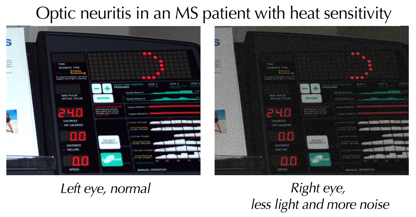
Optic neuritis describes any condition that causes inflammation of the optic nerve; it may be associated with demyelinating diseases, or infectious or inflammatory processes. It is also known as optic papillitis (when the head of the optic nerve is involved), neuroretinitis (when there is a combined involvement of the optic disc and surrounding retina in the macular area) and retrobulbar neuritis (when the posterior part of the nerve is involved). Prelaminar optic neuritis describes involvement of the non-myelinated axons in the retina. It is most often associated with multiple sclerosis, and it may lead to complete or partial loss of vision in one or both eyes. Other causes include: # Leber's hereditary optic neuropathy # Parainfectious optic neuritis (associated with viral infections such as measles, mumps, chickenpox, whooping cough and glandular fever) # Infectious optic neuritis (sinus related or associated with cat-scratch fever, tuberculosis, Lyme disease and cryptoco ... [...More Info...] [...Related Items...] OR: [Wikipedia] [Google] [Baidu] |
Anisocoria
Anisocoria is a condition characterized by an unequal size of the eyes' pupils. Affecting up to 20% of the population, anisocoria is often entirely harmless, but can be a sign of more serious medical problems. Causes Anisocoria is a common condition, defined by a difference of 0.4 mm or more between the sizes of the pupils of the eyes. Anisocoria has various causes: * Physiological anisocoria: About 20% of population has a slight difference in pupil size which is known as physiological anisocoria. In this condition, the difference between pupils is usually less than 1 mm. * Horner's syndrome * Mechanical anisocoria: Occasionally previous trauma, eye surgery, or inflammation (uveitis, angle closure glaucoma) can lead to adhesions between the iris and the lens. * Adie tonic pupil: Tonic pupil is usually an isolated benign entity, presenting in young women. It may be associated with loss of deep tendon reflex (Adie's syndrome). Tonic pupil is characterized by delayed d ... [...More Info...] [...Related Items...] OR: [Wikipedia] [Google] [Baidu] |


