|
Mitral Valve
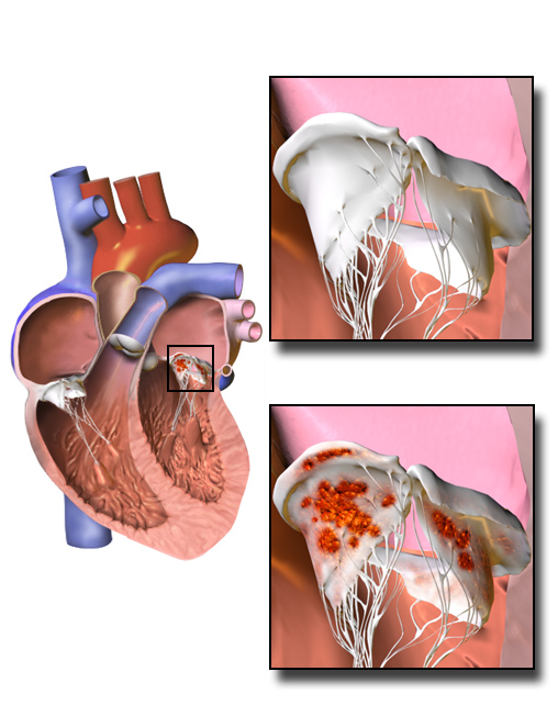
The mitral valve ( ), also known as the bicuspid valve or left atrioventricular valve, is one of the four heart valves. It has two Cusps of heart valves, cusps or flaps and lies between the atrium (heart), left atrium and the ventricle (heart), left ventricle of the heart. The heart valves are all one-way valves allowing blood flow in just one direction. The mitral valve and the tricuspid valve are known as the Heart valve#Atrioventricular valves, atrioventricular valves because they lie between the atria and the ventricles. In normal conditions, blood flows through an open mitral valve during diastole with contraction of the left atrium, and the mitral valve closes during systole with contraction of the left ventricle. The valve opens and closes because of pressure differences, opening when there is greater pressure in the left atrium than ventricle and closing when there is greater pressure in the left ventricle than atrium. In abnormal conditions, blood may flow backward thro ... [...More Info...] [...Related Items...] OR: [Wikipedia] [Google] [Baidu] |
Mitral Regurgitation
Mitral regurgitation (MR), also known as mitral insufficiency or mitral incompetence, is a form of valvular heart disease in which the mitral valve is insufficient and does not close properly when the heart pumps out blood. Section: Valvular Heart Disease Rupture or dysfunction of the papillary muscle are also common causes in acute cases, dysfunction, which can include mitral valve prolapse.VOC=VITIUM ORGANICUM CORDIS, a compendium of the Department of Cardiology at Uppsala Academic Hospital. By Per Kvidal September 1999, with revision by Erik Björklund May 2008 Pathophysiology The pathophysiology of MR can be broken into three phases of the disease process: the acute phase, the chronic compensated phase, and the chronic decompensated phase. Acute phase Acute MR (as may occur due to the sudden rupture of the chordae tendinae or papillary muscle) causes a sudden volume overload of both the left atrium and the left ventricle. The left ventricle develops volume overload because ... [...More Info...] [...Related Items...] OR: [Wikipedia] [Google] [Baidu] |
Bicuspid Aortic Valve
Bicuspid aortic valve (BAV) is a form of heart disease in which two of the leaflets of the aortic valve fuse during development in the womb resulting in a two-leaflet (bicuspid) valve instead of the normal three-leaflet (tricuspid) valve. BAV is the most common cause of heart disease present at birth and affects approximately 1.3% of adults. Normally, the mitral valve is the only bicuspid valve and this is situated between the heart's left atrium and left ventricle. Heart valves play a crucial role in ensuring the unidirectional flow of blood from the atria to the ventricles, or from the ventricle to the aorta or pulmonary trunk. BAV is normally inherited. Signs and symptoms In many cases, a bicuspid aortic valve will cause no problems. People with BAV may become tired more easily than those with normal valvular function and have difficulty maintaining stamina for cardio-intensive activities due to poor heart performance caused by stress on the aortic wall.Maredia A.K., G ... [...More Info...] [...Related Items...] OR: [Wikipedia] [Google] [Baidu] |
Infective Endocarditis
Infective endocarditis is an infection of the inner surface of the heart (endocardium), usually the heart valve, valves. Signs and symptoms may include fever, petechia, small areas of bleeding into the skin, heart murmur, feeling tired, and anemia, low red blood cell count. Complications may include valvular insufficiency, backward blood flow in the heart, heart failure – the heart struggling to pump a sufficient amount of blood to meet the body's needs, Heart block, abnormal electrical conduction in the heart, stroke, and kidney failure. The cause is typically a bacterial infection and less commonly a fungal infection. Risk factors include valvular heart disease, including rheumatic heart disease, rheumatic disease, congenital heart disease, artificial valves, hemodialysis, intravenous drug use, and electronic pacemakers. The bacteria most commonly involved are streptococci or staphylococci. Diagnosis is suspected based on symptoms and supported by blood cultures or Echocardi ... [...More Info...] [...Related Items...] OR: [Wikipedia] [Google] [Baidu] |
Fibrous Rings Of Heart
In cardiology, the cardiac skeleton, also known as the fibrous skeleton of the heart, is a high-density homogeneous structure of connective tissue that forms and anchors the valves of the heart, and influences the forces exerted by and through them. The cardiac skeleton separates and partitions the atria (the smaller, upper two chambers) from the ventricles (the larger, lower two chambers). The heart's cardiac skeleton comprises four dense connective tissue rings that encircle the mitral and tricuspid atrioventricular (AV) canals and extend to the origins of the pulmonary trunk and aorta. This provides crucial support and structure to the heart while also serving to electrically isolate the atria from the ventricles. The unique matrix of connective tissue within the cardiac skeleton isolates electrical influence within these defined chambers. In normal anatomy, there is only one conduit for electrical conduction from the upper chambers to the lower chambers, known as the atriov ... [...More Info...] [...Related Items...] OR: [Wikipedia] [Google] [Baidu] |
Regurgitation (circulation)
Regurgitation is blood flow in the opposite direction from normal, as the backward flowing of blood into the heart or between heart chambers. It is the circulatory equivalent of backflow in engineered systems. It is sometimes called reflux. Types of heart valve regurgitation The various types of heart valve regurgitation via insufficiency are as follows: # Aortic regurgitation: the backflow of blood from the aorta into the left ventricle, owing to insufficiency of the aortic semilunar valve; it may be chronic or acute. # Mitral regurgitation: the backflow of blood from the left ventricle into the left atrium, owing to insufficiency of the mitral valve; it may be acute or chronic, and is usually due to mitral valve prolapse, rheumatic heart disease, or a complication of cardiac dilatation. See also Mitral regurgitation. # Pulmonic regurgitation: the backflow of blood from the pulmonary artery into the right ventricle, owing to insufficiency of the pulmonic semilunar v ... [...More Info...] [...Related Items...] OR: [Wikipedia] [Google] [Baidu] |
Papillary Muscle
The papillary muscles are muscles located in the ventricles of the heart. They attach to the cusps of the atrioventricular valves (also known as the mitral and tricuspid valves) via the chordae tendineae and contract to prevent inversion or prolapse of these valves on systole (or ventricular contraction). Structure There are five total papillary muscles in the heart; three in the right ventricle and two in the left ventricle. The anterior, posterior, and septal papillary muscles of the right ventricle each attach via chordae tendineae to the tricuspid valve. The anterolateral and posteromedial papillary muscles of the left ventricle attach via chordae tendineae to the mitral valve. Blood supply to the left ventricle The mitral valve papillary muscles in the left ventricle are called the anterolateral and posteromedial muscles. * Anterolateral muscle blood supply: left anterior descending artery - diagonal branch (LAD) and left circumflex artery - obtuse marginal bra ... [...More Info...] [...Related Items...] OR: [Wikipedia] [Google] [Baidu] |
Tendon
A tendon or sinew is a tough band of fibrous connective tissue, dense fibrous connective tissue that connects skeletal muscle, muscle to bone. It sends the mechanical forces of muscle contraction to the skeletal system, while withstanding tension (physics), tension. Tendons, like ligaments, are made of collagen. The difference is that ligaments connect bone to bone, while tendons connect muscle to bone. There are about 4,000 tendons in the adult human body. Structure A tendon is made of dense regular connective tissue, whose main cellular components are special fibroblasts called tendon cells (tenocytes). Tendon cells synthesize the tendon's extracellular matrix, which abounds with densely-packed collagen fibers. The collagen fibers run parallel to each other and are grouped into fascicles. Each fascicle is bound by an endotendineum, which is a delicate loose connective tissue containing thin collagen fibrils and elastic fibers. A set of fascicles is bound by an epitenon, whi ... [...More Info...] [...Related Items...] OR: [Wikipedia] [Google] [Baidu] |
Chordae Tendineae
The chordae tendineae (: chorda tendinea) or tendinous cords, colloquially known as the heart strings, are inelastic cords of fibrous connective tissue that connect the papillary muscles to the tricuspid valve and the mitral valve in the heart. Structure The chordae tendineae connect the atrioventricular valves ( tricuspid and mitral), to the papillary muscles within the ventricles. Multiple chordae tendineae attach to each leaflet or cusp of the valves. Chordae tendineae contain elastin in a delicate structure notably at their periphery. Function During atrial systole, blood flows from the atria to the ventricles down the pressure gradient. Chordae tendineae are relaxed because the atrioventricular valves are forced open. When the ventricles of the heart contract in ventricular systole, the increased blood pressures in both chambers push the AV valves to close simultaneously, preventing the backflow of blood into the atria. Since the blood pressure in the atria is much ... [...More Info...] [...Related Items...] OR: [Wikipedia] [Google] [Baidu] |
Heart Left Atrial Appendage Tee View
The heart is a muscular organ found in humans and other animals. This organ pumps blood through the blood vessels. The heart and blood vessels together make the circulatory system. The pumped blood carries oxygen and nutrients to the tissue, while carrying metabolic waste such as carbon dioxide to the lungs. In humans, the heart is approximately the size of a closed fist and is located between the lungs, in the middle compartment of the chest, called the mediastinum. In humans, the heart is divided into four chambers: upper left and right atria and lower left and right ventricles. Commonly, the right atrium and ventricle are referred together as the right heart and their left counterparts as the left heart. In a healthy heart, blood flows one way through the heart due to heart valves, which prevent backflow. The heart is enclosed in a protective sac, the pericardium, which also contains a small amount of fluid. The wall of the heart is made up of three layers: epicardi ... [...More Info...] [...Related Items...] OR: [Wikipedia] [Google] [Baidu] |
Posterior Leaflet Of Left Atrioventricular Valve
Posterior may refer to: * Posterior (anatomy), the end of an organism opposite to anterior ** Buttocks, as a euphemism * Posterior horn (other) * Posterior probability The posterior probability is a type of conditional probability that results from updating the prior probability with information summarized by the likelihood via an application of Bayes' rule. From an epistemological perspective, the posteri ..., the conditional probability that is assigned when the relevant evidence is taken into account * Posterior tense, a relative future tense {{disambiguation ... [...More Info...] [...Related Items...] OR: [Wikipedia] [Google] [Baidu] |
Mitral Valve
The mitral valve ( ), also known as the bicuspid valve or left atrioventricular valve, is one of the four heart valves. It has two Cusps of heart valves, cusps or flaps and lies between the atrium (heart), left atrium and the ventricle (heart), left ventricle of the heart. The heart valves are all one-way valves allowing blood flow in just one direction. The mitral valve and the tricuspid valve are known as the Heart valve#Atrioventricular valves, atrioventricular valves because they lie between the atria and the ventricles. In normal conditions, blood flows through an open mitral valve during diastole with contraction of the left atrium, and the mitral valve closes during systole with contraction of the left ventricle. The valve opens and closes because of pressure differences, opening when there is greater pressure in the left atrium than ventricle and closing when there is greater pressure in the left ventricle than atrium. In abnormal conditions, blood may flow backward thro ... [...More Info...] [...Related Items...] OR: [Wikipedia] [Google] [Baidu] |


