|
Regurgitation (circulation)
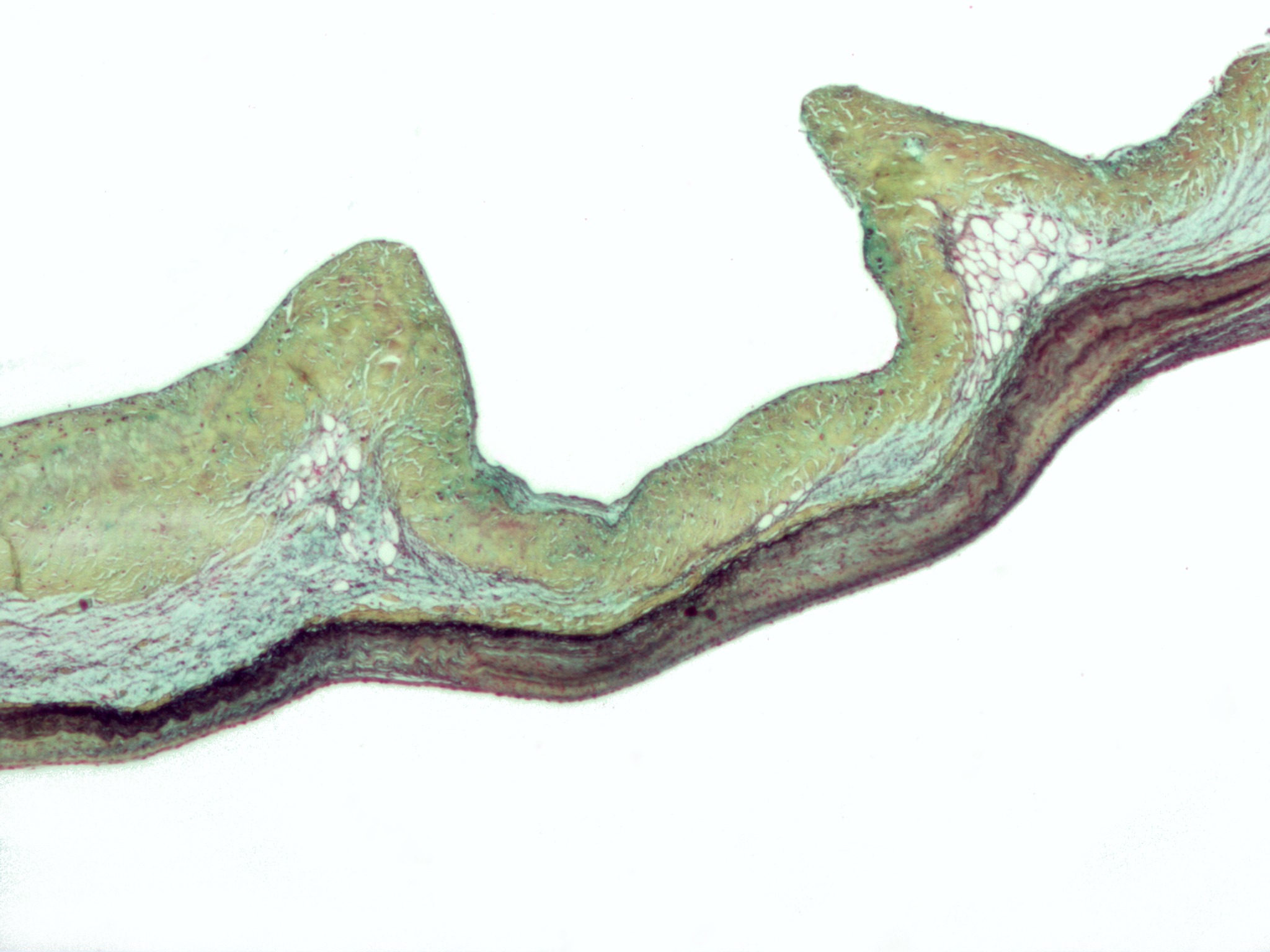
Regurgitation is blood flow in the opposite direction from normal, as the backward flowing of blood into the heart or between heart chambers. It is the circulatory equivalent of backflow in engineered systems. It is sometimes called reflux. Types of heart valve regurgitation The various types of heart valve regurgitation via insufficiency are as follows: # Aortic regurgitation: the backflow of blood from the aorta into the left ventricle, owing to insufficiency of the aortic semilunar valve; it may be chronic or acute. # Mitral regurgitation: the backflow of blood from the left ventricle into the left atrium, owing to insufficiency of the mitral valve; it may be acute or chronic, and is usually due to mitral valve prolapse, rheumatic heart disease, or a complication of cardiac dilatation. See also Mitral regurgitation. # Pulmonic regurgitation: the backflow of blood from the pulmonary artery into the right ventricle, owing to insufficiency of the pulmonic semilunar v ... [...More Info...] [...Related Items...] OR: [Wikipedia] [Google] [Baidu] |
Blood Flow
Hemodynamics American and British English spelling differences#ae and oe, or haemodynamics are the Fluid dynamics, dynamics of blood flow. The circulatory system is controlled by homeostasis, homeostatic mechanisms of autoregulation, just as hydraulic circuits are controlled by control systems. The hemodynamic response continuously monitors and adjusts to conditions in the body and its environment. Hemodynamics explains the physical laws that govern the flow of blood in the blood vessels. Blood flow ensures the transportation of nutrients, hormones, metabolic waste products, oxygen, and carbon dioxide throughout the body to maintain cell-level metabolism, the regulation of the pH, osmotic pressure and temperature of the whole body, and the protection from microbial and mechanical harm. Blood is a non-Newtonian fluid, and is most efficiently studied using rheology rather than hydrodynamics. Because blood vessels are not rigid tubes, classic hydrodynamics and fluids mechanics based o ... [...More Info...] [...Related Items...] OR: [Wikipedia] [Google] [Baidu] |
Tricuspid Valve
The tricuspid valve, or right atrioventricular valve, is on the right dorsal side of the mammalian heart, at the superior portion of the right ventricle. The function of the valve is to allow blood to flow from the right atrium to the right ventricle during diastole, and to close to prevent backflow (Regurgitation (circulation), regurgitation) from the right ventricle into the right atrium during right ventricular contraction (systole). Structure The tricuspid valve usually has three Cusps of heart valves, cusps or leaflets, named the anterior, posterior, and septal cusps. Each leaflet is connected via chordae tendineae to the anterior, posterior, and septal papillary muscles of the right ventricle, respectively. Tricuspid valves may also occur with two or four leaflets; the number may change over a lifetime. Function The tricuspid valve functions as a one-way valve that closes during ventricular systole to prevent regurgitation of blood from the right ventricle back into th ... [...More Info...] [...Related Items...] OR: [Wikipedia] [Google] [Baidu] |
Stenosis
Stenosis () is the abnormal narrowing of a blood vessel or other tubular organ or structure such as foramina and canals. It is also sometimes called a stricture (as in urethral stricture). ''Stricture'' as a term is usually used when narrowing is caused by contraction of smooth muscle (e.g. achalasia, prinzmetal angina); ''stenosis'' is usually used when narrowing is caused by lesion that reduces the space of lumen (e.g. atherosclerosis). The term coarctation is another synonym, but is commonly used only in the context of aortic coarctation. Restenosis is the recurrence of stenosis after a procedure. Examples Examples of vascular stenotic lesions include: * Intermittent claudication (peripheral artery stenosis) * Angina ( coronary artery stenosis) * Carotid artery stenosis which predispose to (strokes and transient ischaemic episodes) * Renal artery stenosis Types In heart valves The types of stenoses in heart valves are: * Pulmonary valve stenosis, which is th ... [...More Info...] [...Related Items...] OR: [Wikipedia] [Google] [Baidu] |
Right Ventricular Volume Overload
Volume overload refers to the state of one of the Heart#Structure, chambers of the heart in which too large a volume of blood exists within it for it to function efficiently. Ventricle (heart), Ventricular volume overload is approximately equivalent to an excessively high Preload (cardiology), preload. It is a cause of Heart failure, cardiac failure. Pathophysiology In accordance with the Frank–Starling law of the heart, the myocardium contracts more powerfully as the end-diastolic volume increases. Stretching of the myofibrils in cardiac muscle causes them to contract more powerfully due to a greater number of cross-bridges being formed between the myofibrils within cardiac myocytes.Klabunde, Richard E. "Cardiovascular Physiology Concepts". Lippincott Williams & Wilkins, 2011, p. 74. This is true up to a point, however beyond this there is a loss of contractile ability due to loss of connection between myofibrils; see figure. Various pathologies, listed below, can lead to vol ... [...More Info...] [...Related Items...] OR: [Wikipedia] [Google] [Baidu] |
Cardiac Muscle
Cardiac muscle (also called heart muscle or myocardium) is one of three types of vertebrate muscle tissues, the others being skeletal muscle and smooth muscle. It is an involuntary, striated muscle that constitutes the main tissue of the wall of the heart. The cardiac muscle (myocardium) forms a thick middle layer between the outer layer of the heart wall (the pericardium) and the inner layer (the endocardium), with blood supplied via the coronary circulation. It is composed of individual cardiac muscle cells joined by intercalated discs, and encased by collagen fibers and other substances that form the extracellular matrix. Cardiac muscle contracts in a similar manner to skeletal muscle, although with some important differences. Electrical stimulation in the form of a cardiac action potential triggers the release of calcium from the cell's internal calcium store, the sarcoplasmic reticulum. The rise in calcium causes the cell's myofilaments to slide past each other i ... [...More Info...] [...Related Items...] OR: [Wikipedia] [Google] [Baidu] |
Framingham Heart Study
The Framingham Heart Study is a long-term, ongoing cardiovascular cohort study of residents of the city of Framingham, Massachusetts. The study began in 1948 with 5,209 adult subjects from Framingham, and is now on its third generation of participants. Prior to the study almost nothing was known about the epidemiology of hypertensive or arteriosclerotic cardiovascular disease. Much of the now-common knowledge concerning heart disease, such as the effects of diet, exercise, and common medications such as aspirin, is based on this longitudinal study. It is a project of the National Heart, Lung, and Blood Institute, in collaboration with (since 1971) Boston University. Various health professionals from the hospitals and universities of Greater Boston staff the project. History In 1948, the study was commissioned by the United States Congress, with multiple communities being considered for study. The final choice was between Framingham, Massachusetts, and Paintsville, Kentucky. ... [...More Info...] [...Related Items...] OR: [Wikipedia] [Google] [Baidu] |
Stroke Volume
In cardiovascular physiology, stroke volume (SV) is the volume of blood pumped from the ventricle (heart), ventricle per beat. Stroke volume is calculated using measurements of ventricle volumes from an Echocardiography, echocardiogram and subtracting the volume of the blood in the ventricle at the end of a beat (called end-systolic volume) from the volume of blood just prior to the beat (called end-diastolic volume). The term ''stroke volume'' can apply to each of the two ventricles of the heart, although when not explicitly stated it refers to the left ventricle and should therefore be referred to as left stroke volume (LSV). The stroke volumes for each ventricle are generally equal, both being approximately 90 mL in a healthy 70-kg man. Any persistent difference between the two stroke volumes, no matter how small, would inevitably lead to venous congestion of either the systemic or the pulmonary circulation, with a corresponding state of hypotension in the other circulatory sys ... [...More Info...] [...Related Items...] OR: [Wikipedia] [Google] [Baidu] |
Mitral Insufficiency
Mitral regurgitation (MR), also known as mitral insufficiency or mitral incompetence, is a form of valvular heart disease in which the mitral valve is insufficient and does not close properly when the heart pumps out blood. Section: Valvular Heart Disease Rupture or dysfunction of the papillary muscle are also common causes in acute cases, dysfunction, which can include mitral valve prolapse.VOC=VITIUM ORGANICUM CORDIS, a compendium of the Department of Cardiology at Uppsala Academic Hospital. By Per Kvidal September 1999, with revision by Erik Björklund May 2008 Pathophysiology The pathophysiology of MR can be broken into three phases of the disease process: the acute phase, the chronic compensated phase, and the chronic decompensated phase. Acute phase Acute MR (as may occur due to the sudden rupture of the chordae tendinae or papillary muscle) causes a sudden volume overload of both the left atrium and the left ventricle. The left ventricle develops volume overload becau ... [...More Info...] [...Related Items...] OR: [Wikipedia] [Google] [Baidu] |
Left Ventricle
A ventricle is one of two large chambers located toward the bottom of the heart that collect and expel blood towards the peripheral beds within the body and lungs. The blood pumped by a ventricle is supplied by an atrium, an adjacent chamber in the upper heart that is smaller than a ventricle. Interventricular means between the ventricles (for example the interventricular septum), while intraventricular means within one ventricle (for example an intraventricular block). In a four-chambered heart, such as that in humans, there are two ventricles that operate in a double circulatory system: the right ventricle pumps blood into the pulmonary circulation to the lungs, and the left ventricle pumps blood into the systemic circulation through the aorta. Structure Ventricles have thicker walls than atria and generate higher blood pressures. The physiological load on the ventricles requiring pumping of blood throughout the body and lungs is much greater than the pressure generated by t ... [...More Info...] [...Related Items...] OR: [Wikipedia] [Google] [Baidu] |
Metonymy
Metonymy () is a figure of speech in which a concept is referred to by the name of something associated with that thing or concept. For example, the word " suit" may refer to a person from groups commonly wearing business attire, such as salespeople or attorneys. Etymology The words ''metonymy'' and ''metonym'' come ; , a suffix that names figures of speech, . Background Metonymy and related figures of speech are common in everyday speech and writing. Synecdoche and metalepsis are considered specific types of metonymy. Polysemy, the capacity for a word or phrase to have multiple meanings, sometimes results from relations of metonymy. Both metonymy and metaphor involve the substitution of one term for another. In metaphor, this substitution is based on some specific analogy between two things, whereas in metonymy the substitution is based on some understood association or contiguity. American literary theorist Kenneth Burke considers metonymy as one of four "master tro ... [...More Info...] [...Related Items...] OR: [Wikipedia] [Google] [Baidu] |
Aortic Regurgitation
Aortic regurgitation (AR), also known as aortic insufficiency (AI), is the leaking of the aortic valve of the heart that causes blood to flow in the reverse direction during ventricular diastole, from the aorta into the left ventricle. As a consequence, the cardiac muscle is forced to work harder than normal. Signs and symptoms Symptoms of aortic regurgitation are similar to those of heart failure and include the following: * Dyspnea on exertion * Orthopnea * Paroxysmal nocturnal dyspnea * Palpitations * Angina pectoris * Cyanosis (in acute cases) Causes In terms of the cause of aortic regurgitation, is often due to the aortic root dilation ('' annuloaortic ectasia''), which is idiopathic in over 80% of cases, but otherwise may result from aging, syphilitic aortitis, osteogenesis imperfecta, aortic dissection, Behçet's disease, reactive arthritis and systemic hypertension.Chapter 1: Diseases of the Cardiovascular system > Section: Valvular Heart Disease in: Aortic root ... [...More Info...] [...Related Items...] OR: [Wikipedia] [Google] [Baidu] |


