|
Divisome
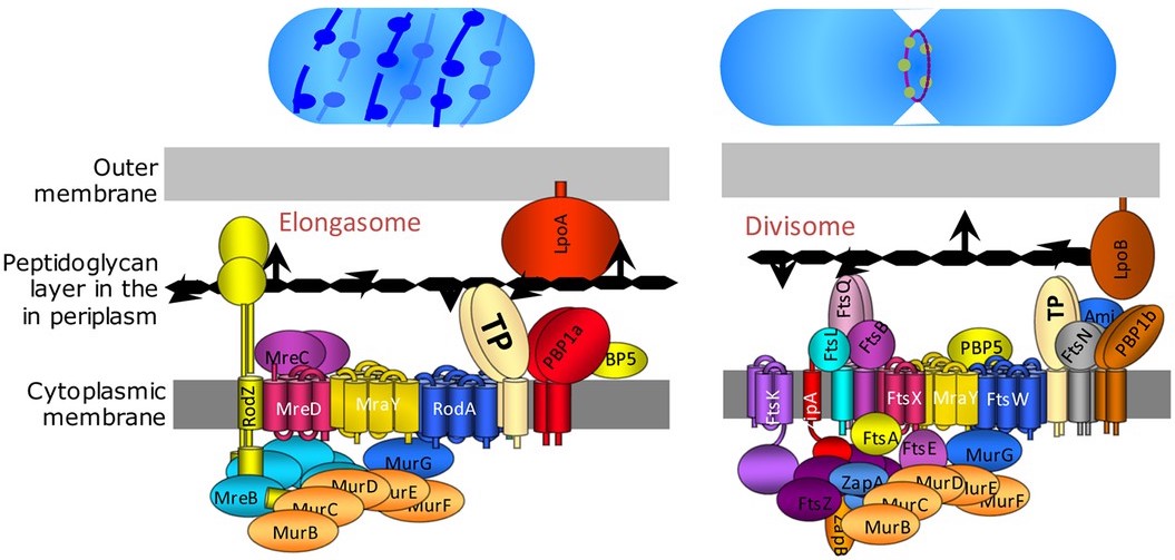
The divisome is a protein complex in bacteria that is responsible for cell division, constriction of inner and outer membranes during division, and peptidoglycan (PG) synthesis at the division site. The divisome is a membrane protein complex with proteins on both sides of the cytoplasmic membrane. In gram-negative cells it is located in the inner membrane. The divisome is nearly ubiquitous in bacteria although its composition may vary between species. The elongasome is a modified version of the divisome, without the membrane-constricting FtsZ-ring and its associated machinery. The elongasome is present only in non-spherical bacteria and directs lateral insertion of PG along the long axis of the cell, thus allowing cylindrical growth (as opposed to spherical growth, as in cocci). History Some of the first cell-division genes of ''Escherichia coli'' were discovered by François Jacob's group in France in the 1960s. They were called ''fts'' genes, because mutants of these gene ... [...More Info...] [...Related Items...] OR: [Wikipedia] [Google] [Baidu] |
FtsK
FtsK is a protein involved in bacterial cell division and chromosome segregation. In ''E. coli'', is one of the largest proteins, consisting of 1329 amino acids. FtsK stands for "Filament temperature sensitive mutant K" because cells expressing a mutant ''ftsK'' allele called ''ftsK44'', which encodes an FtsK variant containing an G80A residue change in the second transmembrane segment, fail to divide at high temperatures and form long filaments instead. FtsK, specifically its C-terminal domain, functions as a DNA translocase, interacts with other cell division proteins, and regulates Xer-mediated recombination. FtsK belongs to the AAA (ATPase Associated with various cellular Activities) superfamily and is present in most bacteria. Structure FtsK is a transmembrane protein composed of three domains: FtsKN, FtsKL, and FtsKC. FtsK functions to coordinate cell division and chromosome segregation through its N-terminal and C-terminal domains. The FtsKN domain is embedded in the c ... [...More Info...] [...Related Items...] OR: [Wikipedia] [Google] [Baidu] |
Cyanidioschyzon Merolae
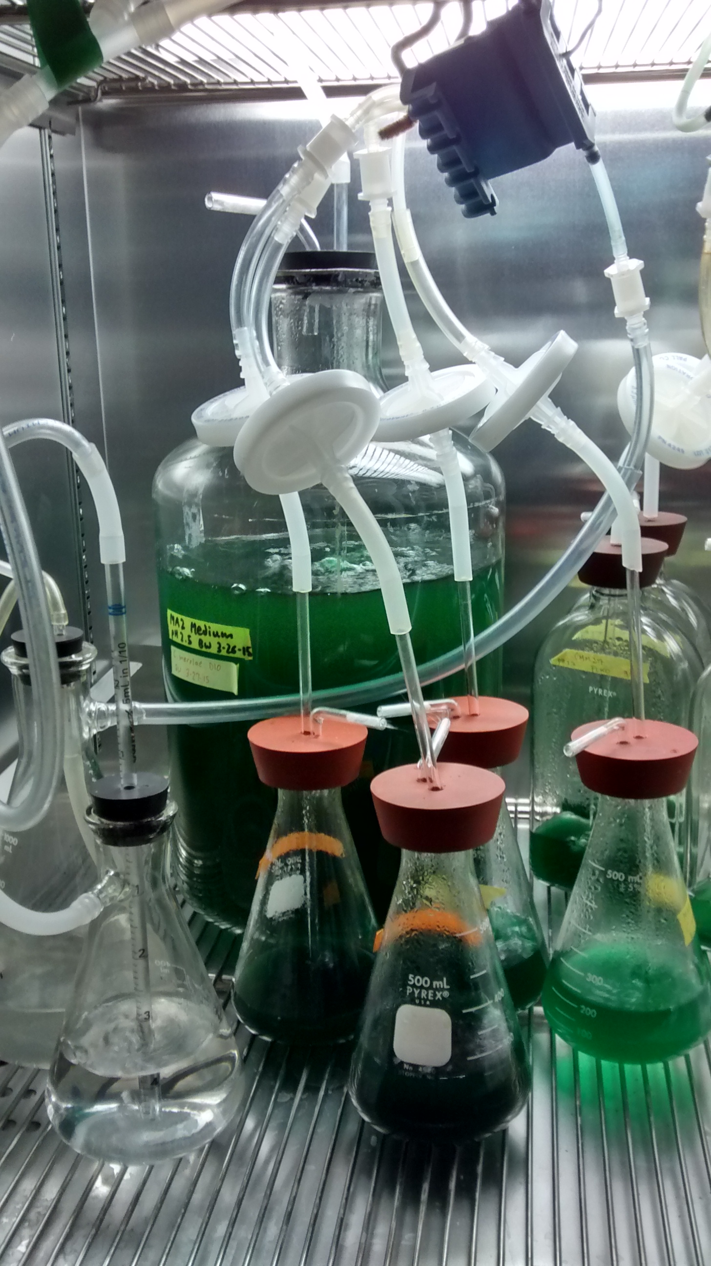
''Cyanidioschyzon merolae'' is a small (2μm), club-shaped, unicellular haploid red alga adapted to high sulfur acidic hot spring environments (pH 1.5, 45 °C). The cellular architecture of ''C. merolae'' is extremely simple, containing only a single chloroplast and a single mitochondrion and lacking a vacuole and cell wall. In addition, the cellular and organelle divisions can be synchronized. For these reasons, ''C. merolae'' is considered an excellent model system for study of cellular and organelle division processes, as well as biochemistry and structural biology. The organism's genome was the first full algal genome to be sequenced in 2004; its plastid was sequenced in 2000 and 2003, and its mitochondrion in 1998. The organism has been considered the simplest of eukaryotic cells for its minimalist cellular organization. Isolation and growth in culture Originally isolated by De Luca in 1978 from the solfatane fumaroles of Campi Flegrei ( Naples, Italy), ''C. merola ... [...More Info...] [...Related Items...] OR: [Wikipedia] [Google] [Baidu] |
Tubulin
Tubulin in molecular biology can refer either to the tubulin protein superfamily of globular proteins, or one of the member proteins of that superfamily. α- and β-tubulins polymerize into microtubules, a major component of the eukaryotic cytoskeleton. It was discovered and named by Hideo Mōri in 1968. Microtubules function in many essential cellular processes, including mitosis. Discovery and development of tubulin inhibitors, Tubulin-binding drugs kill cancerous cells by inhibiting microtubule dynamics, which are required for DNA segregation and therefore cell division. #Eukaryotic, In eukaryotes, there are six members of the tubulin superfamily, although not all are present in all species.Turk E, Wills AA, Kwon T, Sedzinski J, Wallingford JB, Stearns "Zeta-Tubulin Is a Member of a Conserved Tubulin Module and Is a Component of the Centriolar Basal Foot in Multiciliated Cells"Current Biology (2015) 25:2177-2183. Both α and β tubulins have a mass of around 50 kDa and are thus ... [...More Info...] [...Related Items...] OR: [Wikipedia] [Google] [Baidu] |
Cell Division
Cell division is the process by which a parent cell (biology), cell divides into two daughter cells. Cell division usually occurs as part of a larger cell cycle in which the cell grows and replicates its chromosome(s) before dividing. In eukaryotes, there are two distinct types of cell division: a vegetative division (mitosis), producing daughter cells genetically identical to the parent cell, and a cell division that produces Haploidisation, haploid gametes for sexual reproduction (meiosis), reducing the number of chromosomes from two of each type in the diploid parent cell to one of each type in the daughter cells. Mitosis is a part of the cell cycle, in which, replicated chromosomes are separated into two new Cell nucleus, nuclei. Cell division gives rise to genetically identical cells in which the total number of chromosomes is maintained. In general, mitosis (division of the nucleus) is preceded by the S stage of interphase (during which the DNA replication occurs) and is f ... [...More Info...] [...Related Items...] OR: [Wikipedia] [Google] [Baidu] |
Peptidoglycan
Peptidoglycan or murein is a unique large macromolecule, a polysaccharide, consisting of sugars and amino acids that forms a mesh-like layer (sacculus) that surrounds the bacterial cytoplasmic membrane. The sugar component consists of alternating residues of β-(1,4) linked N-Acetylglucosamine, ''N''-acetylglucosamine (NAG) and N-Acetylmuramic acid, ''N''-acetylmuramic acid (NAM). Attached to the ''N''-acetylmuramic acid is an oligopeptide chain made of three to five amino acids. The peptide chain can be cross-linked to the peptide chain of another strand forming the 3D mesh-like layer. Peptidoglycan serves a structural role in the bacterial cell wall, giving structural strength, as well as counteracting the osmotic pressure of the cytoplasm. This repetitive linking results in a dense peptidoglycan layer which is critical for maintaining cell form and withstanding high osmotic pressures, and it is regularly replaced by peptidoglycan production. Peptidoglycan hydrolysis and synthesis ... [...More Info...] [...Related Items...] OR: [Wikipedia] [Google] [Baidu] |
Gram-negative Bacteria
Gram-negative bacteria are bacteria that, unlike gram-positive bacteria, do not retain the Crystal violet, crystal violet stain used in the Gram staining method of bacterial differentiation. Their defining characteristic is that their cell envelope consists of a thin peptidoglycan gram-negative cell wall, cell wall sandwiched between an inner (Cytoplasm, cytoplasmic) Cell membrane, membrane and an Bacterial outer membrane, outer membrane. These bacteria are found in all environments that support life on Earth. Within this category, notable species include the model organism ''Escherichia coli'', along with various pathogenic bacteria, such as ''Pseudomonas aeruginosa'', ''Chlamydia trachomatis'', and ''Yersinia pestis''. They pose significant challenges in the medical field due to their outer membrane, which acts as a protective barrier against numerous Antibiotic, antibiotics (including penicillin), Detergent, detergents that would normally damage the inner cell membrane, and the ... [...More Info...] [...Related Items...] OR: [Wikipedia] [Google] [Baidu] |
FtsZ
FtsZ is a protein encoded by the ''ftsZ'' gene that assembles into a ring at the future site of bacterial cell division (also called the Z ring). FtsZ is a prokaryotic homologue of the eukaryotic protein tubulin. The initials FtsZ mean "Filamenting temperature-sensitive mutant Z." The hypothesis was that cell division mutants of '' E. coli'' would grow as filaments due to the inability of the daughter cells to separate from one another. FtsZ is found in almost all bacteria, many archaea, all chloroplasts and some mitochondria, where it is essential for cell division. FtsZ assembles the cytoskeletal scaffold of the Z ring that, along with additional proteins, constricts to divide the cell in two. History In the 1960s scientists screened for temperature sensitive mutations that blocked cell division at 42 °C. The mutant cells divided normally at 30°, but failed to divide at 42°. Continued growth without division produced long filamentous cells (Filamenting temperature ... [...More Info...] [...Related Items...] OR: [Wikipedia] [Google] [Baidu] |
Coccus
Bacterial cellular morphologies are the shapes that are characteristic of various types of bacteria and often key to their identification. Their direct examination under a light microscope enables the classification of these bacteria (and archaea). Generally, the basic morphologies are spheres (coccus) and round-ended cylinders or rod shaped (bacillus). But, there are also other morphologies such as helically twisted cylinders (example '' Spirochetes''), cylinders curved in one plane (selenomonads) and unusual morphologies (the square, flat box-shaped cells of the Archaean genus '' Haloquadratum)''. Other arrangements include pairs, tetrads, clusters, chains and palisades. Types Coccus A coccus (plural ''cocci'', from the Latin ''coccinus'' (scarlet) and derived from the Greek ''kokkos'' (berry)), is any microorganism (usually bacteria) whose overall shape is spherical or nearly spherical. Coccus refers to the shape of the bacteria and can contain multiple genera, such as st ... [...More Info...] [...Related Items...] OR: [Wikipedia] [Google] [Baidu] |
François Jacob
François Jacob (; 17 June 1920 – 19 April 2013) was a French biologist who, together with Jacques Monod, originated the idea that control of enzyme levels in all cells occurs through regulation of transcription. He shared the 1965 Nobel Prize in Medicine with Jacques Monod and André Lwoff. Early years Jacob was born the only child of Simon, a merchant, and Thérèse (Franck) Jacob, in Nancy, France. An inquisitive child, he learned to read at a young age. Albert Franck, Jacob's maternal grandfather, a four-star general, was Jacob's childhood role model. At seven he entered the Lycée Carnot, where he was schooled for the next ten years; in his autobiography, he describes his impression of it: "a cage". He was antagonized by rightist youth at the Lycée Carnot around 1934. He describes his father as a "conformist in religion", while his mother and other family members important in his childhood were secular Jews; shortly after his bar mitzvah, he became an athe ... [...More Info...] [...Related Items...] OR: [Wikipedia] [Google] [Baidu] |
Protein Complex
A protein complex or multiprotein complex is a group of two or more associated polypeptide chains. Protein complexes are distinct from multidomain enzymes, in which multiple active site, catalytic domains are found in a single polypeptide chain. Protein complexes are a form of protein quaternary structure, quaternary structure. Proteins in a protein complex are linked by non-covalent interactions, non-covalent protein–protein interactions. These complexes are a cornerstone of many (if not most) biological processes. The cell is seen to be composed of modular supramolecular complexes, each of which performs an independent, discrete biological function. Through proximity, the speed and selectivity of binding interactions between Enzyme, enzymatic complex and substrates can be vastly improved, leading to higher cellular efficiency. Many of the techniques used to enter cells and isolate proteins are inherently disruptive to such large complexes, complicating the task of determining ... [...More Info...] [...Related Items...] OR: [Wikipedia] [Google] [Baidu] |
Gram-positive Bacteria
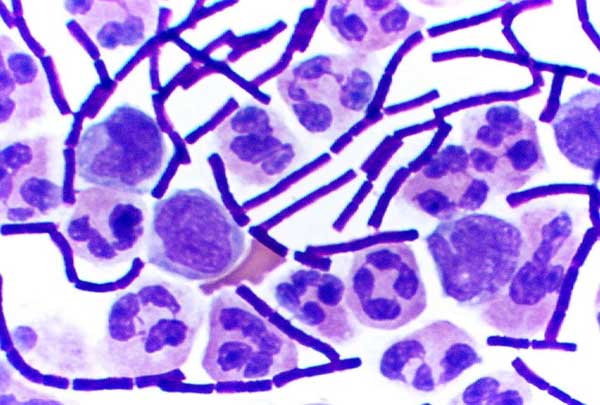
In bacteriology, gram-positive bacteria are bacteria that give a positive result in the Gram stain test, which is traditionally used to quickly classify bacteria into two broad categories according to their type of cell wall. The Gram stain is used by microbiologists to place bacteria into two main categories, gram-positive (+) and gram-negative (−). Gram-positive bacteria have a thick layer of peptidoglycan within the cell wall, and gram-negative bacteria have a thin layer of peptidoglycan. Gram-positive bacteria retain the crystal violet stain used in the test, resulting in a purple color when observed through an optical microscope. The thick layer of peptidoglycan in the bacterial cell wall retains the stain after it has been fixed in place by iodine. During the decolorization step, the decolorizer removes crystal violet from all other cells. Conversely, gram-negative bacteria cannot retain the violet stain after the decolorization step; alcohol used in this stage ... [...More Info...] [...Related Items...] OR: [Wikipedia] [Google] [Baidu] |


