|
Cystadenoma
Cystadenoma is a type of cystic adenoma. When malignant, it is called cystadenocarcinoma. Classification When not otherwise specified, the ICD-O coding is 8440/0. However, the following classifications also exist: By form * serous cystadenoma (8441-8442) * papillary cystadenoma (8450-8451, 8561) * mucinous cystadenoma (8470-8473) By location * Bile duct A bile duct is any of a number of long tube-like structures that carry bile, and is present in most vertebrates. The bile duct is separated into three main parts: the fundus (superior), the body (middle), and the neck (inferior). Bile is requ ... cystadenoma (8161) or biliary cystadenoma is a slow-growing tumour arising from bile ducts of the liver. The presence of endocrine cells in the tumour also indicates its origin from the glands surrounding the bile ducts. The incidence is 1–5 in 100,000 people. Females are affected more than males at 9:1 ratio. Mean age of presentation is at 45 years old. About 30% of biliary c ... [...More Info...] [...Related Items...] OR: [Wikipedia] [Google] [Baidu] |
ICD-O
The International Classification of Diseases for Oncology (ICD-O) is a domain-specific extension of the International Statistical Classification of Diseases and Related Health Problems for tumor diseases. This classification is widely used by cancer registries. It is currently in its third revision (ICD-O-3). ICD-10 includes a list of morphology codes. They stem from ICD-O second edition (ICD-O-2) that was valid at the time of publication. Axes The classification has two axes: topography and morphology. Morphology The morphology axis addresses the microscopic structure (histology) of the tumor. This axis has particular importance because the Systematized Nomenclature of Medicine ("SNOMED") has adopted the ICD-O classification of morphology. SNOMED has been changing continuously, and several different versions of SNOMED are in use. Accordingly, mapping of ICD-O codes to SNOMED requires careful assessment of whether entities are indeed true matches. Topography The topograp ... [...More Info...] [...Related Items...] OR: [Wikipedia] [Google] [Baidu] |
Cystic
A cyst is a closed sac, having a distinct envelope and division compared with the nearby tissue. Hence, it is a cluster of cells that have grouped together to form a sac (like the manner in which water molecules group together to form a bubble); however, the distinguishing aspect of a cyst is that the cells forming the "shell" of such a sac are distinctly abnormal (in both appearance and behaviour) when compared with all surrounding cells for that given location. A cyst may contain air, fluids, or semi-solid material. A collection of pus is called an abscess, not a cyst. Once formed, a cyst may resolve on its own. When a cyst fails to resolve, it may need to be removed surgically, but that would depend upon its type and location. Cancer-related cysts are formed as a defense mechanism for the body following the development of mutations that lead to an uncontrolled cellular division. Once that mutation has occurred, the affected cells divide incessantly and become cancerous, ... [...More Info...] [...Related Items...] OR: [Wikipedia] [Google] [Baidu] |
Micrograph
A micrograph is an image, captured photographically or digitally, taken through a microscope or similar device to show a magnify, magnified image of an object. This is opposed to a macrograph or photomacrograph, an image which is also taken on a microscope but is only slightly magnified, usually less than 10 times. Micrography is the practice or art of using microscopes to make photographs. A photographic micrograph is a photomicrograph, and one taken with an electron microscope is an electron micrograph. A micrograph contains extensive details of microstructure. A wealth of information can be obtained from a simple micrograph like behavior of the material under different conditions, the phases found in the system, failure analysis, grain size estimation, elemental analysis and so on. Micrographs are widely used in all fields of microscopy. Types Photomicrograph A light micrograph or photomicrograph is a micrograph prepared using an optical microscope, a process referred to ... [...More Info...] [...Related Items...] OR: [Wikipedia] [Google] [Baidu] |
Pancreatic Serous Cystadenoma
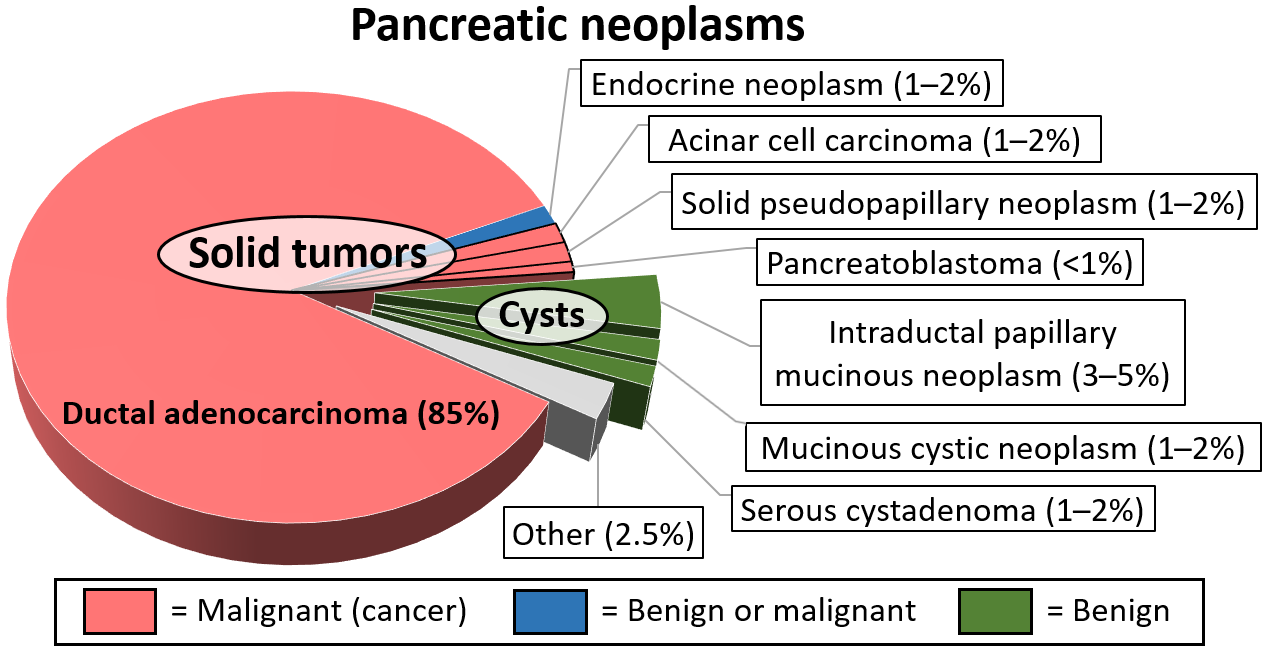
Pancreatic serous cystadenoma is a benign tumour of the pancreas. It is usually solitary and found in the body or tail of the pancreas, and may be associated with von Hippel–Lindau syndrome. In contrast to some of the other cyst-forming tumors of the pancreas (such as the intraductal papillary mucinous neoplasm and the pancreatic mucinous cystadenoma), serous cystic neoplasms are almost always entirely benign. There are some exceptions; rare case reports have described isolated malignant serous cystadenocarcinomas. In addition, serous cystic neoplasms slowly grow, and if they grow large enough they can press on adjacent organs and cause symptoms. Signs and symptoms In most cases, serous cystadenomas of the pancreas are asymptomatic. However, large cysts may cause symptoms related to their size. Classification Pathologists classify serous cystic neoplasms into two broad groups. Those that are benign, that have not spread to other organs, are designated "serous cystadenoma". ... [...More Info...] [...Related Items...] OR: [Wikipedia] [Google] [Baidu] |
H&E Stain
Hematoxylin and eosin stain ( or haematoxylin and eosin stain or hematoxylin–eosin stain; often abbreviated as H&E stain or HE stain) is one of the principal tissue stains used in histology. It is the most widely used stain in medical diagnosis and is often the ''gold standard.'' For example, when a pathologist looks at a biopsy of a suspected cancer, the histological section is likely to be stained with H&E. H&E is the combination of two histological stains: hematoxylin and eosin. The hematoxylin stains cell nuclei a purplish blue, and eosin stains the extracellular matrix and cytoplasm pink, with other structures taking on different shades, hues, and combinations of these colors. Hence a pathologist can easily differentiate between the nuclear and cytoplasmic parts of a cell, and additionally, the overall patterns of coloration from the stain show the general layout and distribution of cells and provides a general overview of a tissue sample's structure. Thus, patte ... [...More Info...] [...Related Items...] OR: [Wikipedia] [Google] [Baidu] |
Adenoma
An adenoma is a benign tumor of epithelium, epithelial tissue with glandular origin, glandular characteristics, or both. Adenomas can grow from many glandular organ (anatomy), organs, including the adrenal glands, pituitary gland, thyroid, prostate, and others. Some adenomas grow from epithelial tissue in nonglandular areas but express glandular tissue structure (as can happen in familial polyposis coli). Although adenomas are benign, they should be treated as pre-cancerous. Over time adenomas may malignant transformation, transform to become malignancy, malignant, at which point they are called adenocarcinomas. Most adenomas do not transform. However, even though benign, they have the potential to cause serious health complications by compressing other structures (mass effect (medicine), mass effect) and by producing large amounts of hormones in an unregulated, non-feedback-dependent manner (causing paraneoplastic syndromes). Some adenomas are too small to be seen macroscopically ... [...More Info...] [...Related Items...] OR: [Wikipedia] [Google] [Baidu] |
Malignant
Malignancy () is the tendency of a medical condition to become progressively worse; the term is most familiar as a characterization of cancer. A ''malignant'' tumor contrasts with a non-cancerous benign tumor, ''benign'' tumor in that a malignancy is not self-limited in its growth, is capable of invading into adjacent tissues, and may be capable of spreading to distant tissues. A benign tumor has none of those properties, but may still be harmful to health. The term benign in more general medical use characterizes a condition or growth that is not cancerous, i.e. does not spread to other parts of the body or invade nearby tissue. Sometimes the term is used to suggest that a condition is not dangerous or serious. Malignancy in cancers is characterized by anaplasia, invasiveness, and metastasis. Malignant tumors are also characterized by genome instability, so that cancers, as assessed by whole genome sequencing, frequently have between 10,000 and 100,000 mutations in their ent ... [...More Info...] [...Related Items...] OR: [Wikipedia] [Google] [Baidu] |
Serous
In physiology, serous fluid or serosal fluid (originating from the Medieval Latin word ''serosus'', from Latin ''serum'') is any of various body fluids resembling serum, that are typically pale yellow or transparent and of a benign nature. The fluid fills the inside of body cavities. Serous fluid originates from serous glands, with secretions enriched with proteins and water. Serous fluid may also originate from mixed glands, which contain both mucous and serous cells. A common trait of serous fluids is their role in assisting digestion, excretion, and respiration. In medical fields, especially cytopathology, serous fluid is a synonym for effusion fluids from various body cavities. Examples of effusion fluid are pleural effusion and pericardial effusion. There are many causes of effusions which include involvement of the cavity by cancer. Cancer in a serous cavity is called a serous carcinoma. Cytopathology evaluation is recommended to evaluate the causes of effusions in thes ... [...More Info...] [...Related Items...] OR: [Wikipedia] [Google] [Baidu] |
Papillary
Papilla (Latin, 'nipple') or papillae may refer to: In animals * Papilla (fish anatomy), in the mouth of fish * Papilla (worms), small bumps on the surface of certain worms * Basilar papilla, a sensory organ of lizards, amphibians and fish * Dental papilla, in a developing tooth * Dermal papillae, part of the skin structure * Major duodenal papilla, in the duodenum * Minor duodenal papilla, in the duodenum * Genital papilla, a feature of the external genitalia of some animals * Mammary papilla or nipple, a raised region of tissue on the surface of the breast * Interdental papilla, part of the gums * Lacrimal papilla, on the bottom eyelid * Lingual papillae, small structures on the upper surface of the tongue * Renal papilla, part of the kidney * Tumor papilla, consisting of tumor cells surrounding a fibrovascular core In plants and fungi * Papilla (mycology), a nipple-shaped protrusion in the center of the cap * Stigmatic papilla, part of the stigma (botany) See also * ... [...More Info...] [...Related Items...] OR: [Wikipedia] [Google] [Baidu] |
Mucinous
Mucus (, ) is a slippery aqueous secretion produced by, and covering, mucous membranes. It is typically produced from cells found in mucous glands, although it may also originate from mixed glands, which contain both serous and mucous cells. It is a viscous colloid containing inorganic salts, antimicrobial enzymes (such as lysozymes), immunoglobulins (especially IgA), and glycoproteins such as lactoferrin and mucins, which are produced by goblet cells in the mucous membranes and submucosal glands. Mucus covers the epithelial cells that interact with outside environment, serves to protect the linings of the respiratory, digestive, and urogenital systems, and structures in the visual and auditory systems from pathogenic fungi, bacteria and viruses. Most of the mucus in the body is produced in the gastrointestinal tract. Amphibians, fish, snails, slugs, and some other invertebrates also produce external mucus from their epidermis as protection against pathogens, to help in move ... [...More Info...] [...Related Items...] OR: [Wikipedia] [Google] [Baidu] |
Bile Duct
A bile duct is any of a number of long tube-like structures that carry bile, and is present in most vertebrates. The bile duct is separated into three main parts: the fundus (superior), the body (middle), and the neck (inferior). Bile is required for the digestion of food and is secreted by the liver into passages that carry bile toward the hepatic duct. It joins the cystic duct (carrying bile to and from the gallbladder) to form the common bile duct which then opens into the intestine. Structure The top half of the common bile duct is associated with the liver, while the bottom half of the common bile duct is associated with the pancreas, through which it passes on its way to the intestine. It opens into the part of the intestine called the duodenum via the ampulla of Vater. Segments The biliary tree (see below) is the whole network of various sized ducts branching through the liver. The path is as follows: bile canaliculi → canals of Hering → interlobular bil ... [...More Info...] [...Related Items...] OR: [Wikipedia] [Google] [Baidu] |
Vermiform Appendix
The appendix (: appendices or appendixes; also vermiform appendix; cecal (or caecal, cæcal) appendix; vermix; or vermiform process) is a finger-like, blind-ended tube connected to the cecum, from which it develops in the embryo. The cecum is a pouch-like structure of the large intestine, located at the junction of the small and the large intestines. The term " vermiform" comes from Latin and means "worm-shaped". The appendix was once considered a vestigial organ, but this view has changed since the early 2000s. Research suggests that the appendix may serve as a reservoir for beneficial gut bacteria. Structure The human appendix averages in length, ranging from . The diameter of the appendix is , and more than is considered a thickened or inflamed appendix. The longest appendix ever removed was long. The appendix is usually located in the lower right quadrant of the abdomen, near the right hip bone. The base of the appendix is located beneath the ileocecal valve tha ... [...More Info...] [...Related Items...] OR: [Wikipedia] [Google] [Baidu] |


