retroperitoneal on:
[Wikipedia]
[Google]
[Amazon]
The retroperitoneal space (retroperitoneum) is the anatomical space (sometimes a potential space) behind (''retro'') the

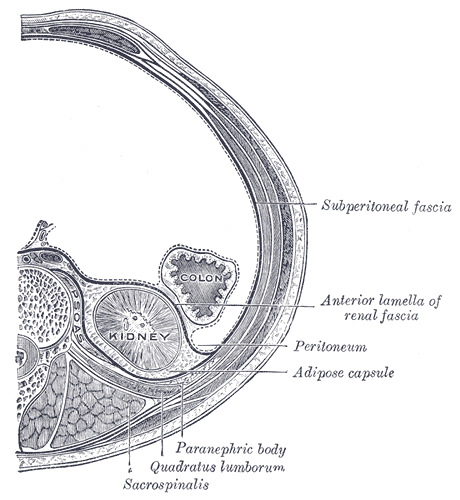
 ;Perirenal space
It is also called the perinephric space. Bounded by the anterior and posterior leaves of the renal fascia. It contains the following structures:
*
peritoneum
The peritoneum is the serous membrane forming the lining of the abdominal cavity or coelom in amniotes and some invertebrates, such as annelids. It covers most of the intra-abdominal (or coelomic) organs, and is composed of a layer of mesotheli ...
. It has no specific delineating anatomical structures. Organs are retroperitoneal if they have peritoneum on their anterior side only. Structures that are not suspended by mesentery
In human anatomy, the mesentery is an Organ (anatomy), organ that attaches the intestines to the posterior abdominal wall, consisting of a double fold of the peritoneum. It helps (among other functions) in storing Adipose tissue, fat and allowi ...
in the abdominal cavity and that lie between the parietal peritoneum and abdominal wall are classified as retroperitoneal.
This is different from organs that are not retroperitoneal, which have peritoneum on their posterior side and are suspended by mesentery in the abdominal cavity.
The retroperitoneum can be further subdivided into the following:
*Perirenal (or perinephric) space
*Anterior pararenal (or paranephric) space
*Posterior pararenal (or paranephric) space
Retroperitoneal structures
Structures that lie behind theperitoneum
The peritoneum is the serous membrane forming the lining of the abdominal cavity or coelom in amniotes and some invertebrates, such as annelids. It covers most of the intra-abdominal (or coelomic) organs, and is composed of a layer of mesotheli ...
are termed "retroperitoneal". Organs that were once suspended within the abdominal cavity by mesentery
In human anatomy, the mesentery is an Organ (anatomy), organ that attaches the intestines to the posterior abdominal wall, consisting of a double fold of the peritoneum. It helps (among other functions) in storing Adipose tissue, fat and allowi ...
but migrated posterior to the peritoneum during the course of embryogenesis
An embryo ( ) is the initial stage of development for a multicellular organism. In organisms that reproduce sexually, embryonic development is the part of the life cycle that begins just after fertilization of the female egg cell by the male ...
to become retroperitoneal are considered to be secondarily retroperitoneal organs.
* Primarily retroperitoneal, meaning the structures were retroperitoneal during the entirety of development:
** urinary
*** adrenal gland
The adrenal glands (also known as suprarenal glands) are endocrine glands that produce a variety of hormones including adrenaline and the steroids aldosterone and cortisol. They are found above the kidneys. Each gland has an outer adrenal corte ...
s
*** kidney
In humans, the kidneys are two reddish-brown bean-shaped blood-filtering organ (anatomy), organs that are a multilobar, multipapillary form of mammalian kidneys, usually without signs of external lobulation. They are located on the left and rig ...
s
*** ureter
The ureters are tubes composed of smooth muscle that transport urine from the kidneys to the urinary bladder. In an adult human, the ureters typically measure 20 to 30 centimeters in length and about 3 to 4 millimeters in diameter. They are lin ...
** circulatory
*** aorta
The aorta ( ; : aortas or aortae) is the main and largest artery in the human body, originating from the Ventricle (heart), left ventricle of the heart, branching upwards immediately after, and extending down to the abdomen, where it splits at ...
*** inferior vena cava
The inferior vena cava is a large vein that carries the deoxygenated blood from the lower and middle body into the right atrium of the heart. It is formed by the joining of the right and the left common iliac veins, usually at the level of the ...
** digestive
***anal canal
The anal canal is the part that connects the rectum to the anus, located below the level of the pelvic diaphragm. It is located within the anal triangle of the perineum, between the right and left ischioanal fossa. As the final functional s ...
* Secondarily retroperitoneal, meaning the structures initially were suspended in mesentery
In human anatomy, the mesentery is an Organ (anatomy), organ that attaches the intestines to the posterior abdominal wall, consisting of a double fold of the peritoneum. It helps (among other functions) in storing Adipose tissue, fat and allowi ...
and later migrated behind the peritoneum during development
** the duodenum
The duodenum is the first section of the small intestine in most vertebrates, including mammals, reptiles, and birds. In mammals, it may be the principal site for iron absorption.
The duodenum precedes the jejunum and ileum and is the shortest p ...
, except for the proximal first segment, which is intraperitoneal
** ascending and descending portions of the colon (but not the transverse colon, sigmoid and the cecum)
** pancreas, except for the tail, which is intraperitoneal
Subdivisions

 ;Perirenal space
It is also called the perinephric space. Bounded by the anterior and posterior leaves of the renal fascia. It contains the following structures:
*
 Adrenal gland
The adrenal glands (also known as suprarenal glands) are endocrine glands that produce a variety of hormones including adrenaline and the steroids aldosterone and cortisol. They are found above the kidneys. Each gland has an outer adrenal corte ...
* Kidney
In humans, the kidneys are two reddish-brown bean-shaped blood-filtering organ (anatomy), organs that are a multilobar, multipapillary form of mammalian kidneys, usually without signs of external lobulation. They are located on the left and rig ...
* Renal vessels
* Perirenal fat (also "perirenal fat capsule", "perinephric fat, or "adipose capsule of the kidney") is external to the fibrous capsule of the kidney, and internal to the renal fascia (which separates it from the pararenal fat); connective tissue trabeculae extend through it to unite the fibrous capsule of the kidney, and the renal fascia. Perirenal fat is most abundant upon the posterior aspect, inferior pole and along the lateral margins of the kidney.
;Anterior pararenal space
Bounded by the posterior layer of peritoneum
The peritoneum is the serous membrane forming the lining of the abdominal cavity or coelom in amniotes and some invertebrates, such as annelids. It covers most of the intra-abdominal (or coelomic) organs, and is composed of a layer of mesotheli ...
and the anterior leaf of the renal fascia. It contains the following structures:
* Pancreas
The pancreas (plural pancreases, or pancreata) is an Organ (anatomy), organ of the Digestion, digestive system and endocrine system of vertebrates. In humans, it is located in the abdominal cavity, abdomen behind the stomach and functions as a ...
* Ascending and descending colon
* Duodenum
The duodenum is the first section of the small intestine in most vertebrates, including mammals, reptiles, and birds. In mammals, it may be the principal site for iron absorption.
The duodenum precedes the jejunum and ileum and is the shortest p ...
;Posterior pararenal space
Bounded by the posterior leaf of the renal fascia and the muscles of the posterior abdominal wall. It contains only fat ("pararenal fat" also known as "pararenal fat body", "paranephric body", or "paranephric fat").
Pararenal fat is a fatty layer situated posterior to the renal compartment, and extending inferiorly into the iliac fossa
The iliac fossa is a large, smooth, concave surface on the internal surface of the Ilium (bone), ilium (part of the three fused bones making the hip bone).
Structure
The iliac fossa is bounded above by the iliac crest, and below by the Arcuate ...
. It is situated posterior to the posterior aspect of renal fascia, and anterior to the aponeuroses of the retrorenal muscles. It is plentiful in the dihedral angle of the iliopsoas muscle and the quadratus lumborum muscle, filling the lumbar fossa posterior and inferior to the kidney.
Clinical significance
Bleeding from a blood vessel or structure in the retroperitoneal area such as theaorta
The aorta ( ; : aortas or aortae) is the main and largest artery in the human body, originating from the Ventricle (heart), left ventricle of the heart, branching upwards immediately after, and extending down to the abdomen, where it splits at ...
or inferior vena cava
The inferior vena cava is a large vein that carries the deoxygenated blood from the lower and middle body into the right atrium of the heart. It is formed by the joining of the right and the left common iliac veins, usually at the level of the ...
into the retroperitoneal space can lead to a retroperitoneal hemorrhage.
* Retroperitoneal fibrosis
* Retroperitoneal lymph node dissection
The portion of the retroperitoneum that is posterior to the wall of the abdomen and superior to the iliac vessels is of importance in gynecologic oncology
''Gynecologic Oncology'' is a Peer review, peer-reviewed medical journal covering all aspects of gynecologic oncology. The journal covers investigations relating to the etiology, diagnosis, and treatment of female cancers, as well as research from ...
. This is the region where para-aortic and paracaval lymphadenectomies take place. The lateral boundary of the retroperitoneum is defined by the ascending and descending colon.
It is also possible to have a neoplasm
A neoplasm () is a type of abnormal and excessive growth of tissue. The process that occurs to form or produce a neoplasm is called neoplasia. The growth of a neoplasm is uncoordinated with that of the normal surrounding tissue, and persists ...
in this area, more commonly a metastasis
Metastasis is a pathogenic agent's spreading from an initial or primary site to a different or secondary site within the host's body; the term is typically used when referring to metastasis by a cancerous tumor. The newly pathological sites, ...
; or very rarely a primary neoplasm. The most common type is a sarcoma
A sarcoma is a rare type of cancer that arises from cells of mesenchymal origin. Originating from mesenchymal cells means that sarcomas are cancers of connective tissues such as bone, cartilage, muscle, fat, or vascular tissues.
Sarcom ...
followed by lymphoma, extragonadal germ cell tumor, and gastrointestinal stromal tumor/GIST. Examples of tumors include:
* Primary peritoneal carcinoma
* Pseudomyxoma peritonei
Examples of sarcomas include:
* Soft-tissue sarcoma
** liposarcoma
** leiomyosarcoma
** Undifferentiated pleomorphic sarcoma, a clinically distinct sarcoma of the area
See also
* Intraperitoneal * Retropubic space * Rectovesical pouch * Vesicouterine pouch * Rectouterine pouch ( Pouch of Douglas)References
{{Authority control Abdomen