|
Renal Fascia
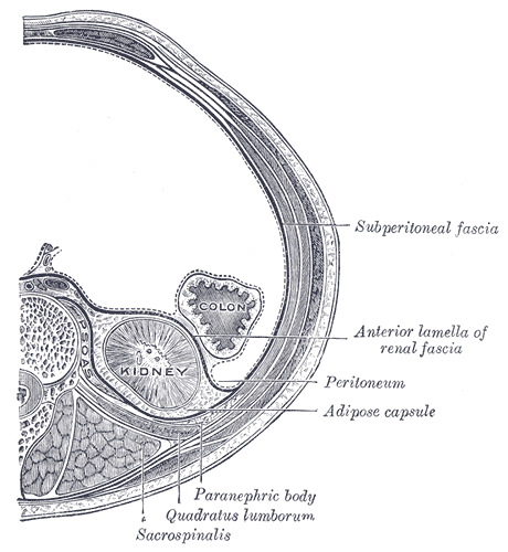
The renal fascia is a dense, elastic connective tissue envelope enclosing the kidney and adrenal gland, together with the layer of perirenal fat surrounding these two. The renal fascia separates the adipose capsule of kidney from the overlying pararenal fat. The deeper layers deep to the renal fascia are, in order, the adipose capsule (or perirenal fat), the renal capsule and finally the parenchyma of the renal cortex. At the renal hilum, the renal capsule extends into the renal sinus The renal sinus is a cavity within the kidney which is occupied by the renal pelvis, renal calyces, blood vessels, nerves and fat. The renal hilum extends into a large cavity within the kidney occupied by the renal vessels, minor renal calyces, .... The renal fascia was originally described as consisting of two distinct structures: the anterior renal fascia (Gerota's fascia), and posterior renal fascia (Zuckerkandl's fascia); these two fasciae were said to fuse laterally to form the lateroco ... [...More Info...] [...Related Items...] OR: [Wikipedia] [Google] [Baidu] |
Transverse Plane
A transverse plane is a plane that is rotated 90° from two other planes. Anatomy The transverse plane is an anatomical plane that is perpendicular to the sagittal plane and the dorsal plane. It is also called the axial plane or horizontal plane, especially in human anatomy, but horizontal plane can be misleading with other animals. The plane splits the body into a cranial (head) side and caudal (tail) side, so in humans the plane will be horizontal (dividing the body into superior and inferior sections) but in quadrupeds it will be vertical. Human anatomy Clinically relevant anatomical planes * Transverse '' thoracic plane'' * '' Xiphosternal plane'' (or xiphosternal junction) * '' Transpyloric plane'' * '' Subcostal plane'' * '' Umbilical plane'' (or transumbilical plane) * '' Supracristal plane'' * '' Intertubercular plane'' (or transtubercular plane) * '' Interspinous plane'' Associated structures * The transverse '' thoracic plane'' ** Plane through T4 & T5 verteb ... [...More Info...] [...Related Items...] OR: [Wikipedia] [Google] [Baidu] |
Urinary System
The human urinary system, also known as the urinary tract or renal system, consists of the kidneys, ureters, urinary bladder, bladder, and the urethra. The purpose of the urinary system is to eliminate waste from the body, regulate blood volume and blood pressure, control levels of Electrolyte, electrolytes and Metabolite, metabolites, and regulate Acid–base homeostasis, blood pH. The urinary tract is the body's drainage system for the eventual removal of urine. The kidneys have an extensive blood supply via the Renal artery, renal arteries which leave the kidneys via the renal vein. Each kidney consists of functional units called nephrons. Following filtration of blood and further processing, waste (in the form of urine) exits the kidney via the ureters, tubes made of smooth muscle fibres that propel urine towards the urinary bladder, where it is stored and subsequently expelled through the urethra during urination. The female and male urinary system are very similar, differin ... [...More Info...] [...Related Items...] OR: [Wikipedia] [Google] [Baidu] |
Elastic Connective Tissue
Elastic fibers (or yellow fibers) are an essential component of the extracellular matrix composed of bundles of proteins (elastin) which are produced by a number of different cell types including fibroblasts, endothelial, smooth muscle, and airway epithelial cells. These fibers are able to stretch many times their length, and snap back to their original length when relaxed without loss of energy. Elastic fibers include elastin, elaunin and oxytalan. Elastic fibers are formed via elastogenesis, a highly complex process involving several key proteins including fibulin-4, fibulin-5, latent transforming growth factor β binding protein 4, and microfibril associated protein 4. In this process tropoelastin, the soluble monomeric precursor to elastic fibers is produced by elastogenic cells and chaperoned to the cell surface. Following excretion from the cell, tropoelastin self associates into ~200 nm particles by coacervation, an entropically driven process involving interacti ... [...More Info...] [...Related Items...] OR: [Wikipedia] [Google] [Baidu] |
Connective Tissue
Connective tissue is one of the four primary types of animal tissue, a group of cells that are similar in structure, along with epithelial tissue, muscle tissue, and nervous tissue. It develops mostly from the mesenchyme, derived from the mesoderm, the middle embryonic germ layer. Connective tissue is found in between other tissues everywhere in the body, including the nervous system. The three meninges, membranes that envelop the brain and spinal cord, are composed of connective tissue. Most types of connective tissue consists of three main components: elastic and collagen fibers, ground substance, and cells. Blood and lymph are classed as specialized fluid connective tissues that do not contain fiber. All are immersed in the body water. The cells of connective tissue include fibroblasts, adipocytes, macrophages, mast cells and leukocytes. The term "connective tissue" (in German, ) was introduced in 1830 by Johannes Peter Müller. The tissue was already recognized as ... [...More Info...] [...Related Items...] OR: [Wikipedia] [Google] [Baidu] |
Kidney
In humans, the kidneys are two reddish-brown bean-shaped blood-filtering organ (anatomy), organs that are a multilobar, multipapillary form of mammalian kidneys, usually without signs of external lobulation. They are located on the left and right in the retroperitoneal space, and in adult humans are about in length. They receive blood from the paired renal artery, renal arteries; blood exits into the paired renal veins. Each kidney is attached to a ureter, a tube that carries excreted urine to the urinary bladder, bladder. The kidney participates in the control of the volume of various body fluids, fluid osmolality, Acid-base homeostasis, acid-base balance, various electrolyte concentrations, and removal of toxins. Filtration occurs in the glomerulus (kidney), glomerulus: one-fifth of the blood volume that enters the kidneys is filtered. Examples of substances reabsorbed are solute-free water, sodium, bicarbonate, glucose, and amino acids. Examples of substances secreted are hy ... [...More Info...] [...Related Items...] OR: [Wikipedia] [Google] [Baidu] |
Adrenal Gland
The adrenal glands (also known as suprarenal glands) are endocrine glands that produce a variety of hormones including adrenaline and the steroids aldosterone and cortisol. They are found above the kidneys. Each gland has an outer adrenal cortex, cortex which produces steroid hormones and an inner Adrenal medulla, medulla. The adrenal cortex itself is divided into three main zones: the zona glomerulosa, the zona fasciculata and the zona reticularis. The adrenal cortex produces three main types of steroid hormones: mineralocorticoids, glucocorticoids, and androgens. Mineralocorticoids (such as aldosterone) produced in the zona glomerulosa help in the regulation of blood pressure and osmoregulation, electrolyte balance. The glucocorticoids cortisol and cortisone are synthesized in the zona fasciculata; their functions include the regulation of metabolism and immune system suppression. The innermost layer of the cortex, the zona reticularis, produces androgens that are converted to ... [...More Info...] [...Related Items...] OR: [Wikipedia] [Google] [Baidu] |
Perirenal Fat
The retroperitoneal space (retroperitoneum) is the anatomical space (sometimes a potential space) behind (''retro'') the peritoneum. It has no specific delineating anatomical structures. Organs are retroperitoneal if they have peritoneum on their anterior side only. Structures that are not suspended by mesentery in the abdominal cavity and that lie between the parietal peritoneum and abdominal wall are classified as retroperitoneal. This is different from organs that are not retroperitoneal, which have peritoneum on their posterior side and are suspended by mesentery in the abdominal cavity. The retroperitoneum can be further subdivided into the following: *Perirenal (or perinephric) space *Anterior pararenal (or paranephric) space *Posterior pararenal (or paranephric) space Retroperitoneal structures Structures that lie behind the peritoneum are termed "retroperitoneal". Organs that were once suspended within the abdominal cavity by mesentery but migrated posterior to the peri ... [...More Info...] [...Related Items...] OR: [Wikipedia] [Google] [Baidu] |
Adipose Capsule Of Kidney
The retroperitoneal space (retroperitoneum) is the anatomical space (sometimes a potential space) behind (''retro'') the peritoneum. It has no specific delineating anatomical structures. Organs are retroperitoneal if they have peritoneum on their anterior side only. Structures that are not suspended by mesentery in the abdominal cavity and that lie between the parietal peritoneum and abdominal wall are classified as retroperitoneal. This is different from organs that are not retroperitoneal, which have peritoneum on their posterior side and are suspended by mesentery in the abdominal cavity. The retroperitoneum can be further subdivided into the following: *Perirenal (or perinephric) space *Anterior pararenal (or paranephric) space *Posterior pararenal (or paranephric) space Retroperitoneal structures Structures that lie behind the peritoneum are termed "retroperitoneal". Organs that were once suspended within the abdominal cavity by mesentery but migrated posterior to the per ... [...More Info...] [...Related Items...] OR: [Wikipedia] [Google] [Baidu] |
Pararenal Fat
The retroperitoneal space (retroperitoneum) is the anatomical space (sometimes a potential space) behind (''retro'') the peritoneum. It has no specific delineating anatomical structures. Organs are retroperitoneal if they have peritoneum on their anterior side only. Structures that are not suspended by mesentery in the abdominal cavity and that lie between the parietal peritoneum and abdominal wall are classified as retroperitoneal. This is different from organs that are not retroperitoneal, which have peritoneum on their posterior side and are suspended by mesentery in the abdominal cavity. The retroperitoneum can be further subdivided into the following: *Perirenal (or perinephric) space *Anterior pararenal (or paranephric) space *Posterior pararenal (or paranephric) space Retroperitoneal structures Structures that lie behind the peritoneum are termed "retroperitoneal". Organs that were once suspended within the abdominal cavity by mesentery but migrated posterior to the peri ... [...More Info...] [...Related Items...] OR: [Wikipedia] [Google] [Baidu] |
Renal Capsule
The renal capsule is a tough fibrous layer surrounding the kidney and covered in a layer of perirenal fat known as the adipose capsule of kidney. The adipose capsule is sometimes included in the structure of the renal capsule. It provides some protection from trauma and damage. The renal capsule is surrounded by the renal fascia. Overlying the renal fascia and between this and the transverse fascia is a region of pararenal fat. The renal capsule resists stretching, limiting renal swelling, with important implications for renal circulation. Stretching of the renal capsule due to swelling of the kidney causes flank pain. Structure The renal capsule surrounds the functional tissue of the kidney, and is itself surrounded by a fatty adipose capsule, fascia, and fat. From the inner part of the kidney to outside the kidney, the positioning of the capsule is: # renal medulla # renal cortex # renal capsule # adipose capsule of kidney (or perirenal fat, or perinephric fat) # rena ... [...More Info...] [...Related Items...] OR: [Wikipedia] [Google] [Baidu] |
Renal Parenchyma
upright=1.6, Lung parenchyma showing damage due to large subpleural bullae. Parenchyma () is the bulk of functional substance in an animal organ such as the brain or lungs, or a structure such as a tumour. In zoology, it is the tissue that fills the interior of flatworms. In botany, it is some layers in the cross-section of the leaf. Etymology The term ''parenchyma'' is Neo-Latin from the Ancient Greek word meaning 'visceral flesh', and from meaning 'to pour in' from 'beside' + 'in' + 'to pour'. Originally, Erasistratus and other anatomists used it for certain human tissues. Later, it was also applied to plant tissues by Nehemiah Grew. Structure The parenchyma is the ''functional'' parts of an organ, or of a structure such as a tumour in the body. This is in contrast to the stroma, which refers to the ''structural'' tissue of organs or of structures, namely, the connective tissues. Brain The brain parenchyma refers to the functional tissue in the brain ... [...More Info...] [...Related Items...] OR: [Wikipedia] [Google] [Baidu] |
Renal Cortex
The renal cortex is the outer portion of the kidney between the renal capsule and the renal medulla. In the adult, it forms a continuous smooth outer zone with a number of projections ( cortical columns) that extend down between the pyramids. It contains the renal corpuscles and the renal tubules except for parts of the loop of Henle which descend into the renal medulla. It also contains blood vessels and cortical collecting ducts. The renal cortex is the part of the kidney where ultrafiltration occurs. Erythropoietin is produced in the renal cortex. Additional images File:Njuren.gif, Kidney File:Kidney-Cortex.JPG, Microscopic cross section of the renal cortex File:Kidney_cd10_ihc.jpg, CD10 immunohistochemical staining of normal kidney In humans, the kidneys are two reddish-brown bean-shaped blood-filtering organ (anatomy), organs that are a multilobar, multipapillary form of mammalian kidneys, usually without signs of external lobulation. They are located on the le ... [...More Info...] [...Related Items...] OR: [Wikipedia] [Google] [Baidu] |


