Electron Cryo-tomography on:
[Wikipedia]
[Google]
[Amazon]
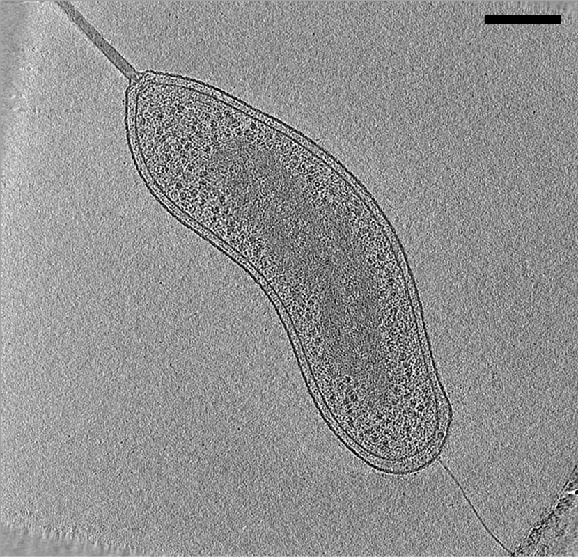
Cryogenic electron tomography (cryoET) is an imaging technique used to reconstruct high-resolution (~1–4 nm) three-dimensional volumes of samples, often (but not limited to) biological
 In
In
 Larger cells, and even tissues, can be prepared for cryoET by thinning, either by cryo-sectioning or by
 The term "missing wedge" originates from the view of the
Getting started in cryo-EM course (Caltech)
{{Electron microscopy Cell biology Electron microscopy techniques
macromolecule
A macromolecule is a "molecule of high relative molecular mass, the structure of which essentially comprises the multiple repetition of units derived, actually or conceptually, from molecules of low relative molecular mass." Polymers are physi ...
s and cells
Cell most often refers to:
* Cell (biology), the functional basic unit of life
* Cellphone, a phone connected to a cellular network
* Clandestine cell, a penetration-resistant form of a secret or outlawed organization
* Electrochemical cell, a d ...
. cryoET is a specialized application of transmission electron cryomicroscopy (CryoTEM) in which samples are imaged as they are tilted, resulting in a series of 2D images that can be combined to produce a 3D reconstruction, similar to a CT scan
A computed tomography scan (CT scan), formerly called computed axial tomography scan (CAT scan), is a medical imaging technique used to obtain detailed internal images of the body. The personnel that perform CT scans are called radiographers or ...
of the human body. In contrast to other electron tomography Electron tomography (ET) is a tomography technique for obtaining detailed 3D structures of sub-cellular, macro-molecular, or materials specimens. Electron tomography is an extension of traditional transmission electron microscopy and uses a trans ...
techniques, samples are imaged under cryogenic
In physics, cryogenics is the production and behaviour of materials at very low temperatures.
The 13th International Institute of Refrigeration's (IIR) International Congress of Refrigeration (held in Washington, DC in 1971) endorsed a univers ...
conditions (< −150 °C). For cellular material, the structure is immobilized in non-crystalline, vitreous ice, allowing them to be imaged without dehydration or chemical fixation
Histopathology (compound of three Greek words: 'tissue', 'suffering', and ''-logia'' 'study of') is the microscopic examination of tissue in order to study the manifestations of disease. Specifically, in clinical medicine, histopathology r ...
, which would otherwise disrupt or distort biological structures.
Description of technique
 In
In electron microscopy
An electron microscope is a microscope that uses a beam of electrons as a source of illumination. It uses electron optics that are analogous to the glass lenses of an optical light microscope to control the electron beam, for instance focusing i ...
(EM), samples are imaged in a high vacuum
A vacuum (: vacuums or vacua) is space devoid of matter. The word is derived from the Latin adjective (neuter ) meaning "vacant" or "void". An approximation to such vacuum is a region with a gaseous pressure much less than atmospheric pressur ...
. Such a vacuum is incompatible with biological samples such as cells; the water would boil off, and the difference in pressure would explode the cell. In room-temperature EM techniques, samples are therefore prepared by fixation and dehydration. Another approach to stabilize biological samples, however, is to freeze them (cryo-electron microscopy
Cryogenic electron microscopy (cryo-EM) is a transmission electron microscopy technique applied to samples cooled to cryogenic temperatures. For biological specimens, the structure is preserved by embedding in an environment of vitreous ice. An ...
or cryoEM). As in other electron cryomicroscopy techniques, samples for cryoET (typically small cells such as Bacteria
Bacteria (; : bacterium) are ubiquitous, mostly free-living organisms often consisting of one Cell (biology), biological cell. They constitute a large domain (biology), domain of Prokaryote, prokaryotic microorganisms. Typically a few micr ...
, Archaea
Archaea ( ) is a Domain (biology), domain of organisms. Traditionally, Archaea only included its Prokaryote, prokaryotic members, but this has since been found to be paraphyletic, as eukaryotes are known to have evolved from archaea. Even thou ...
, or virus
A virus is a submicroscopic infectious agent that replicates only inside the living Cell (biology), cells of an organism. Viruses infect all life forms, from animals and plants to microorganisms, including bacteria and archaea. Viruses are ...
es) are prepared in standard aqueous media and applied to an EM grid. The grid is then plunged into a cryogen, for example liquid ethane, with sufficiently large specific heat that the rate of cooling is rapid enough that water
Water is an inorganic compound with the chemical formula . It is a transparent, tasteless, odorless, and Color of water, nearly colorless chemical substance. It is the main constituent of Earth's hydrosphere and the fluids of all known liv ...
molecule
A molecule is a group of two or more atoms that are held together by Force, attractive forces known as chemical bonds; depending on context, the term may or may not include ions that satisfy this criterion. In quantum physics, organic chemi ...
s do not have time to rearrange into a crystalline
A crystal or crystalline solid is a solid material whose constituents (such as atoms, molecules, or ions) are arranged in a highly ordered microscopic structure, forming a crystal lattice that extends in all directions. In addition, macrosc ...
lattice. The resulting water state is called low-density amorphous ice, or commonly "vitreous ice" for its glass like nature. This form of ice preserves native cellular structures, such as lipid membranes, that would normally be disrupted by hexagonal or other ordered ice upon slower freezing. Plunge-frozen samples are subsequently kept at liquid-nitrogen temperatures through storage and imaging so that the water never warms enough to crystallize.
Samples are imaged in a transmission electron microscope
Transmission electron microscopy (TEM) is a microscopy technique in which a beam of electrons is transmitted through a specimen to form an image. The specimen is most often an ultrathin section less than 100 nm thick or a suspension on a gr ...
(TEM). As in other electron tomography Electron tomography (ET) is a tomography technique for obtaining detailed 3D structures of sub-cellular, macro-molecular, or materials specimens. Electron tomography is an extension of traditional transmission electron microscopy and uses a trans ...
techniques, the sample is tilted to different angles relative to the electron beam (typically every 2-3 degrees from about −60° to +60°), and an image is acquired at each angle. This tilt-series of images can then be computationally reconstructed into a three-dimensional view of the object of interest. This is called a tomogram, or tomographic reconstruction
Tomographic reconstruction is a type of multidimensional inverse problem where the challenge is to yield an estimate of a specific system from a finite number of projection (linear algebra), projections. The mathematical basis for tomographic imag ...
.
Potential for high-resolution ''in situ'' imaging
One of the most commonly cited benefits of cryoET is the ability to reconstruct 3D volumes of individual objects (proteins, cells, ''etc.'') rather than necessitating multiple copies of the sample incrystallographic
Crystallography is the branch of science devoted to the study of molecular and crystalline structure and properties. The word ''crystallography'' is derived from the Ancient Greek word (; "clear ice, rock-crystal"), and (; "to write"). In J ...
methods or in other cryoEM imaging methods like single particle analysis
Single particle analysis is a group of related computerized image processing techniques used to analyze images from transmission electron microscopy (TEM). These methods were developed to improve and extend the information obtainable from TEM ima ...
. CryoET is considered to be an ''in situ
is a Latin phrase meaning 'in place' or 'on site', derived from ' ('in') and ' ( ablative of ''situs'', ). The term typically refers to the examination or occurrence of a process within its original context, without relocation. The term is use ...
'' method when used on an unperturbed cell or other system since plunge-freezing of sufficiently thin samples fixes the specimen in place fast enough to cause minimal changes to atomic positioning. Thick samples, greater than ~500 nm, require additional conditions like high-pressure to promote vitrification throughout the sample.
Considerations
Sample thickness
Intransmission electron microscopy
Transmission electron microscopy (TEM) is a microscopy technique in which a beam of electrons is transmitted through a specimen to form an image. The specimen is most often an ultrathin section less than 100 nm thick or a suspension on a g ...
(TEM), because electron
The electron (, or in nuclear reactions) is a subatomic particle with a negative one elementary charge, elementary electric charge. It is a fundamental particle that comprises the ordinary matter that makes up the universe, along with up qua ...
s interact strongly with matter
In classical physics and general chemistry, matter is any substance that has mass and takes up space by having volume. All everyday objects that can be touched are ultimately composed of atoms, which are made up of interacting subatomic pa ...
, samples must be kept very thin to not cause samples to darken due to multiple elastic scattering
Elastic scattering is a form of particle scattering in scattering theory, nuclear physics and particle physics. In this process, the internal states of the Elementary particle, particles involved stay the same. In the non-relativistic case, where ...
events. Therefore, in cryoET, samples are generally less than ~500 nm thick. For this reason, most cryoET studies have focused on purified macromolecular complexes, viruses, or small cells such as those of many species of Bacteria and Archaea. For example, cryoET has been used to understand encapsulation of 12 nm size protein cage nanoparticle
A nanoparticle or ultrafine particle is a particle of matter 1 to 100 nanometres (nm) in diameter. The term is sometimes used for larger particles, up to 500 nm, or fibers and tubes that are less than 100 nm in only two directions. At ...
s inside 60 nm sized virus-like nanoparticles.
 Larger cells, and even tissues, can be prepared for cryoET by thinning, either by cryo-sectioning or by
 focused ion beam
Focused ion beam, also known as FIB, is a technique used particularly in the semiconductor industry, materials science and increasingly in the biological field for site-specific analysis, deposition, and ablation of materials. A FIB setup is a sc ...
(FIB) milling. In cryo-sectioning, frozen blocks of cells or tissue are sectioned into thin samples with a cryo-microtome
A microtome (from the Greek ''mikros'', meaning "small", and ''temnein'', meaning "to cut") is a cutting tool used to produce extremely thin slices of material known as ''sections'', with the process being termed microsectioning. Important in sc ...
. In FIB-milling, plunge-frozen samples are exposed to a focused beam of ions, typically gallium
Gallium is a chemical element; it has Chemical symbol, symbol Ga and atomic number 31. Discovered by the French chemist Paul-Émile Lecoq de Boisbaudran in 1875,
elemental gallium is a soft, silvery metal at standard temperature and pressure. ...
, that precisely whittle away material from the top and bottom of a sample, leaving a thin lamella suitable for cryoET imaging.
Signal-to-noise ratio
For structures that are present in multiple copies in one or multiple tomograms, higher resolution (even ≤1 nm) can be obtained by subtomogram averaging. Similar to single particle analysis, subtomogram averaging computationally combines images of identical objects to increase thesignal-to-noise ratio
Signal-to-noise ratio (SNR or S/N) is a measure used in science and engineering that compares the level of a desired signal to the level of background noise. SNR is defined as the ratio of signal power to noise power, often expressed in deci ...
.
Limitations
Radiation damage
Electron microscopy is known to swiftly decay biological samples compared to samples inmaterials science
Materials science is an interdisciplinary field of researching and discovering materials. Materials engineering is an engineering field of finding uses for materials in other fields and industries.
The intellectual origins of materials sci ...
and physics
Physics is the scientific study of matter, its Elementary particle, fundamental constituents, its motion and behavior through space and time, and the related entities of energy and force. "Physical science is that department of knowledge whi ...
due to radiation damage
Radiation damage is the effect of ionizing radiation on physical objects including non-living structural materials. It can be either detrimental or beneficial for materials.
Radiobiology is the study of the action of ionizing radiation on living ...
. In most other electron microscopy-based methods for imaging biological samples, combining the signal from many different sample copies has been the general way of surpassing this problem (''e.g.'' crystallography, single particle analysis). In cryoET, instead of taking many images of different sample copies, many images are taken of one area. Consequentially, the fluence
In radiometry, radiant exposure or fluence is the radiant energy ''received'' by a ''surface'' per unit area, or equivalently the irradiance of a ''surface,'' integrated over time of irradiation, and spectral exposure is the radiant exposure per u ...
(number of electrons imparted per unit area) on the sample is around 2-5x more than in single particle analysis. Tomography on materials much more resilient allows drastically higher resolution than typical biological imaging, suggesting that radiation damage is the greatest limitation to cryoET of biological samples.
Depth resolution
The strong interaction of electrons with matter also results in an anisotropic resolution effect. As the sample is tilted during imaging, the electron beam interacts with a thicker apparent sample along theoptical axis
An optical axis is an imaginary line that passes through the geometrical center of an optical system such as a camera lens, microscope or telescopic sight. Lens elements often have rotational symmetry about the axis.
The optical axis defines ...
of the microscope at higher tilt angles. In practice, tilt angles greater than approximately 60–70° do not yield much information and are therefore not used. This results in a "missing wedge" of information in the final tomogram that decreases resolution parallel to the electron beam.
 The term "missing wedge" originates from the view of the
 Fourier transform
In mathematics, the Fourier transform (FT) is an integral transform that takes a function as input then outputs another function that describes the extent to which various frequencies are present in the original function. The output of the tr ...
of the tomogram, where an empty wedge is apparent due to not tilting the sample to 90°. The missing wedge results in a lack of resolution in sample depth, as the missing information is mostly along the z-axis. The missing wedge is also a problem in 3D electron crystallography, where it is usually solved by merging multiple datasets that overlap each other or through symmetry
Symmetry () in everyday life refers to a sense of harmonious and beautiful proportion and balance. In mathematics, the term has a more precise definition and is usually used to refer to an object that is Invariant (mathematics), invariant und ...
expansion where possible. Both of these solutions are due to the nature of crystallography, and so neither can be applied to tomography.
Segmentation
A major obstacle in cryoET is identifying structures of interest within complicated cellular environments. Solutions such as correlated cryo-fluorescence light microscopy, and super-resolution light microscopy (e.g. cryo-PALM) can be integrated with cryoET. In these techniques, a sample containing a fluorescently-tagged protein of interest is plunge-frozen and first imaged in a light microscope equipped with a special stage to allow the sample to be kept at sub-crystallization temperatures (< −150 °C). The location of thefluorescent
Fluorescence is one of two kinds of photoluminescence, the emission of light by a substance that has absorbed light or other electromagnetic radiation. When exposed to ultraviolet radiation, many substances will glow (fluoresce) with color ...
signal is identified and the sample is transferred to the CryoTEM, where the same location is then imaged at high resolution by cryoET.
See also
*Electron microscopy
An electron microscope is a microscope that uses a beam of electrons as a source of illumination. It uses electron optics that are analogous to the glass lenses of an optical light microscope to control the electron beam, for instance focusing i ...
* Electron tomography Electron tomography (ET) is a tomography technique for obtaining detailed 3D structures of sub-cellular, macro-molecular, or materials specimens. Electron tomography is an extension of traditional transmission electron microscopy and uses a trans ...
* Transmission electron cryomicroscopy
* Transmission electron microscopy
Transmission electron microscopy (TEM) is a microscopy technique in which a beam of electrons is transmitted through a specimen to form an image. The specimen is most often an ultrathin section less than 100 nm thick or a suspension on a g ...
References
External links
Getting started in cryo-EM course (Caltech)
{{Electron microscopy Cell biology Electron microscopy techniques