congenital muscular dystrophy on:
[Wikipedia]
[Google]
[Amazon]
Congenital muscular dystrophies are
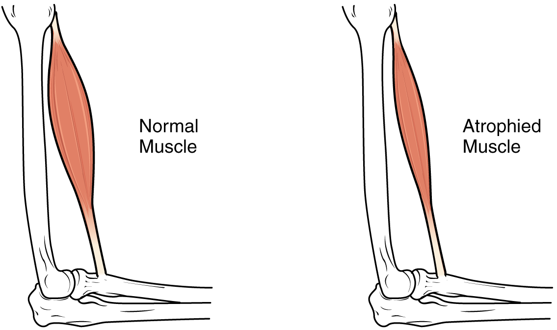
 Most infants with CMD will display some progressive muscle weakness or muscle wasting (
 For the diagnosis of congenital muscular dystrophy, the following tests/exams are done:
* Lab study ( CK levels)
* Muscle
 In terms of the management of congenital muscular dystrophy the American Academy of Neurology recommends that the individuals
need to have monitoring of cardiac function, respiratory, and
PubMed
{{Authority control Muscular dystrophy
autosomal
An autosome is any chromosome that is not a sex chromosome. The members of an autosome pair in a diploid cell have the same morphology, unlike those in allosomal (sex chromosome) pairs, which may have different structures. The DNA in autosome ...
recessively-inherited muscle diseases. They are a group of heterogeneous disorders characterized by muscle weakness which is present at birth and the different changes on muscle biopsy
In medicine, a muscle biopsy is a procedure in which a piece of muscle tissue is removed from an organism and examined microscopically. A muscle biopsy can lead to the discovery of problems with the nervous system, connective tissue, vascular s ...
that ranges from myopathic to overtly dystrophic due to the age at which the biopsy takes place.update 2012
Signs and symptoms
 Most infants with CMD will display some progressive muscle weakness or muscle wasting (
atrophy
Atrophy is the partial or complete wasting away of a part of the body. Causes of atrophy include mutations (which can destroy the gene to build up the organ), malnutrition, poor nourishment, poor circulatory system, circulation, loss of hormone, ...
), although there can be different degrees and symptoms of severeness of progression. The weakness is indicated as ''hypotonia
Hypotonia is a state of low muscle tone (the amount of tension or resistance to stretch in a muscle), often involving reduced muscle strength. Hypotonia is not a specific medical disorder, but it is a potential manifestation of many different dis ...
'', or lack of muscle tone, which can make an infant seem unstable. Eventually, most patients develop joint contractures or fixed joint deformities.
Children may be slow with their motor skills
A motor skill is a function that involves specific movements of the body's muscles to perform a certain task. These tasks could include walking, running, or riding a bike. In order to perform this skill, the body's nervous system, muscles, and b ...
; such as rolling over, sitting up or walking, or may not even reach these milestones of life. Some of the rarer forms of CMD can result in significant learning disabilities.
Genetics
Congenital muscular dystrophies (CMDs) are autosomal recessively inherited, except in some cases of de novo gene mutation and Ullrich congenital muscular dystrophy. This means that in most cases, both parents must be carriers of a CMD gene in order for it to be inherited. CMDs are heterogenous and thus far there have been 35 genes discovered to be involved with different forms of CMD resulting from these mutations.Adam, MP; Mirzaa, GM; Pagon, RA; Wallace, SE; Bean, LJH; Gripp, KW; Amemiya, A; Oliveira, J; Parente Freixo, J; Santos, M; Coelho, T (1993). "LAMA2 Muscular Dystrophy". PMID 22675738.Adam, MP; Mirzaa, GM; Pagon, RA; Wallace, SE; Bean, LJH; Gripp, KW; Amemiya, A; Foley, AR; Mohassel, P; Donkervoort, S; Bolduc, V; Bönnemann, CG (1993). "Collagen VI-Related Dystrophies". PMID 20301676. There are different forms of CMD, often categorized by the protein changes caused by an atypical gene. One group of forms is that for which a patient with affected genes displays defects in genes necessary to the function of theextracellular matrix
In biology, the extracellular matrix (ECM), also called intercellular matrix (ICM), is a network consisting of extracellular macromolecules and minerals, such as collagen, enzymes, glycoproteins and hydroxyapatite that provide structural and bio ...
. One such form is merosin-deficient congenital muscular dystrophy (MDC1A), which accounts for around one-third of all CMD cases and is caused by mutations in the LAMA2
Laminin subunit alpha-2 is a protein that in humans is encoded by the ''LAMA2'' gene.
Function
Laminin, an extracellular matrix protein, is a major component of the basement membrane
The basement membrane, also known as base membrane, is a t ...
gene on the 6q2 chromosome, encoding for the laminin-α2 chain. Laminin-α2 is an essential part of proteins like Laminin
Laminins are a family of glycoproteins of the extracellular matrix of all animals. They are major constituents of the basement membrane, namely the basal lamina (the protein network foundation for most cells and organs). Laminins are vital to bi ...
-2 and Laminin-4 that have important functions in muscle movement, and most patients with a mutated LAMA2 gene have no expression of Laminin-α2 in muscle tissue. Another form in this group is Ullrich congenital muscular dystrophy, which is caused by mutations in the COL6A1, COL6A2 and COL6A3 genes that encode for three of the alpha chains making up Collagen VI. Collagen VI is important in muscle, tendon, and skin tissue, and functions to attach cells to the extracellular matrix. Ullrich CMD can be caused by both autosomal recessive or autosomal dominant mutations, although dominant mutations are usually de novo. Recessive mutations often lead to a complete absence of Collagen VI in the extracellular matrix, while there are different types of dominant mutations that can cause partial function of Collagen V1.
Another form of CMD is rigid spine congenital muscular dystrophy (RSMD1), or rigid spine syndrome, which is caused by mutations in the SELENON gene encoding for selenoprotein In molecular biology a selenoprotein is any protein that includes a selenocysteine (Sec, U, Se-Cys) amino acid residue. Among functionally characterized selenoproteins are five glutathione peroxidases (GPX) and three thioredoxin reductases, (TrxR/TX ...
N. The exact function of selenoprotein N is unknown, but it is expressed in the rough endoplasmic reticulum
The endoplasmic reticulum (ER) is a part of a transportation system of the eukaryotic cell, and has many other important functions such as protein folding. The word endoplasmic means "within the cytoplasm", and reticulum is Latin for "little n ...
of skeletal muscle
Skeletal muscle (commonly referred to as muscle) is one of the three types of vertebrate muscle tissue, the others being cardiac muscle and smooth muscle. They are part of the somatic nervous system, voluntary muscular system and typically are a ...
, heart, brain, lung, and placenta tissues, as well as at high levels in the diaphragm. RSMD1 is characterized by axial and respiratory weakness, spinal rigidity and scoliosis
Scoliosis (: scolioses) is a condition in which a person's Vertebral column, spine has an irregular curve in the coronal plane. The curve is usually S- or C-shaped over three dimensions. In some, the degree of curve is stable, while in others ...
, and muscular atrophy
Muscle atrophy is the loss of skeletal muscle mass. It can be caused by sedentary lifestyle, immobility, aging, malnutrition, medications, or a wide range of injuries or diseases that impact the musculoskeletal or nervous system. Muscle atrophy le ...
, and while it is a rare form of CMD, SEPN1 mutations are observed in other congenital myopathies.
Some of the most common forms of CMDs are dystroglycanopathies caused by glycosylation
Glycosylation is the reaction in which a carbohydrate (or ' glycan'), i.e. a glycosyl donor, is attached to a hydroxyl or other functional group of another molecule (a glycosyl acceptor) in order to form a glycoconjugate. In biology (but not ...
defects of α-dystroglycan
Dystroglycan is a protein that in humans is encoded by the ''DAG1'' gene.
Dystroglycan is one of the dystrophin-associated glycoproteins, which is encoded by a 5.5 kb transcript in ''Homo sapiens'' on chromosome 3. There are two exons that are ...
(α-DG), which helps link the extracellular matrix and the cytoskeleton
The cytoskeleton is a complex, dynamic network of interlinking protein filaments present in the cytoplasm of all cells, including those of bacteria and archaea. In eukaryotes, it extends from the cell nucleus to the cell membrane and is compos ...
. Dystroglycanopathies are caused by mutations in genes encoding for proteins involved in modifying α-DG after translation of the protein, not mutations in the protein itself. 19 genes have been discovered that cause α-DG-related dystrophies, with a wide range of phenotypic effects observed, characterized by brain malformations along with muscular dystrophy. Walker-Warburg syndrome (WWS) is the most severe dystroglycanopathy phenotype, with the POMT1 gene as the first reported causative gene, although there have been 11 additional genes implicated in WWS. These genes include POMT2, FKRP, FKTN, ISPD, CTDC2, TMEM5, POMGnT1, B3GALnT2, GMPPB, B3GnT1, and SGK196, many of which have been identified as involved in other dystroglycanopathies. Patients display muscle weakness and cerebellar and ocular malformations, with a life expectancy of less than 1 year.
An additional dystroglycanopathy phenotype is Fukuyama congenital muscular dystrophy (FCMD) caused by a mutation in the Fukutin (FKTN) gene, which is the second most common type of muscular dystrophy in Japan after Duchenne muscular dystrophy
Duchenne muscular dystrophy (DMD) is a severe type of muscular dystrophy predominantly affecting boys. The onset of muscle weakness typically begins around age four, with rapid progression. Initially, muscle loss occurs in the thighs and pe ...
. The founder mutation of FCMD is a 3- kilo base pair retrotransposon
Retrotransposons (also called Class I transposable elements) are mobile elements which move in the host genome by converting their transcribed RNA into DNA through reverse transcription. Thus, they differ from Class II transposable elements, or ...
insertion in the noncoding region of FKTN, leading to muscle weakness, abnormal eye function, seizures, and intellectual disability. While the exact function of FKTN is unknown, FKTN mRNA is expressed in fetuses in the developing CNS, muscles, and eyes, and is likely necessary for normal development since complete inactivation leads to embryonic death at 7 days. Another phenotype, Muscle-eye-brain disease (MEB) is the dystroglycanopathy most prevalent in Finland, and is caused by mutations in the POMGnT1, FKRP, FKTN, ISPD, and TMEM5 genes. The POMGnT1 gene is expressed in the same tissues as FKTN, and MEB appears to have a similar severity as FCMD. However, symptoms unique to MEB include glaucoma
Glaucoma is a group of eye diseases that can lead to damage of the optic nerve. The optic nerve transmits visual information from the eye to the brain. Glaucoma may cause vision loss if left untreated. It has been called the "silent thief of ...
, atrophy of the optic nerves, and retinal generation. The least severe phenotype of dystroglycanopathies is CMD type 1c (MDC1C), caused by mutations in the FKRP and the LARGE
Large means of great size.
Large may also refer to:
Mathematics
* Arbitrarily large, a phrase in mathematics
* Large cardinal, a property of certain transfinite numbers
* Large category, a category with a proper class of objects and morphisms (o ...
gene, with a phenotype similar to MEB and WWS. MDC1C also includes Limb-Girdle muscular dystrophy.
Mechanism
In terms of the mechanism of congenital muscular dystrophy, one finds that though there are many types of CMD theglycosylation
Glycosylation is the reaction in which a carbohydrate (or ' glycan'), i.e. a glycosyl donor, is attached to a hydroxyl or other functional group of another molecule (a glycosyl acceptor) in order to form a glycoconjugate. In biology (but not ...
of α-dystroglycan and alterations in those genes that are involved are an important part of this conditions pathophysiology
Diagnosis
Musculoskeletal examination of congenital muscular dystrophies
Muscle fibrosis and Joint contractures or fixed deformities are cardinal clinical signs of congenital muscular dystrophies. Muscle fibrosis and shortening eventually lead to joint contractures or fixed deformities. They are important to the diagnosis of CMD. However, some patients initially present with joint laxity. Joint deformities can occur in the extremities and spine. Severe deformities can result in joint dislocation and walking difficulties or gait abnormalities. However, the specific pattern of muscle involvement in each of the CMD subtypes is not fully elucidated. A recent review identified CMD subtype-specific clinical patterns of muscle and Joint involvement which could be of help to the differential diagnosis of CMD subtypes. This was especially true for Merosin-deficient congenital muscular dystrophy (MDC1A) or LAMA2-related CMD subtype. Nonetheless, these muscle and Joint patterns of involvement have to be correlated with other clinical signs, neuro-imaging reports, muscle biopsy immune-staining and molecular or genetic analysis results, whenever available. This comprehensive approach is critical for the correct and timely diagnosis of CMDs.Cardiac examination of congenital muscular dystrophies
The cardiac manifestations of CMD vary greatly. They can range from non-existent or mild to severe and fatal cardiac involvement. Generally, cardiac abnormalities in CMD can manifest in dilated cardiomyopathy, systolic dysfunction, hypertrophic cardiomyopathy, myocardial fibrosis or fatal ventricular arrhythmias. Dystroglycanopathies as Fukuyama Congenital Muscular Dystrophy have a relatively high likelihood for development of significant cardiac manifestations. In Merosin-deficient congenital muscular dystrophy (MDC1A) or LAMA2-related CMD cardiac manifestations are usually asymptomatic. Cardiac manifestations have also been associated with Limb-girdle muscular dystrophy 2I and LMNA-related CMD. Cardiac manifestations may be secondary to severe thoracic spine deformity as in rigid spine syndrome. Cardiac examination and screening are important to patients with CMDs. Surveillance is important to those with a diagnosed cardiac involvement.MRI
Magnetic resonance imaging (MRI) is a medical imaging technique used in radiology to generate pictures of the anatomy and the physiological processes inside the body. MRI scanners use strong magnetic fields, magnetic field gradients, and rad ...
and especially whole body muscle MRI has recently been used to describe muscle abnormalities in patients with primary laminin-α2 (merosin) deficiency subtype of CMD.
* EMG
* Genetic testing
Genetic testing, also known as DNA testing, is used to identify changes in DNA sequence or chromosome structure. Genetic testing can also include measuring the results of genetic changes, such as RNA analysis as an output of gene expression, or ...
(different types of congenital muscular dystrophies)
The subtypes of congenital muscular dystrophy have been established through variations in multiple genes.Phenotype
In genetics, the phenotype () is the set of observable characteristics or traits of an organism. The term covers the organism's morphology (physical form and structure), its developmental processes, its biochemical and physiological propert ...
, as well as, genotype
The genotype of an organism is its complete set of genetic material. Genotype can also be used to refer to the alleles or variants an individual carries in a particular gene or genetic location. The number of alleles an individual can have in a ...
classifications are used to establish the subtypes, in some literature.
One finds that congenital muscular dystrophies can be either autosomal dominant
In genetics, dominance is the phenomenon of one variant (allele) of a gene on a chromosome masking or overriding the Phenotype, effect of a different variant of the same gene on Homologous chromosome, the other copy of the chromosome. The firs ...
or autosomal recessive
In genetics, dominance is the phenomenon of one variant (allele) of a gene on a chromosome masking or overriding the Phenotype, effect of a different variant of the same gene on Homologous chromosome, the other copy of the chromosome. The firs ...
in terms of the inheritance pattern, though the latter is much more common
Individuals with congenital muscular dystrophy fall into one of the following ''types'':
Differential diagnosis
The DDx of congenital muscular dystrophy, in an affected individual, is as follows (non-neuromuscular genetic conditions also exist): * Metabolic myopathies * Dystrophinopathies * Emery-Dreifuss muscular dystrophyManagement
 In terms of the management of congenital muscular dystrophy the American Academy of Neurology recommends that the individuals
need to have monitoring of cardiac function, respiratory, and
 gastrointestinal
The gastrointestinal tract (GI tract, digestive tract, alimentary canal) is the tract or passageway of the digestive system that leads from the mouth to the anus. The tract is the largest of the body's systems, after the cardiovascular system. ...
. Additionally it is believed that therapy in speech, orthopedic
Orthopedic surgery or orthopedics ( alternative spelling orthopaedics) is the branch of surgery concerned with conditions involving the musculoskeletal system. Orthopedic surgeons use both surgical and nonsurgical means to treat musculoskeletal ...
and physical areas, would improve the person's quality of life.
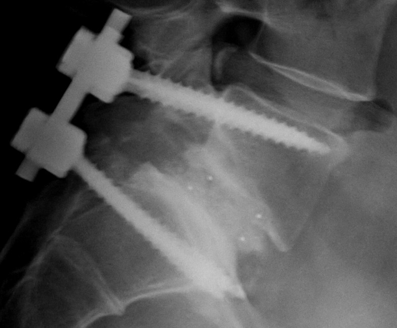
While there is currently no cure available, it is important to preserve muscle activity and any available correction of skeletal abnormalities (as scoliosis). Orthopedic procedures, like spinal fusion
Spinal fusion, also called spondylodesis or spondylosyndesis, is a surgery performed by Orthopedic surgery#Practice, orthopaedic surgeons or neurosurgeons that joins two or more vertebrae. This procedure can be performed at any level in the spine ...
, maintain/increase the individual's prospect for more physical movement.
See also
*Muscular dystrophies
Muscular dystrophies (MD) are a genetically and clinically heterogeneous group of rare neuromuscular diseases that cause progressive weakness and breakdown of skeletal muscles over time. The disorders differ as to which muscles are primarily aff ...
* Ullrich Congenital Muscular Dystrophy
* Fukuyama Congenital Muscular Dystrophy
References
#Further reading
* * * *External links
PubMed
{{Authority control Muscular dystrophy